
Bone tumour
What aspect of bone tumours are you most interested in? · What level is this for?
Which specific bone tumour(s) do you want to cover?
giant cell tumour bone X-ray radiograph
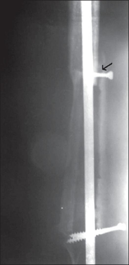
This diagnostic X-ray radiograph demonstrates a post-surgical resection arthrodesis of a long bone, typically the femur or tibia, following treatment for a giant cell tumor. The image shows a long, radio-opaque intramedullary (IM) nail spanning the medullary canal to provide structural stability. Two transverse interlocking screws are visible, securing the nail to the cortical bone. A distinct black arrow points to a localized radiolucent gap between the bone segments, signifying a nonunion of the bone graft at the 1.5-year postoperative follow-up. The surrounding bone shows irregular contours and variable density, indicating unsuccessful bony bridging and attempted remodeling at the graft-host interface. This visual serves as a clinical example of a complication in limb-salvage surgery and the failure of secondary arthrodesis using IM fixation.
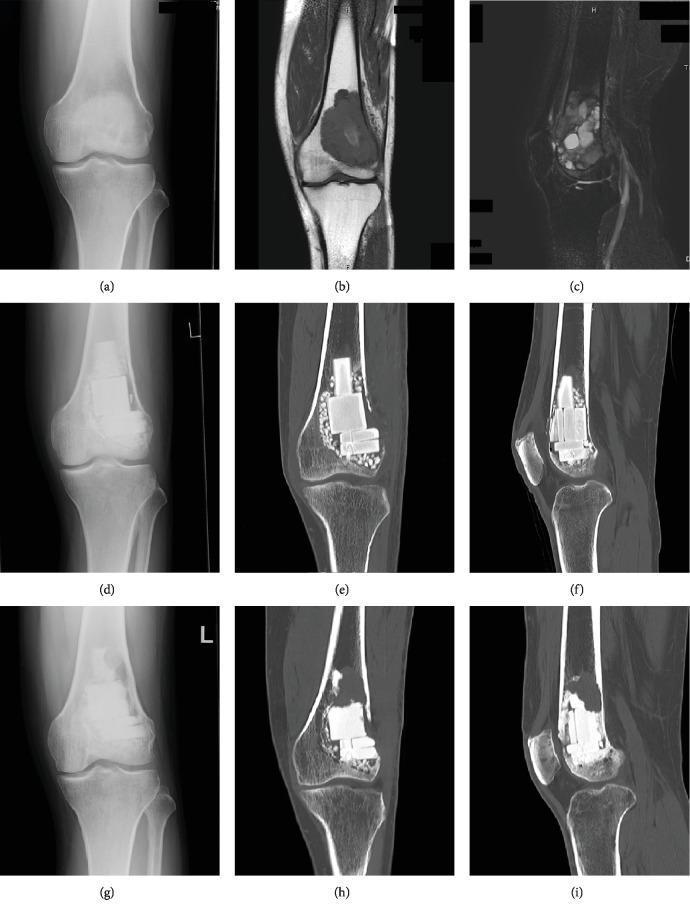
Multi-modal diagnostic imaging sequence (X-ray, MRI, CT) illustrating a Giant Cell Tumor of Bone (GCTB) in the distal femur and its postoperative progression. Top row: (a) Plain radiograph showing an ill-defined osteolytic lesion in the distal femoral metaphysis; (b) T1-weighted MRI showing a homogenous low-signal intensity mass; (c) T2-weighted fat-suppressed MRI showing heterogeneous intermediate-to-high signal intensity. Middle row: (d) Postoperative radiograph and (e, f) coronal/sagittal CT scans at 1 month showing the curetted cavity filled with radiopaque beta-tricalcium phosphate (β-TCP) blocks and surrounding granular particles. Bottom row: (g) Radiograph and (h, i) CT scans at 13 months post-operation demonstrating local recurrence characterized by new osteolysis predominantly within the β-TCP blocks and adjacent thinning of the cortical bone. This sequence serves as a clinical case study for evaluating bone substitute integration versus tumor recurrence using longitudinal multi-modal imaging.
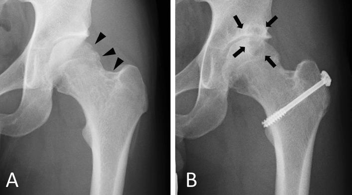
This comparative x-ray study displays two anterior-posterior radiographs of a human left hip joint, illustrating the progression of Tenosynovial Giant Cell Tumor (TSGCT) effects and subsequent post-surgical changes. Radiograph A (initial examination) shows clear osteolytic lesions at the femoral head-neck junction, indicated by black arrowheads; these appear as radiolucent, irregular areas of bone destruction, though the joint space and articular surfaces remain relatively preserved. Radiograph B shows the same hip three years after tumor resection. This follow-up image demonstrates secondary osteoarthritic changes, highlighted by black arrows, including joint space narrowing, subchondral sclerosis, and marginal osteophyte formation (bone spurs) at the superior acetabular and femoral margins. A prominent surgical screw is visible crossing the femoral neck into the head, indicating orthopedic stabilization following the tumor excision. This comparison highlights the clinical challenge of secondary joint degeneration following the management of intra-articular neoplastic processes.
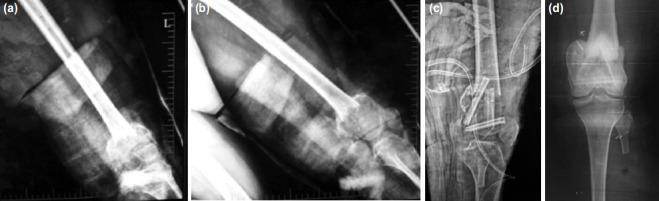
A series of four diagnostic x-ray radiographs documenting the management of a giant cell tumor (GCT) of the distal femur. (a, b) Pre-operative anteroposterior and lateral views showing a large, osteolytic lesion in the distal femoral metaphysis with associated cortical thinning and a pathological fracture, resulting in significant joint misalignment. (c) Immediate post-operative radiograph demonstrating surgical reconstruction following intralesional curettage. The reconstruction utilizes internal fixation with multiple orthopedic screws and cerclage wires alongside bone grafting (fibula struts) to stabilize the fracture site and restore anatomical alignment. (d) Two-year post-operative radiograph showing advanced bone healing and remodeling. There is significant graft consolidation and restoration of the distal femoral shape and bone density. The joint space is maintained, and hardware remains in situ with no evidence of tumor recurrence or subchondral collapse. This sequence illustrates a successful orthopedic oncology intervention for aggressive benign bone tumors complicated by fracture.
"giant cell tumor" bone
Giant Cell Tumour (GCT) of Bone
At a Glance
| Feature | Details |
|---|---|
| Nature | Locally aggressive benign tumour (can rarely metastasize) |
| Age | 20-40 years (80% of cases between 18-45 years) |
| Sex | Slight female predominance (M:F = 2:3) |
| Incidence | ~5% of all bone neoplasms; ~20% of benign bone tumours |
| Common sites | Distal femur > proximal tibia > distal radius (knee = 55% of cases) |
| Location within bone | Epiphysis (subarticular/subchondral), eccentric |
Pathogenesis (Robbins)
- Promotes proliferation of osteoclast precursors
- Drives their differentiation into mature osteoclasts (the actual giant cells)
- Disrupts the normal osteoblast-osteoclast feedback loop
- Results in localized, highly destructive bone resorption
This explains why denosumab (a RANKL inhibitor) works - it targets the driver of osteoclast activity.
Clinical Features
- Progressive pain, initially activity-related, later at rest
- Swelling and tenderness near the joint
- Arthritis-like symptoms due to proximity to joint
- Pathologic fracture in 10-30% of patients at presentation
- Rarely: intraarticular extension
Radiology / Imaging
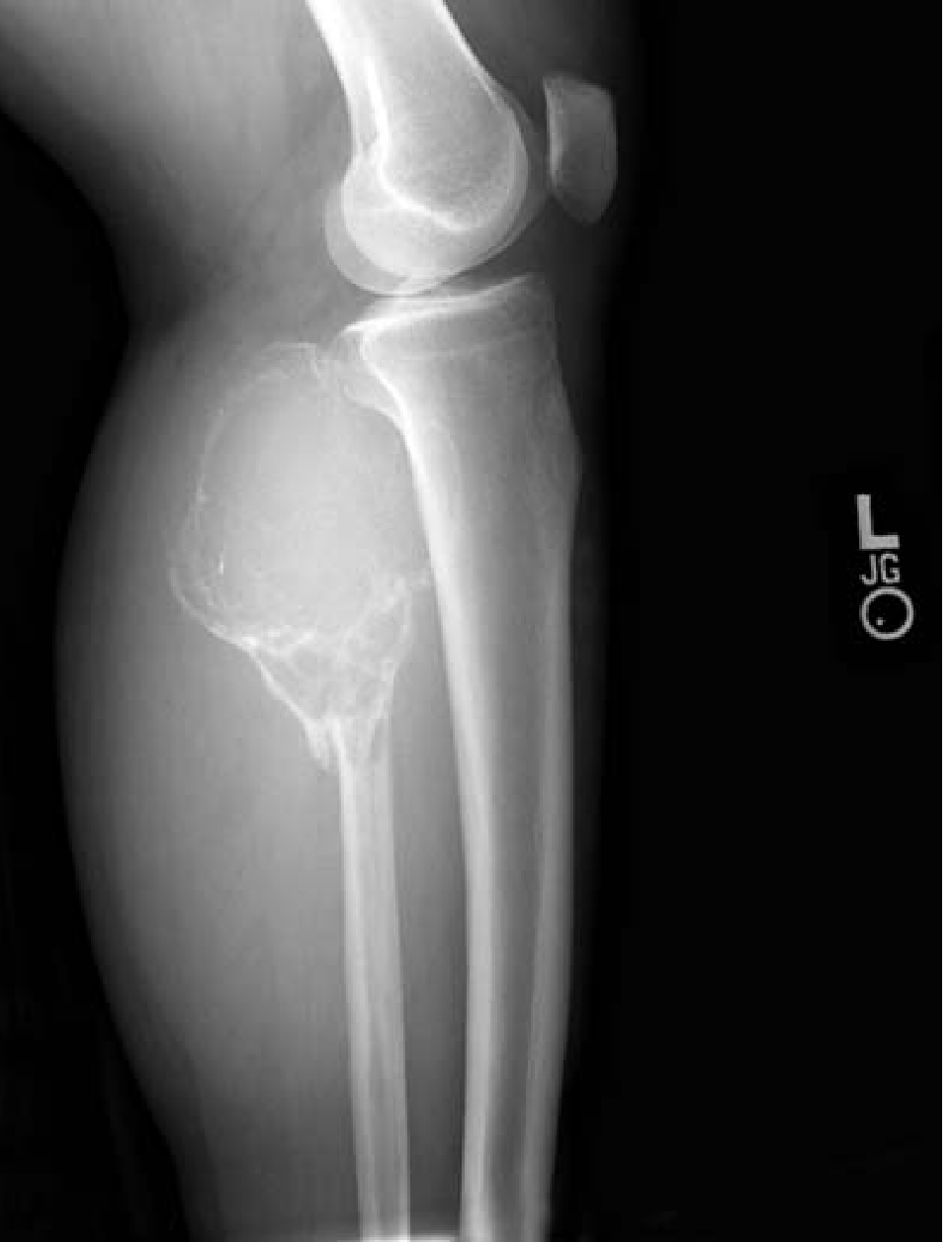
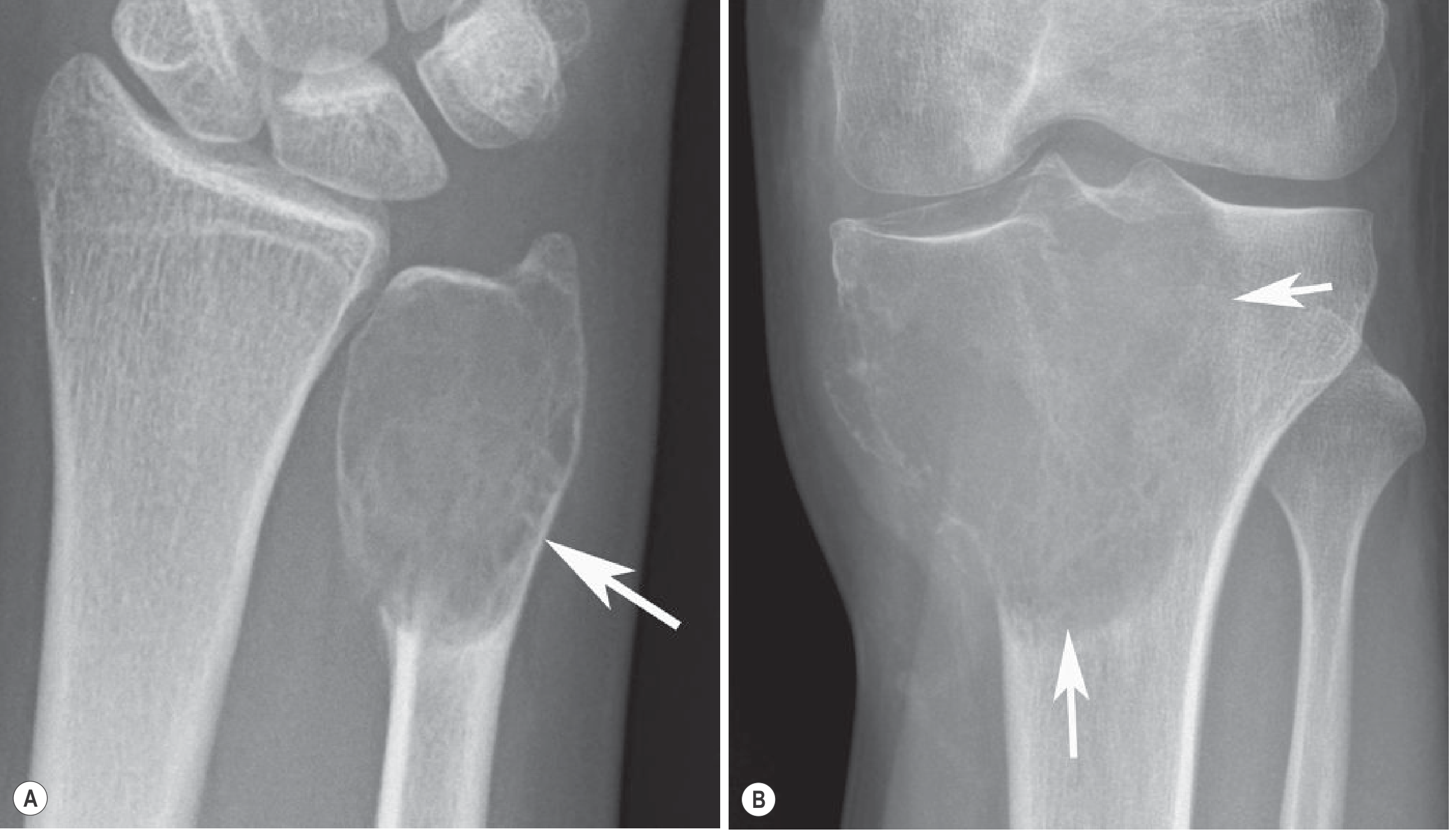
- Purely lytic lesion - no matrix production
- Eccentric, epiphyseal location, abutting subchondral bone
- Geographic destruction with non-sclerotic margin (no rim of reactive bone in most)
- May show cortical expansion, thinning, or destruction
- Soft-tissue extension in up to 50% of cases
- Periosteal reaction in 10-15% (usually indicating a healing pathologic fracture)
- No periosteal new bone or matrix calcification
- T1: Iso- or hypointense (dark)
- T2: Heterogeneous hyperintensity (bright); areas of profound hypointensity reflect haemosiderin deposits
- T1 hyperintensity = subacute haemorrhage
- Fluid-fluid levels on MRI in ~20% of patients - indicates secondary aneurysmal bone cyst (ABC)
- Best modality for soft-tissue and intra-osseous extent
Gross Morphology
- Red-brown mass (due to haemosiderin)
- Destroys overlying cortex
- Produces a bulging soft-tissue mass bounded by a thin shell of reactive bone
- Frequently undergoes cystic degeneration
Histology (Key Exam Feature)
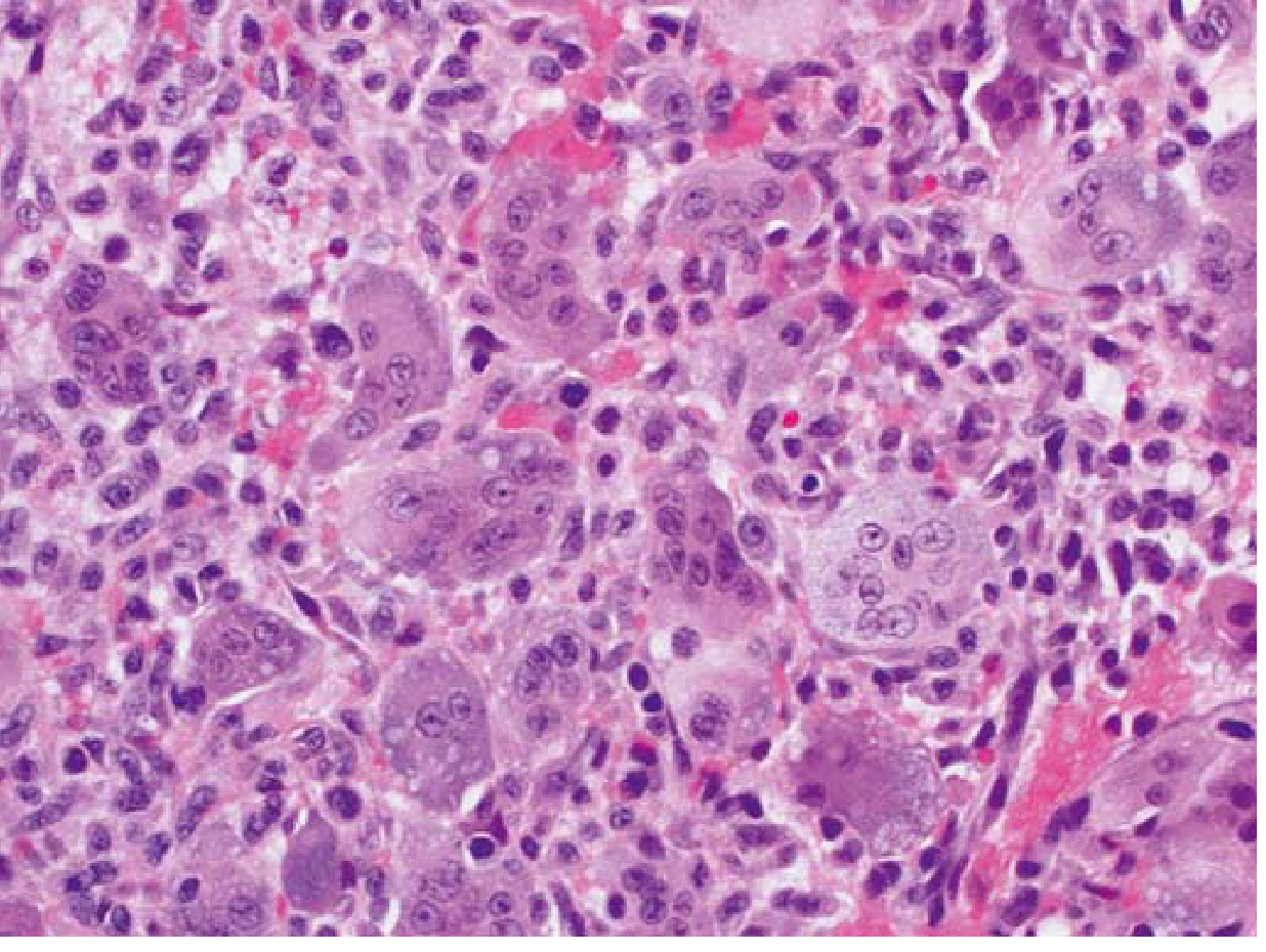
- Multinucleated giant cells with 40-100+ nuclei per cell
- Background of uniform oval mononuclear stromal cells (the actual neoplastic component)
- Critical distinguishing feature: the nuclei of the mononuclear cells are identical to the nuclei within the giant cells - this separates GCT from other giant-cell-containing lesions
- May also see: storiform spindle cells, reactive bone formation, foamy macrophages, secondary ABC areas
- No reliable histologic grading system has shown prognostic significance
Staging (Campanacci / Enneking)
| Stage | Description |
|---|---|
| Stage 1 (Latent) | Inactive, well-defined margin with intact cortex |
| Stage 2 (Active) | Expanded, thin cortex but intact |
| Stage 3 (Aggressive) | Cortical destruction, soft-tissue extension |
Treatment
- Extended curettage with high-speed burr
- Adjuvants used: phenol, liquid nitrogen (cryotherapy), argon beam, hydrogen peroxide
- Cavity filled with bone cement (PMMA) or bone graft
- 40-60% recur locally after curettage alone
- Adjuvants reduce recurrence rates
- Indicated when resection would be deforming or lead to loss of function
- Also used for unresectable/metastatic GCT
- Mechanism: inhibits RANKL, preventing osteoclast differentiation
- Response: causes tumour ossification and hardening, allowing subsequent curettage
Metastasis & Malignant Transformation
- Benign pulmonary metastases: ~3-4% of patients; often behave like the primary tumour; most cured by excision. Mortality ~15% in those with pulmonary spread.
- Malignant GCT (MGCTB): <10% of cases
- Primary MGCTB: sarcomatous cells at initial presentation alongside conventional GCT
- Secondary MGCTB: arises in a previously treated GCT, often years later, frequently after prior radiotherapy
- Most common histologic subtype of malignant transformation: osteosarcoma
- Cannot be distinguished radiologically from aggressive conventional GCT - requires biopsy
Associations / Special Points
- Multifocal GCT (<1% of cases): investigate for hyperparathyroidism (brown tumours of hyperparathyroidism can mimic GCT)
- Paget's disease: GCT rarely complicates familial polyostotic Paget's disease
- GCT of distal radius behaves more aggressively; GCT of hand bones is particularly aggressive with higher recurrence and metastasis risk
- GCT in skeletally immature patients is rare and arises in the metaphysis (not epiphysis)
High-Yield Exam Summary
| Point | Fact |
|---|---|
| Neoplastic cell | Mononuclear stromal cell (osteoblast precursor) |
| Key molecule | RANKL (drives osteoclast formation) |
| Drug target | Denosumab (RANKL inhibitor) |
| Most common site | Distal femur > proximal tibia |
| Location in bone | Epiphysis (eccentric, subarticular) |
| X-ray | Purely lytic, eccentric, no sclerotic rim, no matrix |
| Histology key | Nuclei of mononuclear cells = nuclei of giant cells |
| Local recurrence after curettage | 40-60% |
| Lung metastases | ~3-4% (benign behaviour) |
| Malignant transformation | <10%; osteosarcoma most common |
| Benign but locally aggressive | Yes - can still spread to lungs |