
Septic shock and its pathogenic pathways
septic shock pathogenesis inflammatory cascade cytokines
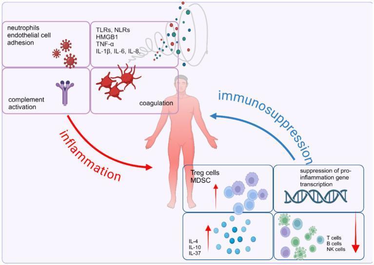
This pathophysiology diagram illustrates the dysregulated host immune response in sepsis, characterized by an imbalance between concurrent pro-inflammatory and anti-inflammatory pathways. Centered on a human figure, the left side (red 'inflammation' arrow) details the acute systemic inflammatory response syndrome (SIRS), featuring neutrophil-endothelial cell adhesion, complement activation, and coagulation cascades. A molecular cloud identifies key mediators including DAMPs/PAMPs (HMGB1, TLRs, NLRs) and pro-inflammatory cytokines (TNF-α, IL-1β, IL-6, IL-8). Conversely, the right side (blue 'immunosuppression' arrow) represents the compensatory anti-inflammatory response syndrome (CARS). This includes the expansion of suppressor cells (Treg cells, MDSCs) and anti-inflammatory cytokines (IL-4, IL-10, IL-37), the suppression of pro-inflammatory gene transcription, and the depletion or exhaustion of effector cells (T cells, B cells, and NK cells). The diagram serves as an educational tool for understanding the complex signaling pathways and cellular interactions that drive multi-organ dysfunction and immune homeostasis failure during septic shock.
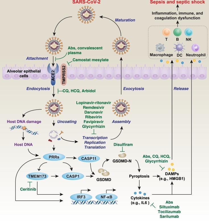
This pathophysiology diagram illustrates the SARS-CoV-2 life cycle and the subsequent host immune response within alveolar epithelial cells. The viral entry phase depicts the attachment of the spike protein to the ACE2 receptor, facilitated by TMPRSS2, followed by endocytosis and uncoating. The replication phase shows viral RNA transcription, translation, and assembly leading to viral maturation and exocytosis. The diagram highlights several pharmacological targets, including Camostat mesylate for protease inhibition, hydroxychloroquine (HCQ) for endocytosis, and Remdesivir or Lopinavir-ritonavir for transcription/translation interference. A secondary pathway describes the inflammatory response: viral components and host DNA damage trigger pattern-recognition receptors (PRRs), activating the TMEM173 and Caspase-1/11 (CASP1/11) pathways. This leads to Gasdermin D (GSDMD)-dependent pyroptosis, releasing damage-associated molecular patterns (DAMPs) and cytokines like IL-6. These mediators activate immune cells—including T cells, B cells, NK cells, macrophages, and neutrophils—potentially resulting in systemic inflammation, coagulation dysfunction, and septic shock. Key transcription factors IRF3 and NF-κB are noted as essential regulators of this hyperinflammatory cascade.
A multi-modal educational graphic combining clinical photographs and a pathophysiology diagram illustrating the pathogenesis of psoriasis. In the top right corner, two clinical photographs demonstrate erythematous, scaly plaques characteristic of psoriasis. The central diagram maps the immunological cascade across the epidermis and dermis. An 'Initiation phase' begins with triggers such as LL37 and self-nucleotides, which activate plasmacytoid dendritic cells (pDC) and macrophages in the dermis to release IFN-α and TNF-α. These cytokines stimulate conventional and inflammatory dendritic cells (CD1c+DC, CD141+DC, and iDC) to produce IL-23. In the 'Maintenance phase,' IL-23 promotes the differentiation and activity of pathogenic Th17 cells. These cells release IL-17A, which acts back upon the epidermis to drive the proliferation and abnormal differentiation of keratinocytes, creating a feed-forward inflammatory loop. The diagram utilizes distinct icons for cytokines and cell types to represent complex neuro-immunological interactions.
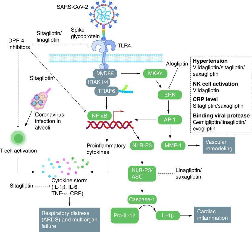
This pathophysiology diagram illustrates the inflammatory signaling pathways initiated by SARS-CoV-2 interaction with the Toll-like receptor 4 (TLR4) and the corresponding therapeutic targets for Dipeptidyl Peptidase-4 (DPP-4) inhibitors. The primary cascade involves the Spike glycoprotein binding to TLR4, activating the MyD88/IRAK1/4/TRAF6 signaling complex. This diverges into two major pathways: the NF-κB pathway, which drives the production of proinflammatory cytokines (IL-1β, IL-6, TNF-α, CRP) leading to a cytokine storm and respiratory distress (ARDS); and the MKKs/ERK/AP-1 pathway, which results in MMP-1 production and vascular remodeling. Additionally, proinflammatory cytokines activate the NLRP3/ASC inflammasome, leading to Caspase-1-mediated processing of Pro-IL-1β to IL-1β, which promotes cardiac inflammation. The diagram highlights specific pharmacological intervention points: Sitagliptin/Linagliptin inhibit TLR4 activation; Alogliptin inhibits ERK; Sitagliptin targets NF-κB and cytokine storm components; and Linagliptin/Saxagliptin inhibit the NLR-P3/ASC complex. A sidebar details associations between specific gliptins and clinical parameters like hypertension, NK cell activation, and viral protease binding.
Septic Shock and Its Pathogenic Pathways
Definition and Epidemiology
- Sepsis with persistent hypotension despite adequate fluid resuscitation, and
- A serum lactate >2 mmol/L, requiring vasopressors to maintain MAP ≥65 mmHg.
Overview of the Pathogenic Cascade
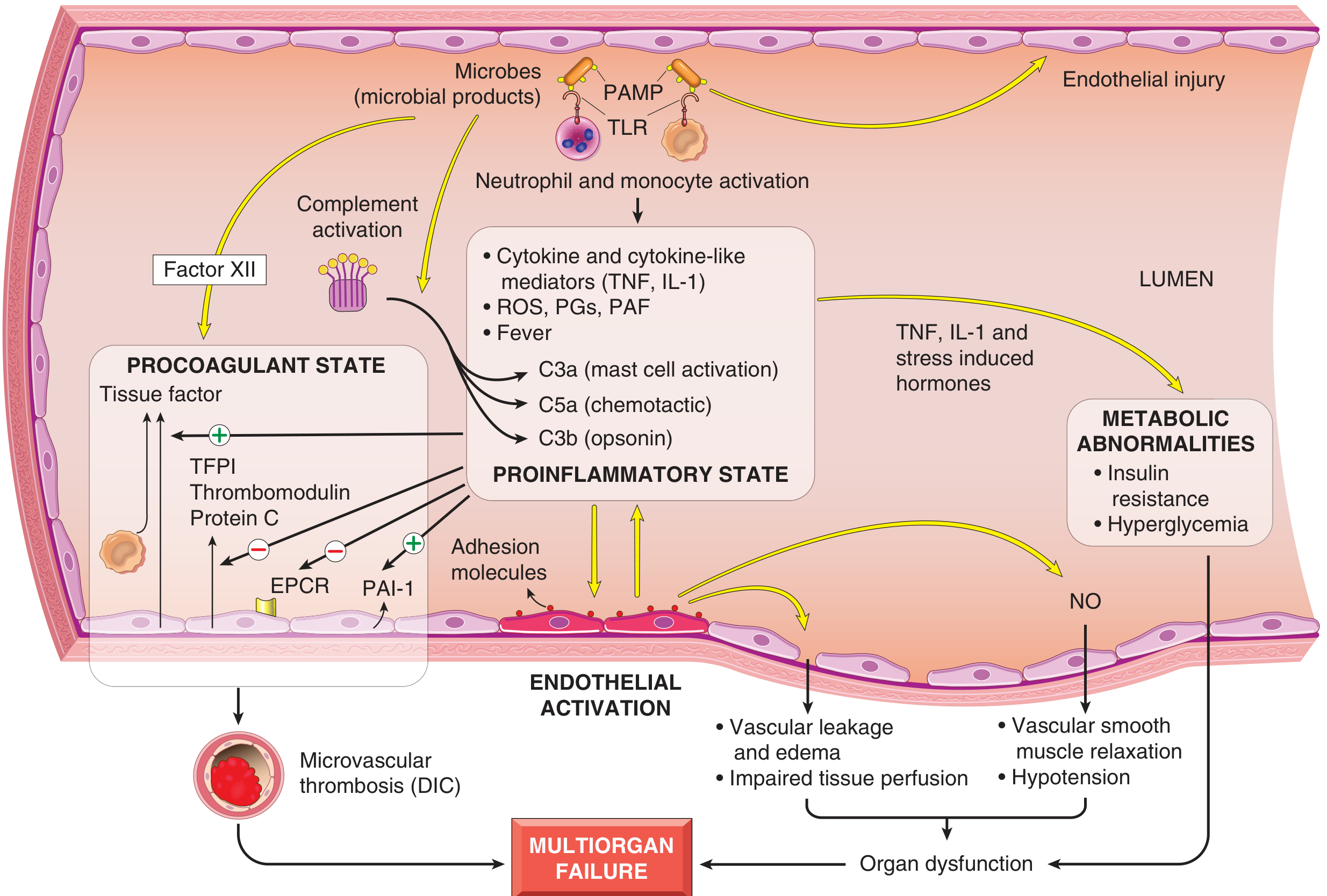
Pathway 1: Innate Immune Activation and the Cytokine Storm
Pattern Recognition
| PRR Type | Ligand | Organism |
|---|---|---|
| TLR-4 | Lipopolysaccharide (LPS/endotoxin) | Gram-negative bacteria |
| TLR-2 | Lipoteichoic acid (LTA) | Gram-positive bacteria |
| C-type lectin receptors (dectins) | Beta-glucans | Fungi |
| G-protein coupled receptors | Bacterial peptides | Various |
| NOD-like receptors (NLRs) | Intracellular PAMPs | Various |
Cytokine Release
- Early mediators: TNF-α, IL-1β, IL-6, IL-8, IL-12, IL-18
- Reactive oxygen species (ROS)
- Lipid mediators: prostaglandins, platelet-activating factor (PAF)
- Late mediators: HMGB-1 (high-mobility group box 1) - released 16-32 hours after initial stimulus; unlike TNF/IL-1, it remains elevated longer and its inhibitors reduce mortality even when given 24 hours after infection onset
Complement Activation
- C3a - mast cell degranulation, anaphylatoxin
- C5a - chemotaxis of neutrophils, powerful anaphylatoxin
- C3b - opsonization
Pathway 2: Endothelial Activation and Injury
- Upregulation of adhesion molecules (ICAM-1, E-selectin, P-selectin) → neutrophil-endothelial adhesion and transendothelial migration
- Loosening of tight junctions → widespread vascular leakage and protein-rich edema throughout the body
- Nitric oxide (NO) overproduction → potent vasodilation, vascular smooth muscle relaxation, and refractory hypotension
- Endothelial injury → increased intestinal permeability → bacterial translocation and endotoxin absorption, which further amplifies the inflammatory phenotype
Pathway 3: Procoagulant State and Disseminated Intravascular Coagulation (DIC)
| Pro-coagulant Factor | Mechanism |
|---|---|
| ↑ Tissue factor | Produced by monocytes and endothelium under cytokine stimulation |
| ↓ Thrombomodulin | Reduced expression → less protein C activation |
| ↓ EPCR (endothelial protein C receptor) | Less anticoagulant protein C pathway |
| ↓ TFPI (tissue factor pathway inhibitor) | Less suppression of the extrinsic coagulation cascade |
| ↑ PAI-1 | Plasminogen activator inhibitor 1 blocks fibrinolysis |
| Factor XII activation | Microbial components activate the contact/intrinsic pathway directly |
| Vascular stasis | Edema and low flow reduce washout of activated clotting factors |
Pathway 4: Counterregulatory Immunosuppression (CARS)
- Shift from Th1 → Th2 cytokine profile
- Production of anti-inflammatory mediators: soluble TNF receptor, IL-1 receptor antagonist, IL-10, IL-4, IL-37
- Lymphocyte apoptosis (T cells, B cells, NK cells)
- Expansion of regulatory T cells (Tregs) and myeloid-derived suppressor cells (MDSCs)
- Suppression of pro-inflammatory gene transcription
Pathway 5: Metabolic Abnormalities
- Insulin resistance and hyperglycemia via:
- Gluconeogenesis stimulation
- Suppressed insulin secretion
- Impaired GLUT-4 transporter expression in liver and peripheral tissues
- Adrenal insufficiency: after an initial cortisol surge, functional glucocorticoid deficiency may develop (especially with Waterhouse-Friderichsen syndrome - adrenal necrosis from DIC)
- Lactic acidosis: cellular hypoxia → impaired oxidative phosphorylation → anaerobic metabolism → lactate accumulation
Pathway 6: Organ Dysfunction and Multiorgan Failure
| Organ | Mechanism of Injury |
|---|---|
| Lung | Endothelial/epithelial injury → protein-rich edema → ARDS |
| Kidney | Hypoperfusion + microvascular thrombosis → AKI, oliguria |
| Heart | Cytokines (TNF-α, IL-1β) suppress myocardial contractility → septic cardiomyopathy |
| Liver | Hypoperfusion + direct cytokine effect → transaminase elevation, jaundice |
| Brain | Hypoperfusion + BBB disruption → septic encephalopathy |
| Gut | Epithelial injury → bacterial translocation → amplification of sepsis |
| Adrenal | Adrenal necrosis (Waterhouse-Friderichsen) → relative cortisol deficiency |
Summary: Integrated Pathogenic Network
Infection (bacteria/fungi)
        ↓
PAMPs detected by TLRs, NLRs, C-type lectins
        ↓
NF-κB activation → Cytokine storm (TNF-α, IL-1β, IL-6...)
    ↙           ↓               ↘
Endothelial   Coagulation      Complement
activation    activation       activation
    ↓               ↓               ↓
NO overproduction  TF↑, TFPI↓    C3a, C5a, C3b
Vascular leak      PAI-1↑        Mast cell/neutrophil
Edema             Thrombin↑      activation
    ↓               ↓
Hypoperfusion    DIC / microvascular thrombosis
         ↘       ↙
      Organ Dysfunction
             ↓
    Multiorgan Failure / Death
         ↕
  CARS (immunosuppression)
  IL-10, Tregs, MDSC, lymphocyte apoptosis
Key Clinical Correlates
- Vasopressors of choice: Norepinephrine (first-line) targets the hypotension driven by NO-mediated vasodilation
- Corticosteroids: Hydrocortisone for refractory septic shock addresses the functional adrenal insufficiency
- Source control + early antibiotics: Interrupts the ongoing PAMP signal driving the cascade; every hour of delay in appropriate antibiotics is associated with increased mortality
- IV fluids: Addresses hypovolemia from vascular leak, but must be balanced against risk of overresuscitation worsening edema
- Lactate clearance: Used as a target because it reflects reversal of anaerobic metabolism and the metabolic pathway of the pathogenesis