
Explain me reticular formation
Reading File
Now let me search the medical library for reticular formation content in parallel.
Finding Sources
Finding Sources
Good hits. Let me now read the main dedicated sections in parallel.
Reading File
Reading File
Reading File
Good content from Neuroanatomy through Clinical Cases. Now let me read more of that section to get the full picture of functions, ARAS, neurotransmitters, and clinical relevance.
Reading File
Reading File
Excellent. I now have very rich content. Let me also fetch the key diagram images to include in the answer.
Loading Image
Loading Image
Reading File
Excellent - I now have very comprehensive content and two great diagrams. Let me fetch the third anatomical figure showing the RF at multiple brainstem levels.
Loading Image
Reading File
I now have all the material I need. Let me compose a thorough, well-structured answer.
Reticular Formation
Definition and Anatomy
The reticular formation (RF) is a central core of nuclei embedded in the tegmentum (dorsal part) of the brainstem, running continuously through the midbrain, pons, and medulla oblongata. It extends caudally into the intermediate zone of the spinal cord and rostrally into the subthalamic region and lateral hypothalamus.
The name "reticular" (meaning net-like or mesh-like) was coined in the late 1800s because conventional histological staining showed no obvious nuclear divisions - just a diffuse network of intermingled fibers and cells. With modern techniques, however, numerous specific nuclei with precisely organized projection patterns have been identified within this region.
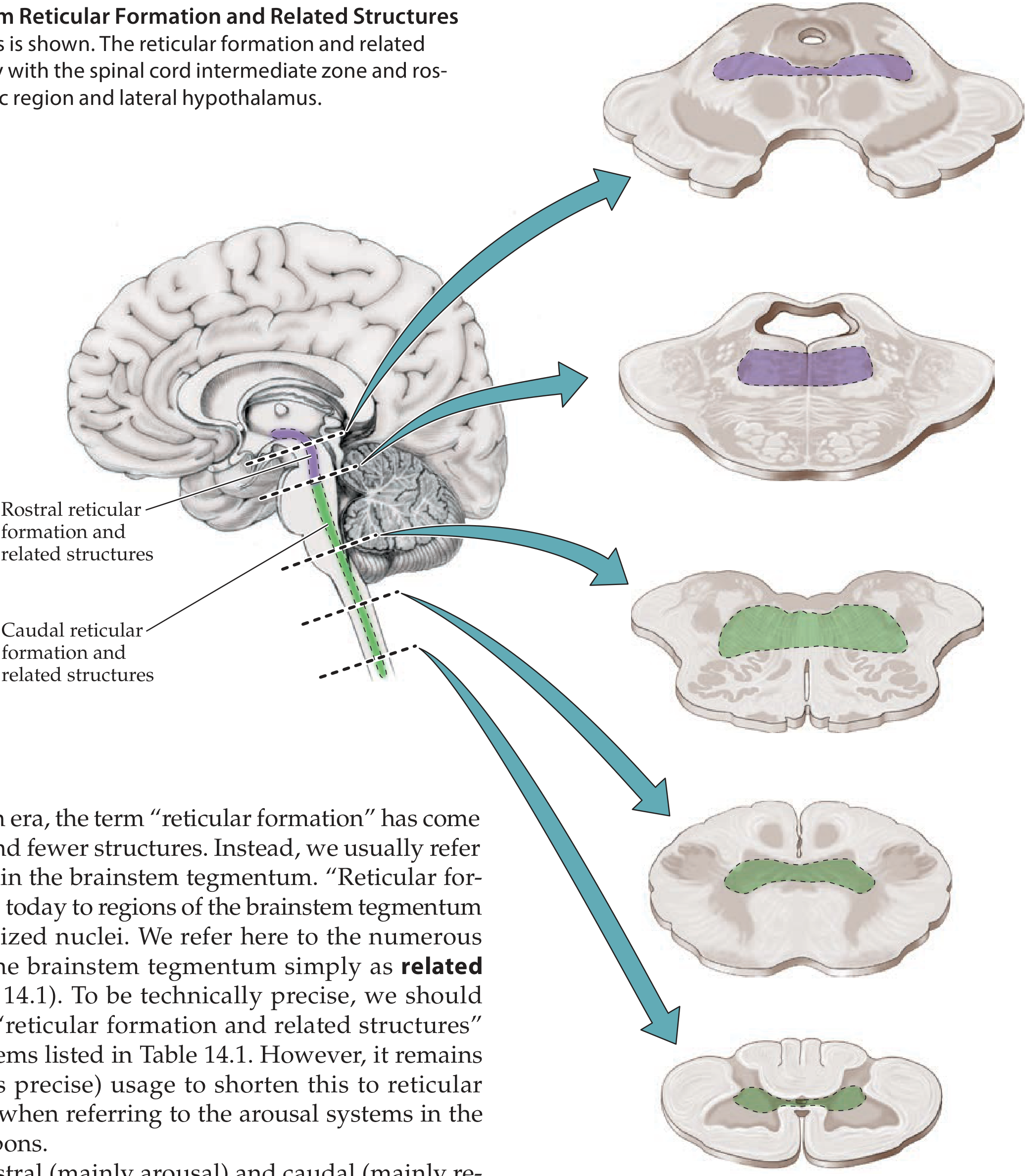
Figure: Reticular formation and related structures at multiple brainstem levels. Rostral RF (purple) in midbrain/upper pons; caudal RF (green) in lower pons/medulla. - Neuroanatomy through Clinical Cases, 3e
Functional Subdivisions
A useful clinical simplification divides the RF into two major parts:
| Division | Location | Main Function |
|---|---|---|
| Rostral RF | Midbrain + upper pons | Arousal, alertness, consciousness |
| Caudal RF | Lower pons + medulla | Motor, reflex, and autonomic control |
1. Rostral Reticular Formation - Arousal and Consciousness
The Ascending Reticular Activating System (ARAS)
Based on animal experiments and human studies in the 1930s-40s, Moruzzi and Magoun demonstrated that:
- Stimulation of the mesencephalic reticular formation produces behavioral and electrographic arousal (even from deep anesthesia)
- Bilateral destruction causes coma
- Unilateral lesions of the mesencephalic RF produce contralateral attention deficits
They named this the ascending reticular activating system (ARAS). Today we understand that there are actually multiple interconnected arousal systems acting in parallel - not a single ARAS - but the concept of a fragile region at the upper brainstem-diencephalic junction where focal lesions can cause coma remains clinically useful.
Where does a lesion cause coma?
- Dysfunction of the upper brainstem reticular formation/related structures, OR
- Dysfunction of extensive bilateral regions of the cerebral cortex
Lesions of the lower pons, medulla, or ventral midbrain (which spare the RF) do not typically cause loss of consciousness.
Arousal Circuit (ARAS pathway)
The pontomesencephalic RF does NOT project directly to the cortex in large numbers. Instead, it projects to relay stations that then activate the cortex:
RF → Thalamic intralaminar nuclei → Cortex + Striatum
Also via:
- Basal forebrain (cholinergic neurons to cortex)
- Hypothalamus (orexin/histamine neurons)
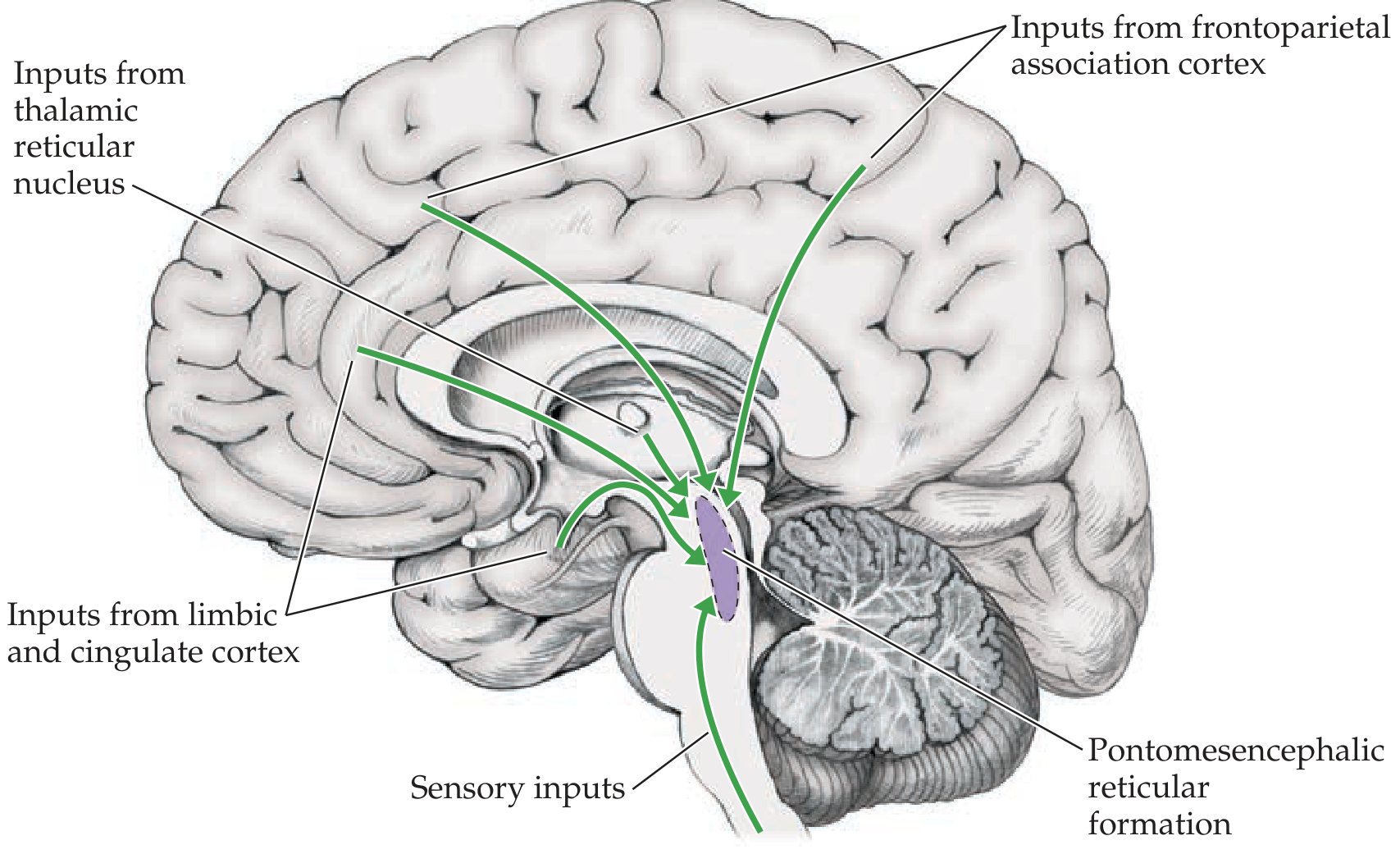
Figure 14.8: Major inputs to the pontomesencephalic RF - including sensory pathways, frontoparietal association cortex, limbic/cingulate cortex, and thalamic reticular nucleus. - Neuroanatomy through Clinical Cases, 3e
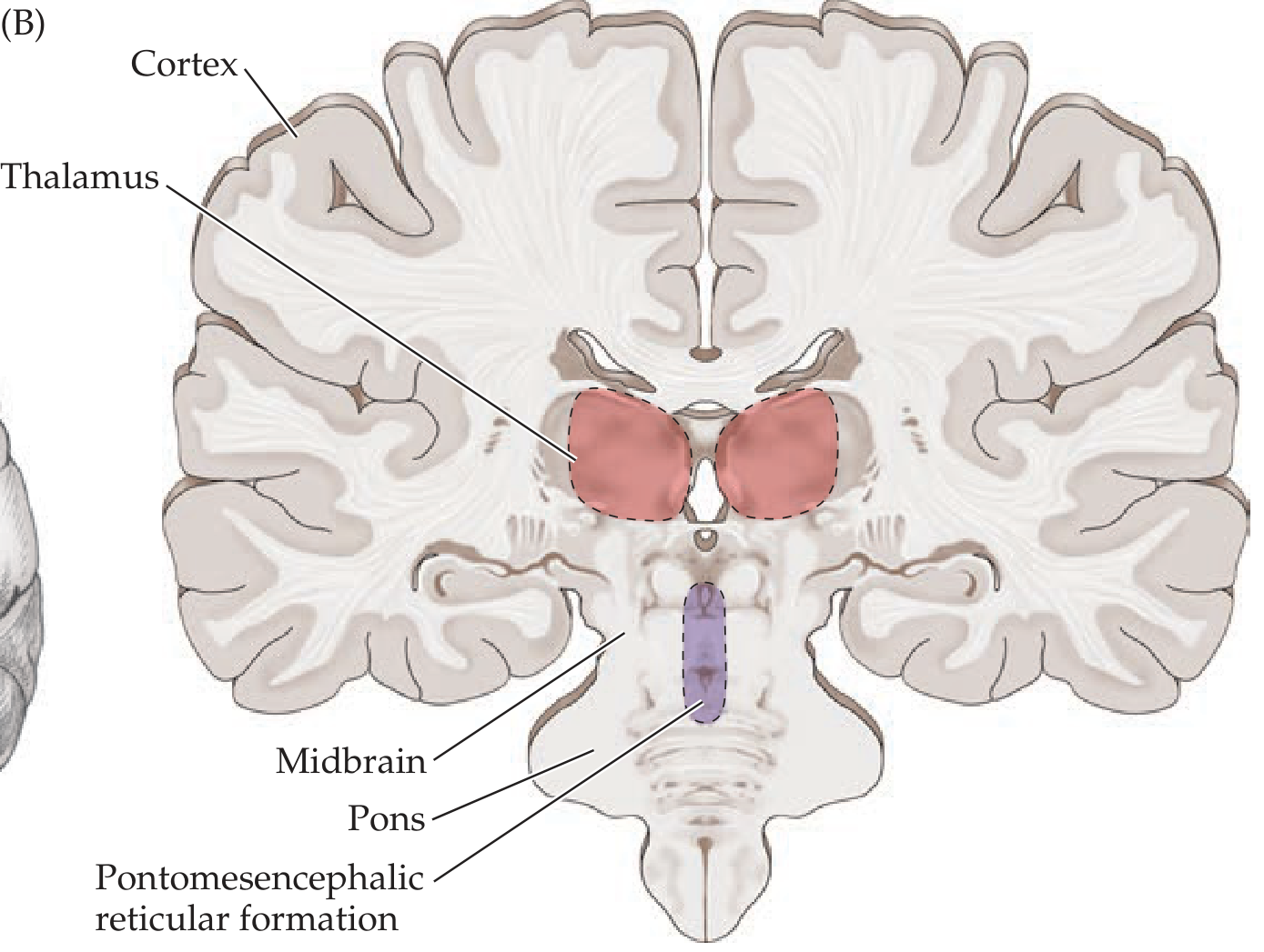
Figure 14.7B: Coronal view. Widespread projections to the cortex arise from the pontomesencephalic RF relayed via thalamic intralaminar nuclei. - Neuroanatomy through Clinical Cases, 3e
Inputs to the RF (what activates it?)
The RF receives:
- Sensory inputs - especially the spinoreticular pathway (anterolateral system, pain)
- Association cortex (frontoparietal) - cognitive processes driving alertness
- Limbic/cingulate cortex - emotional arousal
- Posterior lateral hypothalamus - activates arousal circuits
- Thalamic reticular nucleus - modulatory input
2. Widespread Projection (Neuromodulatory) Systems
The brainstem tegmentum contains nuclei with specific neurotransmitters that have diffuse, widespread projections throughout the CNS - these are the "related structures" now recognized as distinct from the RF proper:
| System | Cell Bodies | Targets | Function |
|---|---|---|---|
| Acetylcholine | Pedunculopontine nucleus (PPN), laterodorsal tegmental nucleus; basal forebrain | Thalamus, cortex, hippocampus | Alertness, memory, REM sleep |
| Norepinephrine | Locus coeruleus (pons) + lateral tegmental area | Entire CNS | Alertness, attention, mood elevation |
| Serotonin | Raphe nuclei (midbrain, pons, medulla) | Entire CNS | Alertness, mood, breathing control |
| Dopamine | Substantia nigra pars compacta + ventral tegmental area (VTA) | Striatum, limbic cortex, prefrontal cortex | Movement, initiative, working memory |
| Histamine | Tuberomammillary nucleus (hypothalamus) + midbrain RF | Entire brain | Alertness |
| Orexin (hypocretin) | Posterior lateral hypothalamus | Entire brain | Alertness, food intake |
| GABA | Basal forebrain, thalamic reticular nucleus, ventrolateral preoptic area (VLPO) | Cortex, thalamus, brainstem | Sleep promotion (inhibits arousal) |
Note: Unlike gross lesions of the pontomesencephalic RF (which readily cause coma), pharmacological blockade or lesions of individual neuromodulatory systems generally produce confusion and drowsiness rather than full coma - highlighting the redundancy of these parallel systems.
3. Caudal Reticular Formation - Motor, Reflex, and Autonomic Functions
The caudal RF (lower pons + medulla) serves "life support" functions:
Respiratory Control
- Circuits in the medulla generate automatic respiratory rhythms
- The pre-Botzinger complex (medulla) functions as a respiratory pacemaker
- Inputs: peripheral chemoreceptors (O₂/pH), lung stretch receptors, central pH-sensing neurons (many serotonergic)
- Outputs: to spinal lower motor neurons (C3-C5 for phrenic nerve - diaphragm)
- Lesions of the medulla can cause respiratory arrest; rostral pontine lesions cause apneusis (prolonged inspiration)
Cardiovascular Control
- The RF contains vasomotor and cardioinhibitory centers
- Lateral tegmental field - vasopressor area (increases BP via sympathetic outflow)
- Nucleus solitarius - receives baroreceptor inputs; coordinates cardiovascular reflexes
- Lesions here can cause severe hemodynamic instability
Motor Functions (Reticulospinal Tracts)
- The RF gives rise to the medial and lateral reticulospinal tracts
- These modulate spinal cord motor neurons - facilitating or inhibiting muscle tone
- The medullary RF is inhibitory (inhibits extensors) and the pontine RF is facilitatory (facilitates extensors)
- In decerebrate rigidity (pontine lesion), the facilitatory tract is unopposed, causing rigid extension
Other Reflex/Autonomic Functions
- Swallowing, vomiting (chemoreceptor trigger zone / area postrema in medulla - important for nausea)
- Salivation (superior/inferior salivatory nuclei lie within the RF)
- Coughing, sneezing
- Micturition and defecation coordination
- Cranial nerve reflex arcs (corneal reflex, gag reflex)
- Pain modulation (periaqueductal gray + RF - inhibitory noradrenergic pathways)
4. Eye Movements
The RF contains specialized gaze centers:
- Pontine reticular formation (PRF) - generates horizontal saccades (paramedian pontine reticular formation / PPRF)
- Mesencephalic reticular formation - generates vertical saccades (rostral interstitial nucleus of the MLF / riMLF)
Lesions of the PPRF cause ipsilateral gaze palsy; lesions at the midbrain level cause vertical gaze palsies (e.g., Parinaud syndrome with dorsal midbrain lesions).
5. Sleep-Wake Regulation
The RF and related structures control the sleep-wake cycle through a "flip-flop switch" mechanism:
- Wake-promoting systems: noradrenergic (locus coeruleus), serotonergic (raphe), histaminergic (tuberomammillary nucleus), cholinergic (PPN/LDT), orexin (lateral hypothalamus) - all mutually excitatory, activate the cortex
- Sleep-promoting: GABA/galanin neurons of the ventrolateral preoptic area (VLPO) of the hypothalamus inhibit all the wake-promoting nuclei
- The switch is binary - you are either awake or asleep, not in between - maintained by mutual inhibition
- Orexin deficiency causes narcolepsy (loss of stability of the wake state)
Clinical Correlations
| Condition | Mechanism |
|---|---|
| Coma | Bilateral lesion of upper brainstem RF (e.g., hemorrhage, infarct at midbrain-diencephalic junction), or bilateral cortical dysfunction |
| Locked-in syndrome | Bilateral ventral pontine lesion - RF spared (consciousness intact) but motor pathways destroyed |
| Vegetative state | Cortex disconnected from RF; ARAS intact (sleep-wake cycles present) but no awareness |
| Narcolepsy | Orexin neuron loss (posterior hypothalamus) - RF arousal state becomes unstable |
| Decerebrate rigidity | Lesion at midbrain - pontine facilitatory RF now unopposed |
| Respiratory failure | Medullary RF lesion (e.g., lateral medullary infarct, central herniation) |
| Midbrain lesion / transtentorial herniation | Blown pupils (CN III compression) + coma (RF compression) |
Summary
The reticular formation is best understood as the brainstem's integrative core - a hub that receives inputs from virtually every sensory and cognitive system and projects widely to maintain:
- Consciousness and arousal (via ARAS, thalamus, and neuromodulators)
- Motor control (via reticulospinal tracts)
- Autonomic homeostasis (breathing, cardiovascular control)
- Sleep-wake cycling
- Reflex coordination (eye movements, swallowing, vomiting, pain modulation)
Sources: Neuroanatomy through Clinical Cases, 3e, p. 654-658; Localization in Clinical Neurology, 8e; Eric Kandel Principles of Neural Science, 6e
This is a shared conversation. Sign in to Orris to start your own chat.