
Cell lines in microbiology
Reading File
Cell lines are a broad microbiology/cell biology topic. Let me search the textbook library and web simultaneously.
Searching sources...
Finding Sources
Finding Sources
Searching the Web
cell lines in microbiology types uses examples HeLa Vero
The medical microbiology textbook (Sherris & Ryan's) has a relevant section. Let me read it along with more content.
Reading File
Finding Sources
Excellent - I have found highly relevant sections in multiple microbiology textbooks. Let me read the key sections from Jawetz and Medical Microbiology and the Quick Compendium.
Reading File
Reading File
Reading File
I now have rich, detailed content from multiple authoritative microbiology textbooks. Let me compile the comprehensive answer.
Reading File
I now have all the information needed for a comprehensive, well-sourced answer. Let me fetch the CPE image as it's highly relevant.
Loading Image
Now I have everything needed. Here is the comprehensive answer:
Cell Lines in Microbiology
What is a Cell Line?
A cell line is a population of cells that can be grown and maintained outside the body (in vitro) under controlled laboratory conditions. In microbiology - particularly virology - cell lines are indispensable tools because viruses are obligate intracellular parasites; they cannot replicate on their own and require living host cells to multiply.
The initial cell population obtained directly from tissue is called a primary culture. To prepare it, tissue is removed from an animal, disaggregated using the proteolytic enzyme trypsin, and seeded into a plastic Petri dish in a nutrient medium containing amino acids, vitamins, minerals, sugars, and animal serum. Cells attach to the dish bottom, divide, and eventually cover the surface (a monolayer). When crowded, they enter a resting state; propagation continues by trypsinizing and reseeding into new plates. - Sherris & Ryan's Medical Microbiology, 8th Ed.
Types of Cell Cultures (The Three Main Categories)
| Type | Description | Passage Limit | Chromosome Pattern | Examples |
|---|---|---|---|---|
| Primary cell cultures | Made by dispersing cells directly from freshly removed tissues; cannot survive more than a few passages | Few passages | Normal | Monkey kidney, human amnion, chick embryo, rabbit kidney |
| Diploid cell lines | Secondary cultures; undergo limited culture but retain normal chromosomes | Up to ~50 passages | Normal (diploid) | MRC-5 (human fetal lung fibroblasts) |
| Continuous (immortal) cell lines | Derived from diploid lines or malignant tumors; capable of indefinite growth; chromosomally altered | Indefinite | Abnormal (aneuploid) | HeLa, HEp-2, A549, Vero |
- Jawetz, Melnick & Adelberg's Medical Microbiology, 28th Ed.
1. Primary Cell Cultures
- Prepared directly from animal or human tissues
- Susceptible to a wider range of viruses (closest to in vivo cells)
- Can carry endogenous viruses already present in the donor tissue (a disadvantage)
- Examples and the viruses they support:
| Primary Cell Culture | Viruses Isolated |
|---|---|
| Monkey kidney cells | Influenza, parainfluenza, enteroviruses |
| Rabbit kidney cells | Herpes simplex virus (HSV) |
| Human embryonic kidney (HEK) | Adenovirus, enterovirus |
| Chick embryo cells | Influenza, mumps |
- Quick Compendium of Clinical Pathology, 5th Ed.
2. Diploid Cell Lines (Semi-Continuous)
- Have undergone a change allowing prolonged but limited culture (50 passages max)
- Retain normal diploid chromosome number - more physiologically representative
- MRC-5 (Medical Research Council strain 5) - diploid fibroblasts from the lung of a human male fetus - is the most classic example
- Predominantly fibroblasts in composition
- Used to isolate: CMV, VZV, HSV, rhinoviruses, adenoviruses, some enteroviruses
- Quick Compendium of Clinical Pathology, 5th Ed.
3. Continuous (Immortal) Cell Lines
These are derived from tumors or from cells transformed by viruses. They have altered, irregular chromosome numbers (aneuploid) and grow indefinitely - hence called "immortal."
How they arise: Most primary cells eventually die; rare survivors may develop into a permanent line. Lines can also be generated directly from tumors or virus-transformed cells. They rarely resemble the tissue of origin but are extremely practical lab workhorses. - Sherris & Ryan's Medical Microbiology, 8th Ed.
Key Examples
| Cell Line | Origin | Used to Isolate/Study |
|---|---|---|
| HeLa | Human cervical adenocarcinoma (Henrietta Lacks, 1951) | First human immortal cell line; used extensively in viral and cancer research |
| HEp-2 | Human epidermoid carcinoma of the larynx | RSV, adenoviruses, HSV |
| A549 | Human lung carcinoma | HSV, adenoviruses |
| Vero | African green monkey kidney | Rabies, rotavirus, measles, many others; widely used in vaccine production |
| HEK-293 | Human embryonic kidney | Adenovirus, recombinant protein production |
| Jurkat | Human T-cell leukemia | T-cell receptor signaling research |
How Cell Cultures Are Used to Detect Viruses
Once a clinical specimen is inoculated onto a cell monolayer, viral growth is detected by:
- Cytopathic effect (CPE) - morphologic changes in cells visible under light microscopy (most important method)
- Immunofluorescence - detecting viral proteins with specific antisera
- Molecular detection - PCR for virus-specific nucleic acid
- Hemadsorption - erythrocytes adsorb to infected cells expressing viral hemagglutinin (used for influenza, parainfluenza)
- Plaque formation - in embryonated eggs, viral growth produces visible pocks on the chorioallantoic membrane
- Jawetz, Melnick & Adelberg's Medical Microbiology, 28th Ed.
Cytopathic Effects (CPE) by Virus
| Virus | Cytopathic Effect |
|---|---|
| Enterovirus | Tear-shaped cells; rapid rounding progressing to complete destruction |
| HSV | Focal swollen, rounded cells (ballooning degeneration) |
| CMV | Focal plaques in human diploid fibroblasts (HDF); "owl eye" inclusions |
| RSV | Syncytia (multinucleated giant cells) |
| Adenovirus | Grape-like clusters of rounded cells |
| Paramyxovirus | Syncytia formation |
The image below shows actual CPE in cell monolayers from different viruses:
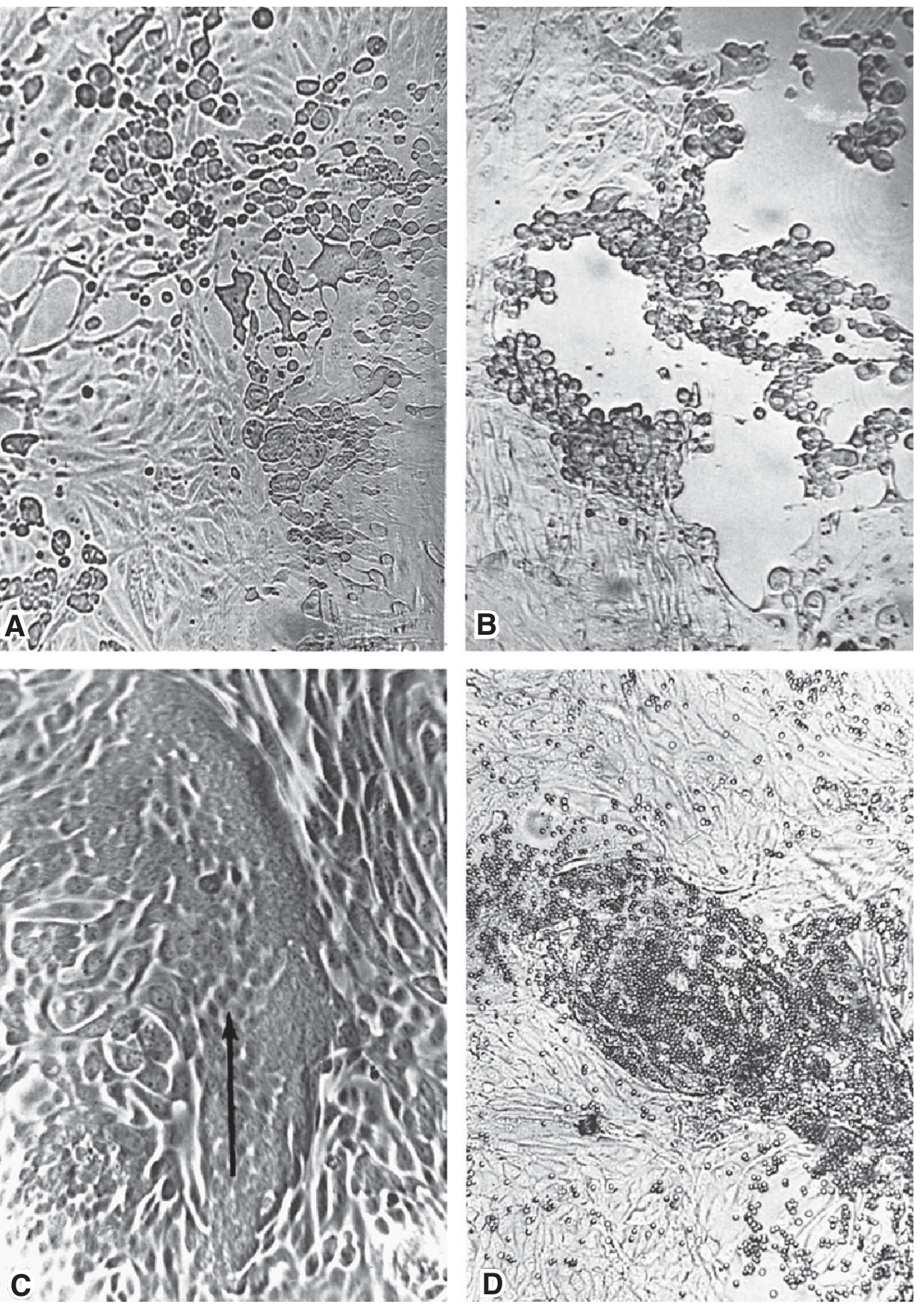
A: Enterovirus - rapid rounding and cell destruction; B: Herpesvirus - focal swollen rounded cells; C: Paramyxovirus - syncytia (fused cells, arrow); D: Hemadsorption (erythrocytes adhering to virus-infected cells). - Jawetz, Melnick & Adelberg's Medical Microbiology, 28th Ed., p.428
Permissive vs. Non-Permissive Cells
An important concept related to cell lines is whether a cell is permissive or non-permissive:
-
Permissive cells - allow complete viral replication and production of progeny virions
-
Non-permissive cells - do not allow viral replication, but may allow viral transformation
-
Semi-permissive cells - support some but not all steps in replication (very inefficient)
-
Medical Microbiology, 9th Ed.
Clinical Applications
- Diagnostic virology - recovering viruses from patient specimens (throat swabs, CSF, stool) to identify the causative agent
- Vaccine production - e.g., Vero cells for rabies, polio, and rotavirus vaccines; human diploid cells (MRC-5) for varicella, hepatitis A, and rabies vaccines
- Antiviral susceptibility testing - the current primary use of traditional tube cultures
- Research - studying viral pathogenesis, replication, and host-pathogen interactions
Summary Table
| Feature | Primary | Diploid | Continuous |
|---|---|---|---|
| Passage limit | Few | ~50 | Indefinite |
| Chromosome number | Normal | Normal | Abnormal |
| Origin | Fresh tissue | Adapted primary | Tumor / transformed |
| Virus sensitivity | Broadest | Moderate | Specific |
| Risk of endogenous virus | Yes | Low | Minimal |
| Examples | Monkey kidney, HEK | MRC-5, WI-38 | HeLa, HEp-2, Vero, A549 |
Sources: Sherris & Ryan's Medical Microbiology 8th Ed. | Jawetz, Melnick & Adelberg's Medical Microbiology 28th Ed. | Quick Compendium of Clinical Pathology 5th Ed. | Medical Microbiology 9th Ed.

photo-1784559769018.jpg
Explain this
Reading File
analyse_media
Now let me search the medical library for deeper content on inclusion bodies, childhood exanthems, and PCR types.
Finding Sources
Finding Sources
Finding Sources
I now have rich, authoritative content from multiple textbooks. Let me compile the full explanation.
Microbiology | Virology - Page 72 Explained
This page covers four major topics: viral replication steps, viral cultivation, inclusion bodies, and childhood exanthems + PCR types. Let me break each down in detail.
1. Viral Replication - The 6 Steps
Viruses cannot replicate on their own - they hijack host cell machinery. The steps are numbered in the diagram (virus approaching the host cell receptor):
Step 1 - Adsorption (Attachment)
The virus binds to specific receptors on the host cell surface. This is highly specific - the receptor determines which cells a virus can infect (called tropism). For example, HIV binds CD4 receptors on T-helper cells.
Step 2 - Penetration
After binding the receptor, the virus enters the cell. This can occur by:
- Membrane fusion (enveloped viruses like influenza)
- Receptor-mediated endocytosis (most viruses)
- Direct injection (some bacteriophages)
Step 3 - Uncoating
The viral capsid (protein coat) is removed inside the cell, releasing the viral genome (DNA or RNA) into the cytoplasm. This is essentially "unwrapping" the virus.
Step 4 - Biosynthesis (Multiplication)
The viral genome is replicated and viral proteins are synthesized using the host's ribosomes, enzymes, and energy supply. This is the most important step with key exceptions:
Where does replication happen?
| Type | Normal Site | Exceptions |
|---|---|---|
| DNA viruses | Nucleus | Poxvirus - replicates in cytoplasm (because it is so large it carries its own enzymes) |
| RNA viruses | Cytoplasm | Influenza and HIV (retrovirus) - replicate in the nucleus |
The handwritten Hindi note confirms: "kyuki size mein bada rehta hai" = "because it is large in size" (for Poxvirus). "Nucleus mein replicate karte hai" = "they replicate in the nucleus" (for Influenza & HIV).
- Sherris & Ryan's Medical Microbiology 8th Ed.: "Most RNA viruses replicate in the cytoplasm - the immediate site of entry, with the exception of influenza viruses and the retroviruses that replicate in the nucleus. All DNA viruses must move from the cytoplasm to the nucleus."
Step 5 - Maturation & Assembly
New viral genomes and structural proteins are assembled into complete virions (mature viral particles).
Step 6 - Release
Two mechanisms:
- Lysis - the host cell bursts and dies, releasing all new virions at once (common in non-enveloped viruses)
- Budding - the virus pushes through the host cell membrane and acquires a lipid envelope derived from the host cell membrane (common in enveloped viruses like influenza, HIV, herpesviruses)
2. Viral Cultivation
Unlike bacteria and fungi, viruses cannot be grown on artificial/synthetic media (like blood agar, MacConkey agar) because they are obligate intracellular parasites - they need living cells to replicate.
Three methods are used instead:
| Method | Details |
|---|---|
| Animal inoculation | Virus injected into lab animals (mice, rabbits); oldest method |
| Cell lines (Tissue culture) | Growing living cells in flasks/tubes, then infecting them; most common in modern labs |
| Egg inoculation | Virus injected into embryonated (fertilized) hen's eggs into specific compartments (allantoic cavity, amniotic cavity, yolk sac, chorioallantoic membrane) |
3. Inclusion Bodies
When a virus multiplies inside a cell, it can leave behind characteristic intracellular deposits called inclusion bodies. These are extremely useful diagnostically - they are seen on histology (H&E staining) and can identify specific viruses.
Jawetz: "In the course of viral multiplication within cells, virus-specific structures called inclusion bodies may be produced. They become far larger than the individual virus particle and often have an affinity for acid dyes."
Intranuclear Inclusion Bodies (inside the nucleus)
| Name | Virus |
|---|---|
| Cowdry Type A | Herpes simplex virus (HSV), Varicella-Zoster Virus (VZV), Yellow fever virus |
| Cowdry Type B | Adenovirus, Poliovirus |
- Cowdry A = Large, eosinophilic, single inclusion surrounded by a clear halo, pushing chromatin to the rim ("owl eye" appearance in some contexts)
- Cowdry B = Small, multiple inclusions, NO halo, NO chromatin margination
Intracytoplasmic Inclusion Bodies (inside the cytoplasm)
| Name | Virus |
|---|---|
| Negri bodies | Rabies virus - eosinophilic cytoplasmic inclusions found in neurons (especially Purkinje cells and hippocampal neurons); pathognomonic of rabies |
| Henderson-Paterson bodies | Molluscum contagiosum - large eosinophilic cytoplasmic inclusions in infected epidermal cells |
Both Intranuclear AND Intracytoplasmic
| Virus | Notes |
|---|---|
| CMV (Cytomegalovirus) | Has both nuclear ("owl eye") AND cytoplasmic inclusions |
| Measles | Has both types - intranuclear (Cowdry A) and intracytoplasmic inclusions; also forms Warthin-Finkeldey giant cells |
4. Childhood Exanthems (The "Disease" Number System)
These are classic childhood viral/bacterial rash illnesses, numbered historically by the order in which they were described/recognized:
| # | Disease | Cause |
|---|---|---|
| 1st | Measles (Rubeola) | Measles virus (Paramyxovirus) - Koplik spots, maculopapular rash, cephalocaudal spread |
| 2nd | Scarlet Fever | Streptococcus pyogenes (Group A Strep) - sandpaper rash, strawberry tongue |
| 3rd | German Measles (Rubella) | Rubella virus (Togavirus) - mild rash, dangerous in pregnancy (congenital rubella) |
| 4th | Duke's disease | Possibly a mild form of Scarlet fever or Staphylococcal exanthem (controversial/outdated) |
| 5th | Erythema Infectiosum (Slapped Cheek) | Parvovirus B19 - "slapped cheek" rash, lacy reticular rash on limbs |
| 6th | Roseola Infantum (Exanthem Subitum) | HHV-6 (Human Herpesvirus 6) - high fever for 3-5 days, then sudden rash when fever breaks |
5. PCR Types for Viral Diagnosis
The right-hand box covers when to use each PCR method:
| Situation | PCR Type | Why |
|---|---|---|
| Unknown causative agent | Multiplex PCR | Tests for multiple pathogens simultaneously in one reaction |
| Low copy number / very small viral load | Nested PCR | Two rounds of amplification for maximum sensitivity |
| RNA virus | RT-PCR (Reverse Transcriptase PCR) | RNA can't be directly amplified - must first convert RNA → DNA (cDNA) using reverse transcriptase enzyme, then amplify. Used for HIV, Influenza, HCV, SARS-CoV-2 |
| Viral load monitoring | Real-time PCR (Quantitative PCR / qPCR) | Measures amount of virus present; used for HIV viral load, CMV, HBV monitoring |
The handwritten note at the bottom is important: "PCR can only detect DNA" - this is why RNA viruses need RT-PCR first (to convert RNA → DNA before PCR can work).
Quick Memory Tricks
- DNA viruses replicate in Nucleus - except Pox (too big, carries its own enzymes)
- RNA viruses replicate in Cytoplasm - except Influenza & HIV (go to nucleus)
- Negri body = Rabies (intracytoplasmic, in neurons)
- Cowdry A = Herpes (intranuclear, with halo)
- 6th disease = Roseola = HHV-6 (high fever → rash when fever breaks)
- RNA virus? → RT first, then PCR
Sources: Jawetz, Melnick & Adelberg's Medical Microbiology 28th Ed. | Sherris & Ryan's Medical Microbiology 8th Ed. | Robbins & Cotran Pathologic Basis of Disease | Quick Compendium of Clinical Pathology 5th Ed.
Hsv-3
Finding Sources
Finding Sources
Good - Medical Microbiology 9e has a dedicated VZV section. Let me read it along with Jawetz and the dermatology text.
Reading File
Finding Sources
Reading File
Reading File
Reading File
Loading Image
Reading File
Loading Image
I now have rich, comprehensive content. Let me compile the complete answer on HSV-3 (VZV).
HSV-3 = Varicella-Zoster Virus (VZV) = HHV-3
HSV-3 is the informal name for Varicella-Zoster Virus (VZV), officially designated Human Herpesvirus 3 (HHV-3). It is an alphaherpesvirus and causes two distinct diseases depending on whether it is a primary or reactivated infection.
Basic Properties
| Feature | Details |
|---|---|
| Family | Herpesviridae |
| Subfamily | Alphaherpesvirinae |
| Genome | Double-stranded DNA (smallest genome among HHVs) |
| Envelope | Yes (enveloped virus - lipid envelope derived from host) |
| Replication site | Nucleus (DNA virus rule) |
| Latency site | Dorsal root ganglia, cranial nerve ganglia |
| Inclusion bodies | Cowdry Type A (intranuclear) |
| CPE | Syncytia formation + Cowdry A inclusions |
| Transmission | Respiratory droplets (primary route), direct contact with skin vesicles |
Two Diseases from One Virus
Disease 1 - Varicella (Chickenpox) = Primary Infection
Disease 2 - Herpes Zoster (Shingles) = Reactivation
Pathogenesis - How the Virus Spreads
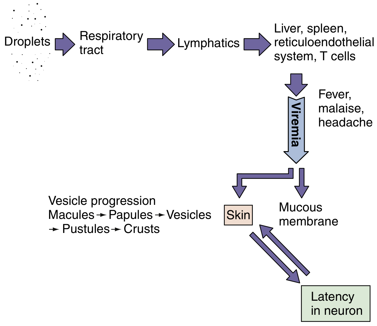
Mechanism of spread of VZV within the body. - Medical Microbiology 9e
Step-by-step:
- Inhaled respiratory droplets reach the tonsils and upper respiratory tract
- Virus enters the lymphatics, then spreads to liver, spleen, and reticuloendothelial system
- Infects T cells → primary viremia (mild, asymptomatic)
- Secondary viremia - T cells carry the virus to the skin; virus transfers from T cells to keratinocytes and epidermal cells
- Vesicular skin lesions form in successive crops
- After primary infection, virus travels retrograde along sensory neurons and establishes latency in dorsal root ganglia
- Medical Microbiology 9e
Disease 1: Varicella (Chickenpox)
Incubation period: ~14 days (range 10-21 days)
Classic hallmark: "Dewdrop on a rose petal" - a thin-walled vesicle on an erythematous (red) base, 2-4 mm in diameter
Rash progression:
Macules → Papules → Vesicles → Pustules → Crusts
Key features:
- Successive crops of lesions over 3-5 days - at any given time, all stages are present simultaneously (pathognomonic)
- Rash is centripetal - more on trunk and head than extremities
- Presence on the scalp distinguishes it from other rashes
- Lesions on mucous membranes (mouth, conjunctivae, vagina)
- Intensely pruritic (itchy) - scratching causes secondary bacterial infection and scarring
Age matters:
- Children (5-9 years): Mild, classic disease
- Adults and teenagers: More severe; pneumonia in 20-30%, potentially fatal
- Immunocompromised / newborns: Risk of life-threatening disseminated disease, encephalitis, pneumonia
Disease 2: Herpes Zoster (Shingles)
Herpes zoster ("zoster" = belt/girdle in Greek) is the reactivation of latent VZV from dorsal root or cranial nerve ganglia.
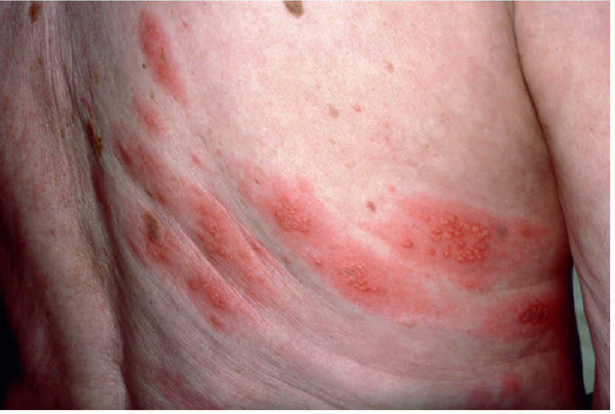
Herpes zoster (shingles) - unilateral dermatomal vesicular rash. - Andrews' Diseases of the Skin
When does reactivation occur? When cell-mediated immunity wanes:
- Old age (most common)
- HIV infection (annual risk ~3%)
- Hematologic malignancy
- Immunosuppressive drugs (steroids, TNF inhibitors, JAK inhibitors, chemotherapy)
Dermatomes affected:
| Site | Frequency |
|---|---|
| Thoracic | 55% (most common) |
| Cranial (trigeminal most common) | 20% |
| Lumbar | 15% |
| Sacral | 5% |
Key clinical features:
- Strictly unilateral - does NOT cross the midline
- Preceded by prodromal pain (burning, shooting, or stabbing) for 1-5 days BEFORE the rash - important diagnostic clue
- Lesions appear in the entire dermatome
- Zoster sine herpete - pain without rash (rare but important)
Complications:
- Postherpetic neuralgia (PHN) - severe, persistent pain lasting weeks to months after rash heals; more common in elderly (most important complication)
- Ramsay Hunt Syndrome - Zoster of geniculate ganglion (CN VII): facial palsy + ear pain + vesicles in external ear canal
- Zoster ophthalmicus - V1 (ophthalmic branch of trigeminal) involvement; risk of corneal ulceration and blindness
- Disseminated zoster - in immunocompromised patients
Immunity
| Type | Role |
|---|---|
| Cell-mediated immunity (CMI) | Essential for resolving acute disease and controlling latency. CMI deficiency → severe/disseminated disease |
| Antibody | Limits viremic spread; passive immunization (VZIg) works within 4 days of exposure |
| IFN-α + NK cells | Limit early tissue spread |
Note: An overzealous CMI response in adults actually causes MORE severe disease (especially pneumonia) compared to children.
Diagnosis
- Clinical - characteristic dermatomal rash + prodromal pain (usually sufficient)
- Tzanck smear - scraping from vesicle base: shows multinucleated giant cells (Warthin-Finkeldey cells); does NOT distinguish VZV from HSV
- PCR - gold standard for confirming VZV (from vesicle fluid, CSF)
- DFA (Direct Fluorescent Antibody) - rapid detection from lesion scraping
- Serology - IgM/IgG (useful for varicella, less so for zoster)
Treatment
| Drug | Use |
|---|---|
| Acyclovir | Drug of choice; phosphorylated by VZV-encoded thymidine kinase (TK), then inhibits viral DNA polymerase |
| Valacyclovir | Pro-drug of acyclovir; better oral bioavailability |
| Famciclovir | Alternative; also requires viral TK activation |
Note: VZV replicates more slowly and in fewer cell types than HSV - this is why acyclovir doses for VZV are higher than for HSV.
Vaccines
| Vaccine | Target | Type |
|---|---|---|
| Varivax (Oka strain) | Varicella (children) | Live attenuated |
| Zostavax | Shingles (adults >60) | Live attenuated (higher dose than Varivax) |
| Shingrix | Shingles (adults >50) | Adjuvanted subunit vaccine (preferred; ~97% efficacy) |
VZIg (Varicella-Zoster Immunoglobulin): Given within 4 days of exposure to high-risk individuals (immunocompromised, newborns of mothers with active varicella within 5 days of delivery).
Quick Summary
| Feature | Varicella (Primary) | Herpes Zoster (Reactivation) |
|---|---|---|
| Population | Children (mainly) | Elderly / Immunocompromised |
| Rash | Widespread, all stages simultaneously | Unilateral, dermatomal |
| Pain | Minimal | Prominent (precedes rash) |
| Transmission | Contagious | Contagious to non-immune contacts only |
| Key complication | Pneumonia (adults) | Postherpetic neuralgia |
Sources: Medical Microbiology 9e | Jawetz, Melnick & Adelberg's Medical Microbiology 28th Ed. | Andrews' Diseases of the Skin, Clinical Dermatology
Images of condyloma acuminatum
Finding Sources
Images found in Andrews' Diseases of the Skin and Robbins. Let me retrieve them along with histology images.
Reading File
Reading File
Found multiple images. Let me fetch the clinical and histology images now.
Loading Image
Loading Image
Finding Sources
Now let me get the histology/microscopy images from Robbins.
Reading File
Reading File
Let me now get the histology image from Robbins and the Buschke-Lowenstein tumor image.
Reading File
Loading Image
Now let me get the histology image from Robbins (Fig 21.12).
Finding Sources
Reading File
I have all the clinical images and detailed textbook content. Let me now look for the histology image URLs in the Robbins block.
Searching Content
"cdn.orris.care" in /textbooks/9780443264528
Reading File
Loading Image
I now have all the clinical and histological images. Here is the complete answer:
Condyloma Acuminatum (Genital Warts) - Images & Overview
Condyloma acuminatum (plural: condylomata acuminata) is a benign sexually transmitted wart caused by Human Papillomavirus (HPV) - predominantly low-risk types HPV 6 and HPV 11. It can occur on any moist mucocutaneous surface of the external genitalia in either sex.
Clinical Images
Figure 1 - Condylomata Acuminata in a Male (penile lesions)
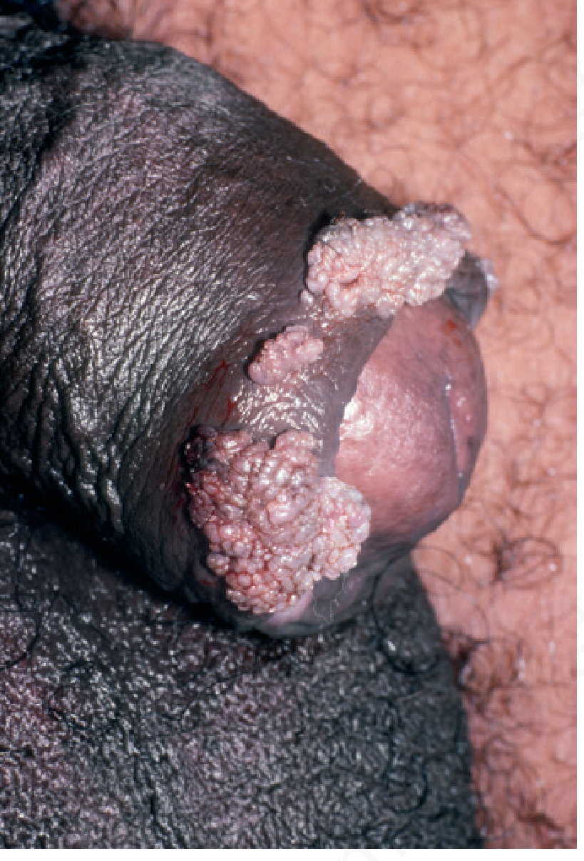
Fig. 19.49 Condylomata acuminata (male). Cauliflower-like lobulated papules at the coronal sulcus and prepuce. - Andrews' Diseases of the Skin
Figure 2 - Condylomata Acuminata in a Female (vulvar lesions)
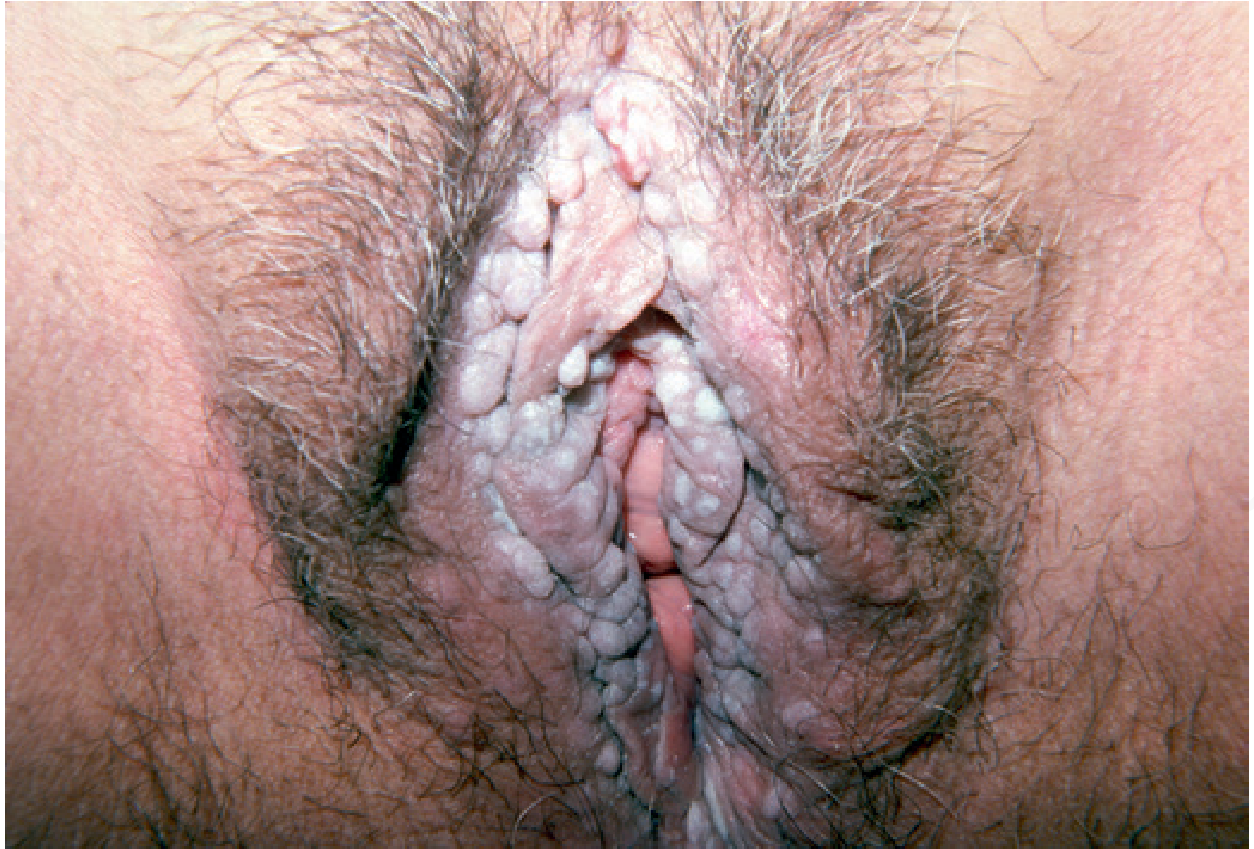
Fig. 19.50 Condylomata acuminata (female). Extensive confluent warty growths on the vulva. - Andrews' Diseases of the Skin
Figure 3 - Giant Condyloma / Buschke-Lowenstein Tumor
This is the extreme end of the spectrum - a massive, locally invasive verrucous growth also caused by HPV-6:
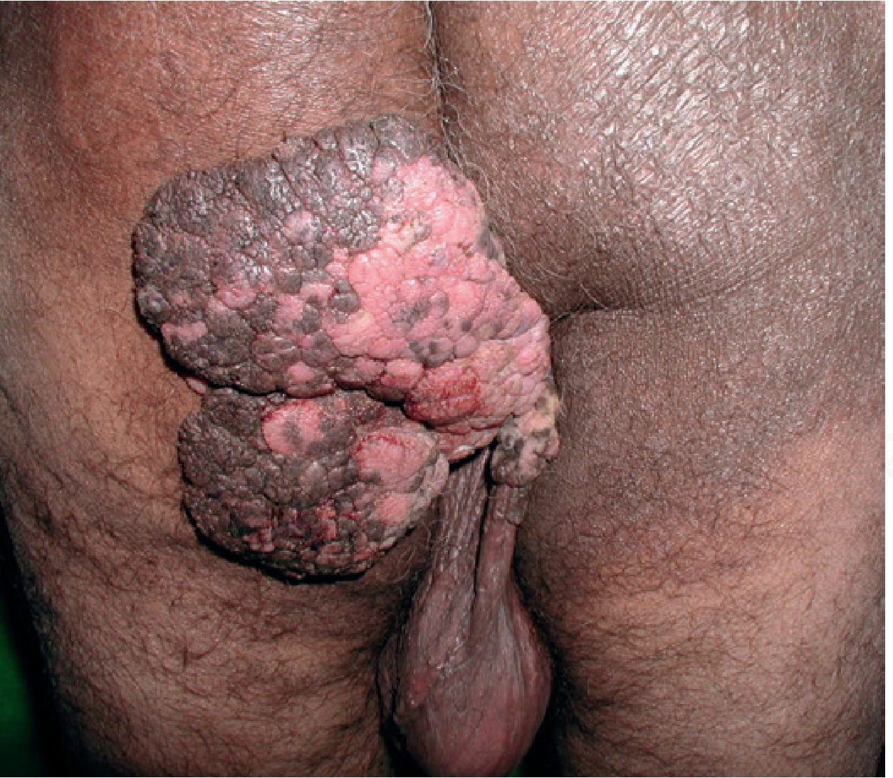
Fig. 19.52 Giant condyloma acuminatum (Buschke-Lowenstein tumor). A massive verrucous carcinoma. - Andrews' Diseases of the Skin
Histology Images (Microscopy)
Figure 4 - Histopathology of Condyloma Acuminatum (H&E)
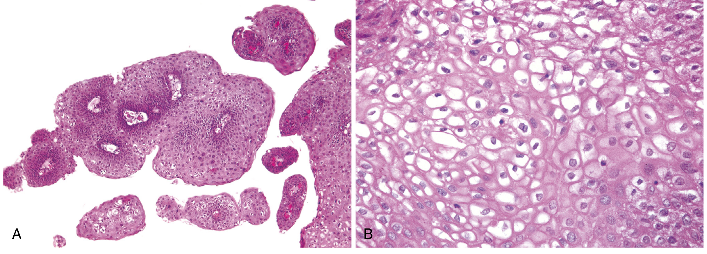
Fig. 21.12 Condyloma acuminatum of the penis.
- (A) Low magnification: Papillary/villous architecture with branching fronds; thickening of the epidermis (acanthosis); surface hyperkeratosis
- (B) High magnification: Koilocytes - cells with perinuclear cytoplasmic vacuolization (clearing around the nucleus with a wrinkled/raisin-like nucleus) - this is pathognomonic of HPV infection
- Robbins, Cotran & Kumar Pathologic Basis of Disease
Key Features to Know
| Feature | Detail |
|---|---|
| Cause | HPV 6 and HPV 11 (low-risk strains) |
| Appearance | Lobulated, cauliflower-like papules; 2-5 mm (range: microscopic to several cm) |
| Color | Gray, pale yellow, or pink |
| Locations | Penis (coronal sulcus, prepuce), vulva, cervix, perineum, perianal skin, inguinal folds |
| Histology hallmark | Koilocytosis (perinuclear vacuolization) + acanthosis + papillary stroma |
| Malignant potential | Benign; HPV 6/11 are low-risk; NOT the same strains as cervical cancer (HPV 16/18) |
| Giant variant | Buschke-Lowenstein tumor (HPV 6) - locally invasive, rarely metastasizes |
Sources: Robbins, Cotran & Kumar Pathologic Basis of Disease | Andrews' Diseases of the Skin, 12th Ed.
Laryngeal warts image
Finding Sources
Finding Sources
Reading File
Reading File
Found two excellent endoscopic images of laryngeal papillomas. Let me fetch both now.
Loading Image
Loading Image
Excellent endoscopic images retrieved. Here is the complete answer:
Laryngeal Warts (Recurrent Respiratory Papillomatosis - RRP)
Laryngeal warts, formally called Recurrent Respiratory Papillomatosis (RRP), are squamous papillomas caused by HPV 6 and HPV 11 (the same low-risk strains as genital condylomata). They are the most common benign neoplasm seen by laryngologists, making up ~84% of all benign laryngeal tumors.
Endoscopic Image 1 - Papilloma on the Vocal Fold (Before & After Treatment)
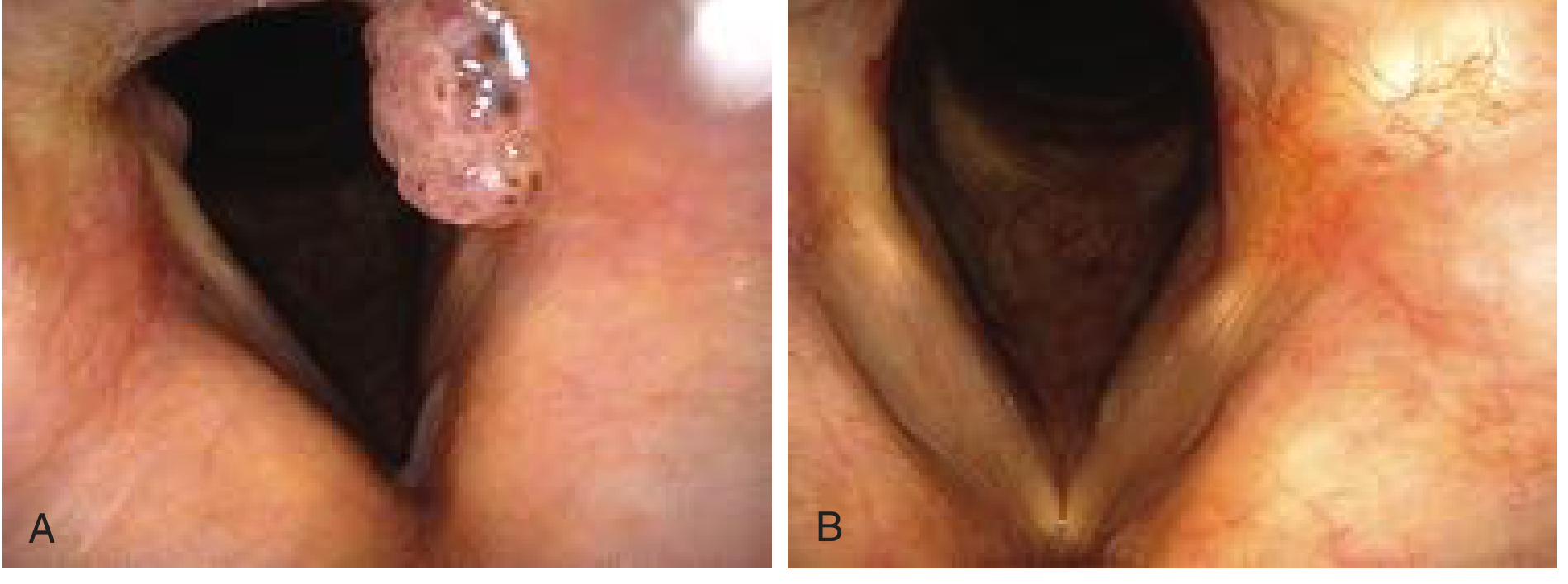
(A) - Large exophytic, lobulated papilloma (cauliflower-like mass) at the posterior left vocal fold, significantly obstructing the glottis.
(B) - Same patient 2 weeks after microsurgical debridement + cidofovir injection. Vocal folds appear normal; voice returned.
Fig. 60.34 - Cummings Otolaryngology Head and Neck Surgery
Endoscopic Image 2 - "Carpet Variant" Papillomatosis (Standard Light vs. Narrow-Band Imaging)
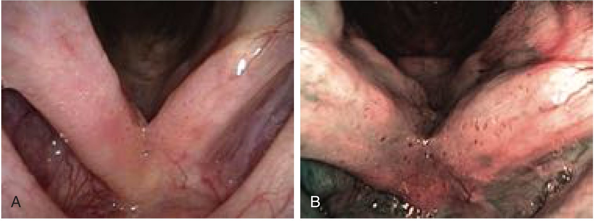
(A) Standard white light - Subtle, faint stippled vascularity and mild pink inflammation. These patients are frequently misdiagnosed with acid reflux disease.
(B) Narrow-band imaging (NBI) - The same area reveals the characteristic stippled "carpet variant" papillomatosis - distinctive dot-like vessels from HPV infection become clearly apparent.
Fig. 60.35 - Cummings Otolaryngology Head and Neck Surgery
Key Facts
| Feature | Detail |
|---|---|
| Cause | HPV 6 (milder) and HPV 11 (more aggressive, higher risk of tracheopulmonary spread) |
| Most common site | Vocal folds (true cords) at the anterior commissure |
| Age distribution | Bimodal - children <5 years (juvenile-onset) and adults >15 years |
| Transmission | Vertical - from mother with genital condylomata during vaginal delivery |
| Presentation | Hoarseness (most common symptom), stridor, voice changes, rarely airway obstruction |
| Recurrence | Very high - hallmark of the disease; requires repeated surgeries |
| Risk of malignancy | ~14% develop carcinoma (higher with prior radiation therapy) |
| HPV 16/18 | When present, carry a higher risk of malignant transformation |
Treatment
- CO₂ laser ablation - gold standard for surgical debulking
- Microdebrider (laryngeal shaver) - removes bulky exophytic papillomas
- Intralesional cidofovir - adjuvant antiviral; reduces recurrence intervals
- HPV vaccination (Gardasil) - combined with surgery; shows reduced recurrences; vaccination of young women is expected to reduce the prevalence of RRP in children
Source: Cummings Otolaryngology Head and Neck Surgery | Andrews' Diseases of the Skin, 12th Ed.
This is a shared conversation. Sign in to Orris to start your own chat.