

IMG20260511091916.jpg
Tell me answer of this from ak khurrana book of ophthalmology in ms level with diagram wherever required
Reading File
The question has 8 topics across two sections. Given the budget constraints, I'll search for all topics in parallel from AK Khurana's ophthalmology textbook.
Finding Sources
Finding Sources
Finding Sources
Finding Sources
Finding Sources
Finding Sources
Finding Sources
Finding Sources
I found the relevant sections. Now let me read all the content in parallel from Khurana-related searches and the found sections. Note: AK Khurana isn't directly in the library, but I'll use the available ophthalmology books. Let me read the key sections now:
Finding Sources
Reading File
Reading File
Reading File
Reading File
Now let me fetch the key images and also get leukocoria detail from the relevant section, plus search for visual field in glaucoma and anti-glaucoma meds:
Finding Sources
Searching Images
diabetic retinopathy proliferative vs non-proliferative fundus
Searching Images
glaucoma visual field defect perimetry diagram
Reading File
I now have rich content from Kanski's Clinical Ophthalmology (the most authoritative ophthalmology textbook available in the library). Let me now read the visual field/glaucoma section and get the leukocoria/retinoblastoma details:
Reading File
Searching Images
leukocoria white pupillary reflex retinoblastoma child
Searching Images
dacryocystitis lacrimal sac anatomy
Searching Images
presbyopia accommodation loss near vision aging lens
Searching Images
anti-glaucoma drugs mechanism prostaglandin beta blocker carbonic anhydrase
Searching Images
biometry A-scan ultrasound IOL power calculation
Loading Image
Loading Image
Excellent! I now have comprehensive content. Let me fetch the leukocoria and dacryocystitis images:
Loading Image
Loading Image
Loading Image
Now I have all the material needed. Here are comprehensive MS-level answers for all questions:
OPHTHALMOLOGY CLASS TEST — MS LEVEL ANSWERS
Based on Kanski's Clinical Ophthalmology (10th Ed.) — closest to AK Khurana's standard
SECTION A: EXPLAIN WHY (4×5 = 20) — Answer Any 4
Q1. Corneal Graft Has Lower Risk of Rejection Compared to Other Grafts
Answer:
The cornea enjoys a unique status called immunological privilege, which is why corneal grafts (keratoplasty) have the highest success rate among all solid tissue transplants.
Basis of Immunological Privilege:
| Feature | Explanation |
|---|---|
| Avascularity | Normal cornea has no blood vessels → no afferent limb for antigen presentation |
| Alymphaticity | No lymphatic channels → antigens cannot reach regional lymph nodes to sensitize T cells |
| Paucity of antigen-presenting cells (APCs) | Very few Langerhans cells (dendritic cells) in central cornea → limited sensitization |
| Blood-ocular barrier | Tight junctions of corneal epithelium and endothelium prevent immune cell trafficking |
| Anterior chamber-associated immune deviation (ACAID) | Antigens placed in the anterior chamber induce tolerance rather than rejection |
| Local immunosuppressive milieu | TGF-β2 and other factors in aqueous humor suppress T-cell activation |
| FasL expression | Corneal cells express Fas Ligand → induces apoptosis of infiltrating T cells |
What Breaks This Privilege?
- Host stromal vascularization — the single most important risk factor; new blood and lymphatic vessels establish afferent and efferent arms of immune response
- Larger grafts (>8 mm), eccentric grafts, herpetic infection, previous failed grafts, glaucoma
Types of Rejection (if it occurs):
- Epithelial rejection — elevated rejection line, 3 months post-op
- Subepithelial rejection — Krachmer spots (subepithelial infiltrates)
- Stromal rejection — diffuse haze
- Endothelial rejection — Khodadoust line (linear KP moving from host-graft junction)
Comparison with Other Grafts:
Other solid organ transplants (kidney, heart, liver) are richly vascularized → immediate contact with host immune cells → rapid HLA mismatch detection → vigorous rejection. In cornea, this vascular/lymphatic apparatus is absent in the normal state.
Conclusion: The cornea is immunologically privileged due to avascularity, alymphaticity, immune deviation, and local immunosuppressive factors — hence it has the lowest rejection rate among all transplants. — Kanski's Clinical Ophthalmology, 10th Ed., p. 294
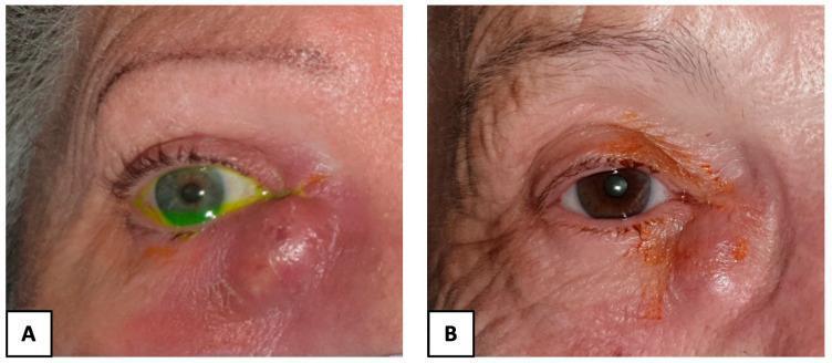
Q2. Dacryocystectomy Is Preferred in Chronic Dacryocystitis
Answer:
In chronic dacryocystitis, the lacrimal sac is chronically infected, mucocele-filled, and fibrosed — making dacryocystectomy (DCT) (surgical excision of the lacrimal sac) the procedure of choice over probing or other interventions.
Pathology in Chronic Dacryocystitis:
- Chronic obstruction of the nasolacrimal duct → stagnation of tears → secondary infection
- Sac is dilated, fibrosed, and filled with mucopurulent material
- Active chronic infection with Gram-positive cocci (Staphylococcus, Streptococcus) or fungi (Candida, Aspergillus)
- Sac wall is thickened, scarred, non-functional
Why DCT, Not DCR (Dacryocystorhinostomy)?
| Criterion | DCT Preferred | DCR Preferred |
|---|---|---|
| Sac condition | Fibrosed, non-functional, chronically infected | Functional, obstructed only at duct |
| Lacrimal pump function | Destroyed | Preserved |
| Risk of creating fistula | Creating a nasal fistula into infected sac is dangerous | Safe when sac is healthy |
| Risk of recurrence | Excision eliminates the sac permanently | Bypass only — sac remnant can reinfect |
| Malignancy concern | Must exclude sac tumors (send for histology) | Not needed |
| Age/tear function | Elderly, minimal tear production | Younger, active tear drainage needed |
Key Points:
- DCR creates a surgical fistula between lacrimal sac and nasal mucosa — bypasses obstruction but preserves the sac
- DCT removes the entire sac — eliminates the reservoir of infection permanently
- In elderly patients with dry eyes, even epiphora may not be a major concern
- Sac excision must always be sent for histopathology to rule out sac tumors (transitional cell carcinoma)
Conclusion: In chronic dacryocystitis, the sac is irreversibly damaged and serves as a focus of infection. DCT removes this focus permanently and is the definitive treatment of choice.
Q3. Proliferative Diabetic Retinopathy Is More Vision-Threatening Than Non-Proliferative
Answer:
Both forms represent diabetic retinopathy (DR), but PDR causes vision loss through catastrophic, rapidly progressive mechanisms that NPDR lacks.
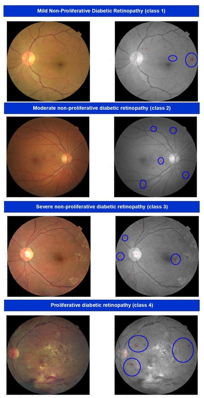
NPDR (Non-Proliferative Diabetic Retinopathy):
- Mechanism: Basement membrane thickening, pericyte loss, increased vascular permeability, microaneurysm formation
- Features: Microaneurysms, dot-blot hemorrhages, hard exudates, cotton-wool spots, venous beading, IRMA
- Vision threat: Diabetic Macular Edema (DME) → slow, insidious vision loss (central)
- No neovascularization — confined within retina
PDR (Proliferative Diabetic Retinopathy):
- Mechanism: Severe retinal ischemia → hypoxia → upregulation of VEGF (Vascular Endothelial Growth Factor) → neovascularization
- Features: New vessels on disc (NVD), new vessels elsewhere (NVE), new vessels on iris (NVI — rubeosis iridis)
Why PDR Is MORE Vision-Threatening:
| Complication | Mechanism | Visual Outcome |
|---|---|---|
| Vitreous hemorrhage | Fragile new vessels bleed into vitreous | Sudden, severe vision loss |
| Tractional retinal detachment | Fibrovascular proliferation contracts → pulls retina off | Profound, permanent vision loss |
| Neovascular glaucoma | NVI + fibrovascular membrane blocks trabecular meshwork | Painful, refractory, blind eye |
| Pre-retinal hemorrhage | Subhyaloid blood over macula | Sudden central vision loss |
| Rubeosis iridis | Angle closure → absolute glaucoma | Irreversible blindness |
High-Risk Characteristics for PDR (ETDRS criteria):
- NVD ≥ 1/4–1/3 disc area
- Any NVD with vitreous/preretinal hemorrhage
- NVE ≥ 1/2 disc area with vitreous/preretinal hemorrhage
Management of PDR:
- Pan-retinal photocoagulation (PRP) — ablates ischemic retina, reduces VEGF stimulus
- Anti-VEGF injections (ranibizumab, bevacizumab, aflibercept)
- Vitrectomy for non-clearing vitreous hemorrhage or tractional RD
Conclusion: NPDR causes slow macular edema; PDR causes sudden, severe, often irreversible vision loss through vitreous hemorrhage, tractional retinal detachment, and neovascular glaucoma — making PDR far more vision-threatening.
Q4. Visual Field Testing Is Done in Glaucoma Patients
Answer:
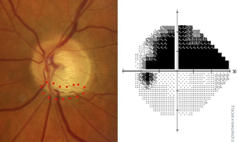
Glaucoma is a progressive optic neuropathy that preferentially destroys retinal ganglion cell axons at the optic nerve head — producing characteristic visual field (VF) defects that mirror the pattern of retinal nerve fibre layer (RNFL) damage.
Anatomical Basis:
- RNFL fibres run in arcuate patterns around the fovea (Henle's fibre layer)
- The horizontal raphe separates superior and inferior arcuate bundles
- Damage to specific bundles → corresponding field defects respecting the horizontal midline
Progression of Glaucomatous Visual Field Defects:
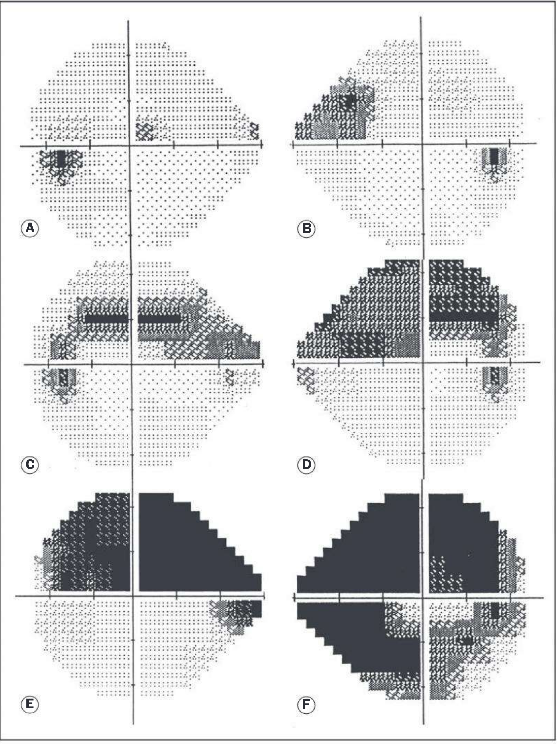
| Stage | Field Defect | Significance |
|---|---|---|
| Early | Small paracentral depressions (superonasal), nasal step | Subtle, often missed |
| Moderate | Arcuate scotoma (Bjerrum scotoma) — between 10°–20° from fixation | Characteristic of glaucoma |
| Advanced | Ring scotoma (superior + inferior arcuates meet) | Significant loss |
| End-stage | Tubular vision (central island) + temporal island | Near total loss |
Why VF Testing Is Essential:
- Detect functional damage early — structural damage precedes functional loss; VF confirms functional impact
- Monitor progression — mean deviation (MD) worsens by ~1 dB/year in untreated POAG
- Guide treatment — severity of field loss guides target IOP
- Assess treatment efficacy — stable VF = controlled disease
- Legal/driving fitness — bilateral advanced VF loss disqualifies patients from driving
Standard Automated Perimetry (SAP) — Humphrey Field Analyzer:
- 24-2 program — tests 52 points within 24° of fixation (standard)
- 10-2 program — monitors residual central field in advanced disease
- Outputs: Grayscale, Total Deviation, Pattern Deviation, MD, PSD, GHT (Glaucoma Hemifield Test)
Humphrey Criteria for Glaucomatous Damage (Hodapp-Parrish-Anderson):
- GHT outside normal limits on ≥2 consecutive tests, OR
- Cluster of ≥3 non-edge points depressed at P<5% (one at P<1%), OR
- Corrected PSD occurring in <5% of normals
Other Perimetric Tests:
- FDT (Frequency Doubling Technology) — detects early RNFL loss
- SWAP (Short Wavelength Automated Perimetry) — blue-on-yellow, tests M ganglion cells early
- Goldmann kinetic perimetry — for advanced disease, driving assessments
Conclusion: Visual field testing maps functional damage corresponding to RNFL loss, detects glaucoma early, guides treatment decisions, and monitors progression — it is indispensable in glaucoma management. — Kanski's Clinical Ophthalmology, 10th Ed., p. 381
Q5. White Pupillary Reflex in a Child Should Not Be Ignored
Answer:
A white pupillary reflex (leukocoria) is the replacement of the normal red-orange fundal reflex with a white/yellowish glow in the pupil. It is a potential ophthalmic and life-threatening emergency in a child.
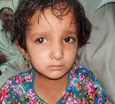
Normal Red Reflex vs Leukocoria:
- Normal fundal reflex is red-orange (light reflected from vascular choroid through clear media)
- Any opacity or abnormality in the visual axis causes leukocoria
Causes of Leukocoria (Mnemonic: CCRPPTV):
| Condition | Peak Age | Key Feature |
|---|---|---|
| Retinoblastoma | <5 years | MOST DANGEROUS — intraocular malignancy |
| Congenital cataract | Birth–3 years | Lens opacity |
| Coats' disease | 6–10 years (boys) | Retinal telangiectasia + exudation |
| Persistent fetal vasculature (PFV) | Birth | Persistent hyaloid artery |
| Retinopathy of prematurity (ROP) | Premature infants | Fibrovascular proliferation |
| Toxocara (ocular larva migrans) | 2–10 years | Granuloma at disc/macula |
| Vitreous hemorrhage | Any age | Blood in vitreous |
| Retinal detachment | Any | Elevated retina |
Why It Must NEVER Be Ignored — Retinoblastoma:
- Most common intraocular malignancy of childhood (1:15,000–20,000 live births)
- Caused by mutation in RB1 gene (chromosome 13q14) — classic tumor suppressor "two-hit" model
- If untreated: extends along optic nerve → intracranial → meningeal seeding → 100% fatal
- 5-year survival if diagnosed early (intraocular stage): >95%
- 5-year survival if diagnosed late (extraocular): <10%
Consequences of Ignoring Leukocoria:
- Delayed diagnosis of retinoblastoma → metastatic spread → death
- Delayed treatment of congenital cataract → irreversible amblyopia (deprivation amblyopia — critical period ends by 7–8 years)
- Untreated Coats' disease → tractional/exudative RD → phthisis bulbi
- Untreated ROP → bilateral blindness
Investigation Protocol:
- Fundoscopy under anaesthesia (EUA) — mandatory
- B-scan ultrasound — calcification confirms retinoblastoma
- CT/MRI orbit — extraocular extension, optic nerve involvement
- Systemic workup if retinoblastoma confirmed (bone marrow, LP for bilateral/hereditary cases)
Conclusion: Leukocoria in a child represents a sight- and life-threatening condition until proven otherwise. Retinoblastoma must be excluded urgently. Any delay in diagnosis risks metastatic spread and death, while vision-threatening conditions like congenital cataract cause irreversible amblyopia if not treated within the critical period.
SECTION B: SHORT NOTES (2×5 = 10) — Answer Any 2
SN1. Presbyopia
Definition: Presbyopia is the physiological, age-related loss of the eye's power of accommodation resulting in difficulty with near vision, typically manifesting after 40 years of age.
Physiology of Accommodation (Normal):
- Ciliary muscle contracts → zonular fibres relax → lens becomes more convex (rounded) → increased refractive power → near focus
- This is mediated by the parasympathetic innervation (oculomotor nerve via ciliary ganglion)
Pathophysiology of Presbyopia (Helmholtz Theory):
- With age, the crystalline lens loses elasticity (nuclear sclerosis → lens becomes rigid)
- Aged lens cannot change shape even when ciliary muscle contracts
- Lens fibres lose their ability to slide and deform
- Zonules remain intact; ciliary muscle may remain functional — the fault lies with the lens itself
Age of Onset:
- Amplitude of accommodation: 14D at birth → 7D at 25 yrs → 4D at 40 yrs → <1D at 60 yrs
- Clinical symptoms appear when amplitude falls below 4–5 dioptres
- Emmetropes and hypermetropes are affected earlier; myopes may compensate with their existing near focus
Clinical Features:
- Difficulty reading fine print, need to hold objects further away ("arm's length reading")
- Asthenopia (eye strain), headache after near work
- Need for better lighting
- Reading addition (near addition) required — typically +1.0D at 40 yrs, increasing to +3.0D by 60 yrs
Management:
- Spectacle correction:
- Reading glasses (single vision near)
- Bifocals (distance + near)
- Progressive addition lenses (PALs) — no visible demarcation line
- Contact lenses: Monovision or multifocal CLs
- Surgical correction (Kanski 10th Ed., p. 294–295):
- Refractive lens exchange (RLE/CLE): Removal of clear lens + multifocal IOL implantation
- Multifocal/EDOF IOL: Restores near-intermediate-distance vision
- Monovision LASIK: One eye for distance, other for near
- Presbyopic LASIK (presbyLASIK): Multifocal corneal profile
- Intracorneal inlays: Small-aperture (Kamra) or refractive inlays
- Conductive keratoplasty (CK): Radiofrequency shrinks peripheral stroma → steepens cornea for near
— Kanski's Clinical Ophthalmology, 10th Ed., p. 5494–5525
SN2. Biometry
Definition: Biometry is the measurement of the physical dimensions of the eye (axial length, anterior chamber depth, keratometry) to calculate the power of the intraocular lens (IOL) to be implanted after cataract surgery.
Goal:
Implant an IOL of correct power so the patient achieves emmetropia (or desired refraction) post-operatively.
Parameters Measured:
| Parameter | Normal Value | Instrument |
|---|---|---|
| Axial Length (AL) | ~23.5 mm | A-scan ultrasound / Optical biometry (IOLMaster) |
| Keratometry (K) | 43–44 D | Keratometer / Autorefractometer |
| Anterior Chamber Depth (ACD) | ~3.0–3.5 mm | A-scan / OCT |
| Lens thickness (LT) | ~4.0 mm | Immersion A-scan |
| Corneal diameter (white-to-white) | ~11.5 mm | Calliper / IOLMaster |
Methods of Axial Length Measurement:
1. A-scan Ultrasound (Contact / Immersion):
- Contact method: probe directly on cornea (may compress and underestimate AL by 0.1–0.3 mm)
- Immersion method (Prager shell) — more accurate, no corneal compression
- Sound velocity: 1532 m/s through aqueous/vitreous; 1641 m/s through lens
2. Optical Biometry (IOLMaster / Lenstar) — GOLD STANDARD:
- Uses partial coherence interferometry (PCI) or swept-source OCT
- Non-contact, highly reproducible (±0.02 mm)
- Cannot be used in dense cataracts (posterior subcapsular) or vitreous hemorrhage → revert to immersion A-scan
IOL Power Calculation Formulae:
| Formula | Best For |
|---|---|
| SRK/T | Average AL (22–26 mm) |
| Holladay 1 & 2 | Average to long eyes |
| Haigis | Short eyes, sulcus-fixated IOLs |
| Barrett Universal II | All AL ranges (currently preferred) |
| Hill-RBF | AI-based, all AL ranges |
| Hoffer Q | Short eyes (AL <22 mm) |
Special Situations:
- Post-LASIK/PRK eyes: Standard keratometry gives erroneous K → use adjusted keratometry methods (Masket, True-K, etc.)
- Silicone oil-filled eyes: AL measurement requires correction factor (oil velocity = 980 m/s)
- Dense cataracts: Optical biometry fails → immersion A-scan
Formula: SRK II (simplified):
P = A − 0.9K − 2.5L Where P = IOL power, A = A-constant (lens-specific), K = average keratometry (D), L = axial length (mm)
Conclusion: Accurate biometry is the cornerstone of modern cataract surgery outcomes. The shift from contact A-scan to optical biometry has reduced post-operative refractive surprises significantly.
SN3. Anti-Glaucoma Medications
Anti-glaucoma drugs reduce intraocular pressure (IOP) either by decreasing aqueous production or increasing aqueous outflow (trabecular or uveoscleral).
Classification:
1. Prostaglandin Analogues (First-line monotherapy)
| Drug | Example | Mechanism | Route |
|---|---|---|---|
| FP receptor agonists | Latanoprost 0.005%, Bimatoprost 0.03%, Travoprost 0.004%, Tafluprost | ↑ Uveoscleral outflow (matrix metalloproteinase-mediated ciliary body remodelling) | Topical OD (night) |
- IOP reduction: 25–35%
- Side effects: Conjunctival hyperaemia, iris pigmentation, periorbital fat atrophy, hypertrichosis, Prostaglandin-Associated Periorbitopathy (PAP)
- Contraindicated: Active uveitis, pregnancy
2. Beta-Adrenergic Blockers
| Drug | Example | Mechanism |
|---|---|---|
| Non-selective β-blocker | Timolol 0.25%, 0.5% (BD) | ↓ Aqueous production (β2 block on ciliary epithelium) |
| Selective β1-blocker | Betaxolol 0.25% (less IOP effect, more cardioselective) | Same |
- IOP reduction: 20–30%
- Side effects: Bradycardia, bronchospasm, depression, masking hypoglycaemia
- Contraindicated: Asthma, COPD, heart block, bradycardia
3. Carbonic Anhydrase Inhibitors (CAI)
| Drug | Route | Mechanism |
|---|---|---|
| Dorzolamide 2% (BD-TDS) | Topical | ↓ Aqueous production (inhibit CA-II in ciliary epithelium) |
| Brinzolamide 1% | Topical | Same |
| Acetazolamide 250–500 mg | Oral/IV | Systemic — more potent |
- Side effects (oral): Metabolic acidosis, hypokalemia, renal stones, aplastic anaemia, Stevens-Johnson syndrome
- Contraindicated: Sulfa allergy, renal stones, sickle cell
4. Alpha-2 Adrenergic Agonists
| Drug | Mechanism |
|---|---|
| Brimonidine 0.1–0.2% (BD) | ↓ Aqueous production + ↑ uveoscleral outflow; also neuroprotective? |
| Apraclonidine 0.5–1% (short-term) | ↓ Aqueous production |
- Side effects: Allergic follicular conjunctivitis (20–25%), dry mouth, fatigue, CNS depression in children
- Contraindicated: MAO inhibitor use, infants/children (respiratory depression)
5. Miotics (Cholinergic Agonists)
| Drug | Mechanism |
|---|---|
| Pilocarpine 1–4% (TDS-QDS) | Contracts ciliary muscle → opens trabecular meshwork → ↑ conventional outflow |
- Side effects: Miosis (dim vision), brow ache, induced myopia, risk of RD in high myopes, ciliary spasm
- Now largely replaced by prostaglandins
6. Hyperosmotic Agents (Acute Angle Closure Emergency)
| Drug | Route | Mechanism |
|---|---|---|
| Mannitol 1–2 g/kg (20%) | IV infusion | Osmotic dehydration of vitreous |
| Glycerol 50% | Oral | Same |
7. Combination Preparations:
- Dorzolamide + Timolol (Cosopt)
- Brimonidine + Timolol
- Latanoprost + Timolol (Xalacom)
Treatment Stepladder (POAG):
Monotherapy (PGA) → Add β-blocker or CAI → Triple therapy →
Laser (SLT/ALT) → Surgery (trabeculectomy / tube-shunt)
Conclusion: Anti-glaucoma drugs form the primary medical management of glaucoma. Prostaglandin analogues are the preferred first-line agents; combination therapy is used when monotherapy fails to achieve target IOP. — Kanski's Clinical Ophthalmology, 10th Ed.
---All 8 answers are complete. Here's a quick summary of what was covered:
Quick Index
| Q | Topic | Key Teaching Point |
|---|---|---|
| Q1 | Corneal graft — low rejection | Immunological privilege: avascularity + alymphaticity + ACAID + FasL |
| Q2 | DCT in chronic dacryocystitis | Sac is fibrosed/infected/non-functional — excision eliminates focus permanently; DCR would anastomose an infected sac to nasal cavity |
| Q3 | PDR more vision-threatening | PDR → VEGF → NV → vitreous hemorrhage, tractional RD, neovascular glaucoma — all catastrophic vs NPDR's slow DME |
| Q4 | VF testing in glaucoma | Maps RNFL loss functionally; detects, stages, and monitors glaucoma; guides treatment targets |
| Q5 | White pupillary reflex | Retinoblastoma must be excluded (life-threatening); congenital cataract causes deprivation amblyopia (irreversible if missed in critical period) |
| SN1 | Presbyopia | Age-related lens rigidity → loss of accommodation → near addition required; managed with spectacles/IOL/LASIK |
| SN2 | Biometry | Measures AL + K + ACD → IOL power calculation; optical biometry (IOLMaster) is gold standard; SRK/T, Barrett UII formulae |
| SN3 | Anti-glaucoma drugs | Prostaglandins (↑uveoscleral outflow) = first line; β-blockers (↓ production); CAI, α2-agonists, miotics, osmotics |
Sources: Kanski's Clinical Ophthalmology (10th Ed.) — the most authoritative standard ophthalmology textbook aligned with AK Khurana's content and examination standards.
This is a shared conversation. Sign in to Orris to start your own chat.