
Can you connect to previous chats ?
- Each session starts fresh - I only know what's in the current conversation window.
- No persistent memory - I can't recall your name, preferences, or prior questions from other sessions.
- No cross-session linking - Even if you've chatted with me many times, I can't retrieve any of that history.
Create a summary PDF of top 50 high-yield topics with one-liners for quick revision for NET PG EXAM with detailed explanation
Which subject(s) should this cover? NET PG can span many domains. · Which NET PG exam specifically?
mkdir -p /tmp/workspace/neet-pg-revision && ls /tmp/workspace/
~/neet-pg-revision/generate_pdf.py
from reportlab.lib.pagesizes import A4
from reportlab.lib import colors
from reportlab.lib.styles import getSampleStyleSheet, ParagraphStyle
from reportlab.lib.units import cm
from reportlab.platypus import (
    SimpleDocTemplate, Paragraph, Spacer, Table, TableStyle,
    HRFlowable, PageBreak, KeepTogether
)
from reportlab.lib.enums import TA_CENTER, TA_LEFT, TA_JUSTIFY
from reportlab.platypus import BaseDocTemplate, Frame, PageTemplate
from reportlab.lib.colors import HexColor

# ── Color palette ─────────────────────────────────────────────────────────────
C_DARK_BLUE   = HexColor("#1A237E")
C_MED_BLUE    = HexColor("#1565C0")
C_LIGHT_BLUE  = HexColor("#E3F2FD")
C_ACCENT      = HexColor("#F57F17")
C_GREEN       = HexColor("#1B5E20")
C_GREEN_LIGHT = HexColor("#E8F5E9")
C_RED         = HexColor("#B71C1C")
C_RED_LIGHT   = HexColor("#FFEBEE")
C_GREY_BG     = HexColor("#F5F5F5")
C_WHITE       = colors.white
C_BLACK       = colors.black

# ── Document setup ─────────────────────────────────────────────────────────────
OUTPUT = "/tmp/workspace/neet-pg-revision/NEET_PG_Top50_High_Yield.pdf"
doc = SimpleDocTemplate(
    OUTPUT,
    pagesize=A4,
    rightMargin=1.8*cm, leftMargin=1.8*cm,
    topMargin=2*cm, bottomMargin=2*cm,
    title="NEET PG – Top 50 High-Yield Topics",
    author="Orris AI"
)

styles = getSampleStyleSheet()

# Custom styles
TITLE_STYLE = ParagraphStyle("TitleS", parent=styles["Title"],
    fontSize=26, textColor=C_WHITE, alignment=TA_CENTER,
    spaceAfter=6, fontName="Helvetica-Bold")

SUBTITLE_STYLE = ParagraphStyle("SubtitleS", parent=styles["Normal"],
    fontSize=12, textColor=C_LIGHT_BLUE, alignment=TA_CENTER,
    spaceAfter=4, fontName="Helvetica")

SUBJECT_HEADER = ParagraphStyle("SubjH", parent=styles["Heading1"],
    fontSize=14, textColor=C_WHITE, fontName="Helvetica-Bold",
    spaceAfter=2, spaceBefore=10, alignment=TA_LEFT,
    leftIndent=6)

TOPIC_NUM = ParagraphStyle("TopicNum", parent=styles["Normal"],
    fontSize=11, textColor=C_WHITE, fontName="Helvetica-Bold",
    alignment=TA_CENTER)

TOPIC_NAME = ParagraphStyle("TopicName", parent=styles["Normal"],
    fontSize=13, textColor=C_DARK_BLUE, fontName="Helvetica-Bold",
    spaceAfter=3, spaceBefore=2)

ONE_LINER_LABEL = ParagraphStyle("OLL", parent=styles["Normal"],
    fontSize=9, textColor=C_ACCENT, fontName="Helvetica-Bold",
    spaceAfter=1)

ONE_LINER_TEXT = ParagraphStyle("OLT", parent=styles["Normal"],
    fontSize=11, textColor=C_RED, fontName="Helvetica-Bold",
    spaceAfter=6, leftIndent=8)

DETAIL_LABEL = ParagraphStyle("DL", parent=styles["Normal"],
    fontSize=9, textColor=C_MED_BLUE, fontName="Helvetica-Bold",
    spaceAfter=1)

DETAIL_TEXT = ParagraphStyle("DT", parent=styles["Normal"],
    fontSize=10, textColor=C_BLACK, fontName="Helvetica",
    spaceAfter=4, leftIndent=8, leading=14, alignment=TA_JUSTIFY)

BULLET_STYLE = ParagraphStyle("BS", parent=styles["Normal"],
    fontSize=10, textColor=C_BLACK, fontName="Helvetica",
    leftIndent=16, spaceAfter=2, leading=13,
    bulletIndent=8)

TOC_STYLE = ParagraphStyle("TOCS", parent=styles["Normal"],
    fontSize=10, textColor=C_DARK_BLUE, fontName="Helvetica",
    spaceAfter=2, leftIndent=8)

PAGE_NOTE = ParagraphStyle("PN", parent=styles["Normal"],
    fontSize=8, textColor=HexColor("#757575"), fontName="Helvetica",
    alignment=TA_CENTER)

# ── Topic data ─────────────────────────────────────────────────────────────────
# Each entry: (number, subject, topic_name, one_liner, detailed_explanation, bullets)
TOPICS = [
    # ── ANATOMY ──────────────────────────────────────────────────────────────
    (1, "ANATOMY", "Dermatomes & Myotomes",
     "C5 – shoulder abduction; L4 – knee extension; S1 – plantar flexion.",
     "Dermatomes are skin areas supplied by a single spinal nerve. Myotomes are muscle groups controlled by one spinal segment. Clinical testing maps neurological deficits to cord levels, guiding diagnosis of disc prolapses and cord injuries.",
     ["C5: Deltoid (abduction) | Biceps reflex",
      "C6: Wrist extension | Brachioradialis reflex",
      "C7: Wrist/finger extension | Triceps reflex",
      "L3-L4: Quadriceps (knee extension) | Knee jerk",
      "S1: Plantar flexion, eversion | Ankle jerk",
      "L4-L5: Dorsiflexion of foot (L4 = tibialis anterior)"]),

    (2, "ANATOMY", "Structures Passing Through Foramina",
     "Foramen ovale: V3; Foramen rotundum: V2; Foramen spinosum: middle meningeal artery.",
     "Knowledge of foraminal contents is high-yield for skull base anatomy questions. Damage to specific foramina produces predictable cranial nerve palsies.",
     ["Superior orbital fissure: CN III, IV, V1, VI + ophthalmic veins",
      "Jugular foramen: CN IX, X, XI + sigmoid sinus → IJV",
      "Hypoglossal canal: CN XII",
      "Foramen magnum: medulla, vertebral arteries, CN XI (spinal root)",
      "Internal acoustic meatus: CN VII, VIII"]),

    (3, "ANATOMY", "Femoral Triangle",
     "Contents (lateral → medial): NAVY – Nerve, Artery, Vein, Y-fronts (empty space/canal).",
     "The femoral triangle is bounded by the inguinal ligament (superiorly), sartorius (laterally), and adductor longus (medially). The femoral sheath encloses artery, vein, and canal (not the nerve). The femoral nerve lies outside the sheath.",
     ["Femoral nerve – most lateral, outside femoral sheath",
      "Femoral artery – central in sheath",
      "Femoral vein – medial to artery",
      "Femoral canal – most medial, contains lymphatics (Cloquet's node)",
      "Femoral hernia passes through the femoral canal"]),

    # ── PHYSIOLOGY ────────────────────────────────────────────────────────────
    (4, "PHYSIOLOGY", "Cardiac Action Potential Phases",
     "Phase 0=rapid depolarisation (Na+); Phase 2=plateau (Ca2+); Phase 3=repolarisation (K+).",
     "The ventricular action potential has 5 phases. Phase 0 rapid Na+ influx; Phase 1 early repolarisation (K+ out, Cl- in); Phase 2 plateau sustained by L-type Ca2+ channels balanced by K+ efflux; Phase 3 rapid repolarisation by K+ efflux; Phase 4 resting membrane potential (−90 mV). SA node lacks phase 1 and has automatic phase 4 depolarisation.",
     ["Longest phase is Phase 2 (plateau) – prevents tetany of heart",
      "Absolute refractory period = Phase 0, 1, 2, and most of Phase 3",
      "SA node pacemaker: funny current (If) – Na+ mediated",
      "Drugs targeting phases: Class I (Na+), Class III (K+), Class IV (Ca2+)",
      "QT interval correlates with Phase 2 + Phase 3"]),

    (5, "PHYSIOLOGY", "Starling's Law of the Heart",
     "Increased venous return → increased sarcomere stretch → increased stroke volume.",
     "Frank-Starling mechanism states that stroke volume is proportional to end-diastolic volume (preload). Myosin-actin overlap is optimised at sarcomere length 2.2 µm. Beyond this, force generation falls (descending limb). This mechanism allows the heart to eject whatever venous blood it receives without external nervous input.",
     ["Preload ↑ → EDV ↑ → SV ↑",
      "Afterload ↑ (e.g. hypertension) → SV ↓",
      "Contractility (inotropy) shifts the Starling curve upward",
      "Clinical: heart failure – operating on descending limb",
      "Optimal sarcomere length ≈ 2.2 µm"]),

    (6, "PHYSIOLOGY", "Spirometry Values (Normal Adult)",
     "FEV1/FVC >0.8 normal; <0.7 = obstruction; Normal FEV1/FVC with low FVC = restriction.",
     "Forced Vital Capacity (FVC) is the total air forcibly expelled. FEV1 is the volume in 1 second. The ratio distinguishes obstructive (asthma, COPD) from restrictive (fibrosis, obesity) disease. TLC is increased in obstruction and decreased in restriction.",
     ["FVC: 4–5 L (male), 3–4 L (female)",
      "FEV1: ~80% of FVC normally",
      "Obstructive: ↓ FEV1, normal/↑ FVC, ↓ FEV1/FVC, ↑ TLC",
      "Restrictive: ↓ FVC, normal FEV1/FVC, ↓ TLC",
      "Dead space (anatomical): ~150 mL",
      "Closing volume: small airway closure – ↑ with age"]),

    # ── BIOCHEMISTRY ──────────────────────────────────────────────────────────
    (7, "BIOCHEMISTRY", "Enzyme Inhibition Types",
     "Competitive: raises Km, same Vmax. Non-competitive: same Km, lowers Vmax.",
     "Enzyme kinetics are tested extensively. In competitive inhibition, the inhibitor resembles the substrate and competes for the active site; increasing substrate concentration can overcome it. Non-competitive inhibitors bind allosteric sites and cannot be overcome by substrate. Uncompetitive inhibitors only bind the enzyme-substrate complex, lowering both Km and Vmax.",
     ["Competitive: ↑ apparent Km, unchanged Vmax",
      "Non-competitive: unchanged Km, ↓ Vmax",
      "Uncompetitive: ↓ Km and ↓ Vmax (parallel line shift on Lineweaver-Burk)",
      "Methotrexate: competitive inhibitor of DHFR",
      "Aspirin: irreversible (covalent) inhibitor of COX",
      "Lineweaver-Burk plot: 1/v vs 1/[S]"]),

    (8, "BIOCHEMISTRY", "Glycolysis Key Enzymes",
     "Rate-limiting enzyme of glycolysis is Phosphofructokinase-1 (PFK-1).",
     "Glycolysis converts glucose (6C) to 2 pyruvate (3C), generating net 2 ATP and 2 NADH. Three irreversible steps: Hexokinase (glucose→G6P), PFK-1 (F6P→F1,6BP), and Pyruvate kinase. PFK-1 is allosterically inhibited by ATP/citrate and activated by AMP/F2,6BP.",
     ["Net ATP yield: 2 ATP per glucose",
      "PFK-1 inhibited by: ATP, citrate (+ = AMP, F2,6BP)",
      "Pyruvate kinase deficiency → hemolytic anemia",
      "G6PD deficiency → Heinz bodies, bite cells on smear",
      "Fructose bypasses PFK-1 (dietary excess → fat)",
      "Warburg effect: cancer cells prefer aerobic glycolysis"]),

    (9, "BIOCHEMISTRY", "Collagen Synthesis Steps",
     "Hydroxyproline and hydroxylysine synthesis requires Vitamin C (ascorbic acid).",
     "Collagen is the most abundant protein in the body. Synthesis: transcription → pre-procollagen → procollagen (in RER, hydroxylation of Pro/Lys by prolyl/lysyl hydroxylase requiring Vit C and Fe2+) → triple helix formation → extracellular cleavage → fibrillogenesis (cross-linking by lysyl oxidase requiring Cu2+).",
     ["Vit C deficiency → scurvy: perifollicular hemorrhages, poor wound healing",
      "Osteogenesis imperfecta: Type I collagen mutation → brittle bones",
      "Ehlers-Danlos: collagen structural/processing defect → hyperextensible skin",
      "Menkes disease: Cu2+ deficiency → lysyl oxidase failure → kinky hair",
      "Type I: bone, skin, tendon | Type II: cartilage | Type III: blood vessels | Type IV: basement membrane"]),

    # ── PATHOLOGY ─────────────────────────────────────────────────────────────
    (10, "PATHOLOGY", "Cell Injury & Necrosis Types",
     "Coagulative necrosis = most organs; Liquefactive = brain/abscess; Caseous = TB.",
     "Cell injury mechanisms: ATP depletion, free radical injury, Ca2+ influx, membrane damage. Reversible: cell swelling, fatty change. Irreversible: nuclear changes (pyknosis→karyorrhexis→karyolysis). Necrosis types are determined by which enzymes and tissue architecture prevail.",
     ["Coagulative: ischemic infarcts (except brain) – architecture preserved",
      "Liquefactive: brain infarcts, bacterial abscesses (proteases dominate)",
      "Caseous: TB/fungi –'cottage cheese', surrounded by granuloma",
      "Fat necrosis: pancreatitis/breast trauma – saponification (Ca soap deposits)",
      "Fibrinoid: immune complex vasculitis, malignant hypertension",
      "Gangrenous: limb ischemia; Wet gangrene = superimposed infection"]),

    (11, "PATHOLOGY", "Inflammatory Cells & Their Roles",
     "Neutrophils first (0-2 days); Macrophages replace them (days 2-7); Lymphocytes in chronic.",
     "Acute inflammation hallmarks: rubor, tumor, calor, dolor, functio laesa. Mediators: histamine (early), prostaglandins, leukotrienes, cytokines. Chronic inflammation: macrophages, lymphocytes, plasma cells, granuloma formation.",
     ["Neutrophils: first responders, pus cells, multilobed nucleus",
      "Eosinophils: parasites, allergic reactions, Charcot-Leyden crystals",
      "Mast cells: degranulate on IgE crosslinking → histamine release",
      "Macrophages: CD68+, phagocytosis, IL-1/TNF/IL-12 production",
      "Giant cells: Langhans (TB), Foreign body, Touton (fat necrosis)",
      "Plasma cells: produce antibodies, clock-face chromatin, Russell bodies"]),

    (12, "PATHOLOGY", "Tumour Markers",
     "AFP: HCC/germ cell; CEA: colorectal; PSA: prostate; CA-125: ovarian; CA 19-9: pancreatic.",
     "Tumour markers are used for monitoring treatment response, not screening (low specificity). They are produced by tumour cells or host response. A rise after treatment suggests recurrence.",
     ["AFP: Hepatocellular carcinoma, Yolk sac tumour, Neural tube defects (maternal AFP)",
      "CEA: Colorectal, lung, breast, pancreatic cancer",
      "PSA: Prostate cancer – elevated also in BPH, prostatitis",
      "CA-125: Epithelial ovarian cancer, endometriosis",
      "CA 19-9: Pancreatic, biliary cancer",
      "Beta-hCG: Choriocarcinoma, hydatidiform mole, testicular (non-seminoma)",
      "LDH: Non-specific – Seminoma, lymphoma, haemolysis",
      "S-100: Melanoma, schwannoma, Langerhans cell histiocytosis"]),

    (13, "PATHOLOGY", "Granuloma Forming Conditions",
     "SARCOIDOSIS – non-caseating; TB – caseating granuloma with central necrosis.",
     "A granuloma is a collection of activated macrophages (epithelioid cells) ± multinucleate giant cells, typically surrounded by lymphocytes. Formation requires T-cell mediated (Type IV) hypersensitivity and persistent antigen.",
     ["Non-caseating: Sarcoidosis, Crohn's disease, Berylliosis, foreign body",
      "Caseating: TB (Mycobacterium), Histoplasma, Coccidioides",
      "Sarcoid granuloma: Schaumann bodies (calcified), asteroid bodies",
      "Langhans giant cell (horseshoe nuclei) vs Foreign body giant cell (random nuclei)",
      "Leprosy: Virchow cells in lepromatous; Granulomas in tuberculoid"]),

    # ── MICROBIOLOGY ──────────────────────────────────────────────────────────
    (14, "MICROBIOLOGY", "Gram Positive vs Gram Negative Bacteria",
     "Gram +ve: thick peptidoglycan, no outer membrane. Gram −ve: thin PG, outer membrane with LPS.",
     "Gram staining differentiates bacteria based on cell wall composition. Gram +ve retain crystal violet (appear purple); Gram −ve decolorize and take safranin (appear red/pink). LPS (endotoxin) in Gram −ve bacteria mediates septic shock via TLR-4.",
     ["Gram +ve cocci: Staph aureus, Strep pyogenes, Strep pneumoniae, Enterococcus",
      "Gram +ve rods: Bacillus, Clostridium, Listeria, Corynebacterium",
      "Gram −ve cocci: Neisseria meningitidis, N. gonorrhoeae",
      "Gram −ve rods: E. coli, Klebsiella, Pseudomonas, Salmonella, Shigella",
      "LPS = Lipid A = endotoxin → fever, hypotension, DIC",
      "SOME bugs are Gram variable or poorly staining: Mycobacteria, Treponema"]),

    (15, "MICROBIOLOGY", "Virulence Factors & Toxins",
     "Cholera toxin: ADP-ribosylates Gs (↑cAMP → rice-water diarrhea). Botulinum: cleaves SNARE.",
     "Bacterial toxins are key NEET PG targets. Exotoxins are proteins (heat-labile, antigenic); endotoxin (LPS) is heat-stable, part of outer membrane.",
     ["Cholera toxin: Gs stimulation → ↑cAMP → Cl- secretion → watery diarrhea",
      "E. coli LT toxin: similar mechanism to cholera",
      "Pertussis toxin: Gi inhibition → ↑cAMP → lymphocytosis",
      "Anthrax toxin: Edema factor (adenylate cyclase) + Lethal factor (protease)",
      "Botulinum toxin: blocks ACh release at NMJ → flaccid paralysis",
      "Tetanus toxin (Tetanospasmin): blocks GABA/glycine at inhibitory interneurons → spastic paralysis",
      "Diphtheria toxin: ADP-ribosylates EF-2 → halts protein synthesis → myocarditis"]),

    (16, "MICROBIOLOGY", "TORCH Infections in Pregnancy",
     "TORCH: Toxoplasma, Other, Rubella, CMV, Herpes/HIV – cause congenital infections.",
     "TORCH infections cross the placenta or infect during delivery. CMV is the most common congenital viral infection. Rubella in first trimester causes the classic triad. Toxoplasma: cat litter. Herpes: neonatal infection during vaginal delivery.",
     ["Toxoplasma: Chorioretinitis, hydrocephalus, intracranial calcifications (periventricular)",
      "Rubella (1st trimester): Cataracts, PDA, sensorineural deafness (classic triad)",
      "CMV: Most common; periventricular calcifications, 'blueberry muffin rash', SNHL",
      "Herpes HSV-2: Neonatal encephalitis, skin/eye/mouth lesions",
      "Syphilis (Treponema): Saddle nose, sabre tibia, Hutchinson teeth, interstitial keratitis",
      "HIV: Mother-to-child transmission; Zidovudine reduces transmission"]),

    # ── PHARMACOLOGY ──────────────────────────────────────────────────────────
    (17, "PHARMACOLOGY", "Beta-Blocker Pharmacology",
     "Beta-blockers: negative inotropy/chronotropy; 1st line in HF (bisoprolol), post-MI, HTN.",
     "Beta-adrenergic blockers competitively block beta-1 (heart) and/or beta-2 (lung, peripheral vessels) receptors. Cardioselective agents (beta-1 selective) preferred in asthmatics, diabetics (mask hypoglycemia symptoms except sweating).",
     ["Cardioselective (beta-1): Metoprolol, Bisoprolol, Atenolol (MBA)",
      "Non-selective: Propranolol (also used for thyrotoxicosis, essential tremor, migraine prophylaxis)",
      "With ISA: Pindolol (partial agonist – less bradycardia at rest)",
      "Alpha+Beta: Carvedilol, Labetalol (used in hypertensive emergencies, pregnancy)",
      "Contraindications: Asthma, AV block, cardiogenic shock",
      "Abrupt withdrawal: rebound hypertension, angina, arrhythmia"]),

    (18, "PHARMACOLOGY", "Antibiotic Mechanisms & Resistance",
     "Penicillin: inhibits transpeptidase; Aminoglycosides: 30S; Fluoroquinolones: DNA gyrase.",
     "Antibiotics are categorized by their target. Resistance mechanisms: beta-lactamase production, efflux pumps, target modification, reduced permeability.",
     ["Cell wall synthesis: Beta-lactams (penicillin, cephalosporins, carbapenems), Vancomycin (binds D-Ala-D-Ala)",
      "30S ribosome: Aminoglycosides (irreversible), Tetracyclines (reversible)",
      "50S ribosome: Chloramphenicol, Macrolides (erythromycin), Linezolid, Clindamycin",
      "DNA gyrase/topoisomerase IV: Fluoroquinolones (ciprofloxacin)",
      "RNA polymerase: Rifampicin",
      "Folate synthesis: Sulfonamides (DHPS), Trimethoprim (DHFR)",
      "Cell membrane: Polymyxin B, Daptomycin (Gram +ve)"]),

    (19, "PHARMACOLOGY", "Drug-Induced Side Effects (High Yield)",
     "Aminoglycosides: ototoxicity + nephrotoxicity. Chloroquine: retinopathy. Rifampicin: orange urine.",
     "Specific drug toxicities are classic MCQ fodder. Always link drug → unique side effect → mechanism.",
     ["Statins: Myopathy/rhabdomyolysis (↑ CK); Hepatotoxicity",
      "Metformin: Lactic acidosis (contraindicated in renal failure/contrast)",
      "Cisplatin: Nephrotoxicity, ototoxicity, peripheral neuropathy",
      "Amiodarone: Pulmonary fibrosis, thyroid dysfunction, corneal deposits, photosensitivity",
      "Isoniazid: Peripheral neuropathy (Vit B6 depletion), hepatitis, SLE-like",
      "Clozapine: Agranulocytosis (monitor CBC weekly)",
      "Lithium: Tremor, polyuria (nephrogenic DI), hypothyroidism",
      "Tetracyclines: Teeth discoloration, photosensitivity (avoid <8 yr, pregnancy)"]),

    # ── MEDICINE ──────────────────────────────────────────────────────────────
    (20, "MEDICINE", "Myocardial Infarction – Diagnosis & Management",
     "STEMI: ST elevation + troponin rise; primary PCI within 90 min is gold standard.",
     "Acute MI results from plaque rupture and coronary artery thrombosis. Irreversible injury begins after 20–40 min ischemia. Biomarkers: Troponin I/T (most sensitive and specific), CK-MB (early re-infarction marker), Myoglobin (earliest, least specific).",
     ["STEMI: ST elevation ≥1 mm in ≥2 contiguous leads; LBBB pattern",
      "NSTEMI: Troponin +ve without ST elevation",
      "Unstable angina: no troponin rise, ischemia at rest/minimal exertion",
      "Complication timeline: Arrhythmia (1st day) > Pericarditis (days 2-4) > Dressler syndrome (weeks)",
      "Papillary muscle rupture (day 3-5): new MR murmur, acute pulmonary edema",
      "Acute management: MONA – Morphine, O2 (if SpO2 <90%), Nitrates, Aspirin (loading 300 mg) + Clopidogrel/Ticagrelor",
      "Long-term: ACEi, Beta-blocker, Statin, Aspirin, Clopidogrel"]),

    (21, "MEDICINE", "Heart Failure Classification & Management",
     "HFrEF (EF <40%): ACEi + Beta-blocker + spironolactone reduce mortality.",
     "Heart failure is inability to maintain adequate cardiac output. Systolic (HFrEF) vs Diastolic (HFpEF). NYHA classification grades functional capacity. BNP/NT-proBNP is the diagnostic biomarker of choice.",
     ["HFrEF drugs with mortality benefit: ACEi/ARB, Beta-blockers (bisoprolol/carvedilol), MRA (spironolactone), ARNI (sacubitril/valsartan), SGLT2i (dapagliflozin)",
      "Acute pulmonary edema: Furosemide + GTN + sit upright + O2",
      "NYHA I: No symptoms; II: Symptoms with moderate exertion; III: Symptoms with minimal exertion; IV: Symptoms at rest",
      "BNP >100 pg/mL supports HF diagnosis",
      "CXR: Bat-wing pulmonary edema, cardiomegaly, Kerley B lines, pleural effusion"]),

    (22, "MEDICINE", "Diabetes Mellitus – Diagnosis & Management",
     "Diagnosis: Fasting glucose ≥126 mg/dL, or 2hr OGTT ≥200, or HbA1c ≥6.5%.",
     "Type 1 DM: absolute insulin deficiency, autoimmune (anti-GAD, anti-IA2, anti-insulin Ab). Type 2: insulin resistance + progressive beta-cell failure. HbA1c reflects average glucose over 2-3 months.",
     ["Metformin: 1st line T2DM; inhibits hepatic gluconeogenesis; CI: eGFR <30",
      "SGLT2 inhibitors (gliflozins): ↓ cardiovascular mortality in HF + T2DM",
      "GLP-1 agonists (liraglutide): weight loss + CV benefit",
      "DKA: T1DM; pH <7.3, ketones +, glucose >250; Tx: fluids + insulin + K+",
      "HHS: T2DM; hyperosmolar, minimal ketosis, very high glucose (>600), high mortality",
      "Hypoglycemia: Whipple's triad – symptoms + low BG + relief with glucose",
      "Target HbA1c: <7% (ADA), <53 mmol/mol (NICE)"]),

    (23, "MEDICINE", "Hypertension Classification & Management",
     "HTN: BP ≥140/90 mmHg on two occasions; Stage 2: ≥160/100; Hypertensive emergency: end-organ damage.",
     "Essential hypertension (95%): no identifiable cause. Secondary causes: CKD (most common secondary cause), primary aldosteronism, renal artery stenosis, phaeochromocytoma, Cushing's.",
     ["1st line for most: ACEi/ARB (especially in DM + CKD), CCB (amlodipine), thiazide",
      "CCBs preferred in elderly, Afro-Caribbean patients",
      "ACEi: C/I in pregnancy → bilateral RAS → dry cough (switch to ARB)",
      "Hypertensive urgency (no organ damage): oral labetalol/amlodipine",
      "Hypertensive emergency: IV labetalol/nitroprusside; reduce MAP by 25% in first hour",
      "Malignant hypertension: >180/120 + papilloedema/retinal haemorrhages"]),

    (24, "MEDICINE", "Thyroid Disorders",
     "Hypothyroidism: TSH ↑, T4 ↓; Hyperthyroidism: TSH ↓, T4 ↑. TSH is the best screening test.",
     "The hypothalamic-pituitary-thyroid axis regulates thyroid hormone production. TRH → TSH → T3/T4. T4 is prohormone; T3 is active form. Negative feedback at pituitary.",
     ["Hypothyroidism Sx: cold intolerance, constipation, weight gain, bradycardia, non-pitting edema (myxedema), delayed DTRs",
      "Hashimoto's thyroiditis: most common cause hypothyroidism; anti-TPO Ab, anti-Tg Ab",
      "Hyperthyroidism Sx: heat intolerance, diarrhea, weight loss, palpitations, tremor, exophthalmos (Graves)",
      "Graves disease: TSH receptor Ab (TSI); autoimmune; pretibial myxedema",
      "Treatment Graves: Carbimazole/PTU, Radioiodine, Surgery",
      "Thyroid storm: Propranolol + PTU + iodine (Lugol's) + hydrocortisone",
      "Sick euthyroid syndrome: ↓ T3, normal/↓ T4, normal/↓ TSH – seen in critically ill"]),

    # ── SURGERY ───────────────────────────────────────────────────────────────
    (25, "SURGERY", "Surgical Causes of Acute Abdomen",
     "Guarding + rigidity = peritonitis; Shifting dullness + absent bowel sounds = obstruction.",
     "Acute abdomen requires rapid diagnosis. Murphy's sign (cholecystitis), Rovsing's sign (appendicitis), Cullen's sign (umbilical bruising – haemoperitoneum), Grey-Turner sign (flank bruising – retroperitoneal hemorrhage).",
     ["Appendicitis: McBurney's point tenderness, Psoas sign, Obturator sign; WBC ↑",
      "Acute pancreatitis: epigastric pain radiating to back, Serum amylase/lipase >3× normal",
      "Cholecystitis: Murphy's sign +, fever, RUQ pain after fatty meal, USS gold standard",
      "Peptic ulcer perforation: sudden-onset severe epigastric pain, 'free air' under diaphragm on erect CXR",
      "Mesenteric ischemia: pain out of proportion to exam, atrial fibrillation risk factor",
      "Intestinal obstruction: colicky pain, vomiting, distension, constipation; central dilated loops (small bowel) vs peripheral (large bowel) on AXR"]),

    (26, "SURGERY", "Fluid & Electrolyte Management",
     "Hartmann's (Ringer's lactate) is the resuscitation fluid of choice in surgery/trauma.",
     "IV fluids replace lost volume and correct electrolyte imbalances. Crystalloids vs colloids. 0.9% NaCl can cause hyperchloraemic acidosis if large volumes given. Dextrose distributes into intracellular compartment.",
     ["Normal saline (0.9%): 154 mEq/L Na and Cl; hyperchloraemic acidosis with large volumes",
      "Hartmann's: Na 131, K 5, Ca 2, Cl 111, lactate 29 mEq/L – most physiological",
      "5% Dextrose: distributes to all body water (TBW); not for volume resuscitation",
      "Daily requirements: Na 1 mmol/kg, K 1 mmol/kg, fluid 30 mL/kg/day",
      "Hyponatraemia: correct slowly (<10-12 mEq/L per 24h) to prevent central pontine myelinolysis",
      "Hyperkalaemia >6.5 mEq/L with ECG changes: IV calcium gluconate (membrane stabilisation) first"]),

    # ── OBSTETRICS & GYNAECOLOGY ──────────────────────────────────────────────
    (27, "OBG", "Pre-eclampsia & Eclampsia",
     "Pre-eclampsia: BP ≥140/90 + proteinuria after 20 weeks. Only cure is delivery.",
     "Pre-eclampsia results from abnormal placentation and maternal endothelial dysfunction. HELLP syndrome (Haemolysis, Elevated Liver enzymes, Low Platelets) is a severe complication. Risk factors: nulliparity, multiple pregnancy, CKD, prior PE.",
     ["Diagnosis: BP ≥140/90 on 2 occasions 4h apart + proteinuria ≥300 mg/24h after 20 wk",
      "Severe PE: BP ≥160/110, severe headache, visual disturbances, epigastric pain, oliguria",
      "HELLP: haemolysis (↑LDH, ↑bilirubin, schistocytes), AST/ALT ↑, platelets <100,000",
      "Eclampsia: seizures in setting of pre-eclampsia; Tx: IV MgSO4 (anticonvulsant of choice)",
      "MgSO4 toxicity: loss of DTRs (first sign, at ~7 mEq/L), respiratory depression (>12 mEq/L); antidote: IV calcium gluconate",
      "Antihypertensives safe in pregnancy: Labetalol, Hydralazine, Nifedipine, Methyldopa"]),

    (28, "OBG", "Normal Labour & Stages",
     "Stage 1: effacement/dilation (latent <6cm, active >6cm); Stage 2: delivery; Stage 3: placenta.",
     "Labour is defined as regular uterine contractions with progressive cervical effacement and dilation. Active management reduces PPH. CTG monitoring helps detect fetal distress.",
     ["Latent phase: 0–6 cm; Active phase: 6–10 cm (rate ≥1 cm/hr expected)",
      "Stage 2: full dilation to delivery of baby; ≤2 hr nullipara, ≤1 hr multipara",
      "Stage 3: delivery of placenta; active management with oxytocin reduces PPH",
      "PPH: blood loss >500 mL (vaginal) or >1000 mL (caesarean); 4 Ts – Tone (most common), Trauma, Tissue, Thrombin",
      "CTG: Normal FHR 110–160 bpm; decelerations – Early (head compression), Variable (cord compression), Late (placental insufficiency)"]),

    (29, "OBG", "Gynaecological Cancers – Key Features",
     "Endometrial cancer: most common; postmenopausal bleeding. Cervical: HPV 16/18; Pap smear screen.",
     "Gynaecological malignancies differ in risk factors, screening, and treatment. Know the classic presentation for each.",
     ["Cervical cancer: HPV 16/18 (squamous cell – 80%); Pap smear/colposcopy; LEEP/cone biopsy",
      "Endometrial cancer: unopposed oestrogen (obesity, PCOS, tamoxifen), Lynch syndrome; D&C/hysteroscopy biopsy; Tx: hysterectomy + BSO",
      "Ovarian cancer: 'silent killer' – CA-125 + pelvic USS; BRCA1/2 mutation risk; serous cystadenocarcinoma most common",
      "Vulvar cancer: SCC, postmenopausal, pruritus, white plaque (lichen sclerosus)",
      "CIN grading: CIN 1 (mild dysplasia) → CIN 3 (carcinoma in situ)"]),

    # ── PAEDIATRICS ───────────────────────────────────────────────────────────
    (30, "PAEDIATRICS", "Developmental Milestones",
     "Smile: 6 wk; Sit unsupported: 6 mo; Walk: 12–15 mo; 2-word phrases: 24 mo.",
     "Developmental assessment covers gross motor, fine motor, speech/language, and social domains. Regression of milestones is always abnormal. Delays prompt investigation for metabolic/structural causes.",
     ["3 months: holds head up, social smile (6 weeks), follows past midline",
      "6 months: rolls over, sits with support, babbles",
      "9 months: sits unsupported, pincer grasp emerging, stranger anxiety",
      "12 months: walks with support/alone, 1-2 words with meaning, waves bye-bye",
      "18 months: walks well, 10+ words, points to body parts",
      "24 months: runs, 2-word phrases, 50+ words; parallel play",
      "Red flags: no social smile by 3 mo, no babble by 12 mo, no words by 16 mo, any regression"]),

    (31, "PAEDIATRICS", "Childhood Vaccination Schedule (India – UIP)",
     "BCG: birth; OPV+Penta: 6,10,14 wk; Measles: 9 mo; MMR: 15 mo; DPT booster: 18 mo.",
     "India's Universal Immunisation Programme (UIP) protects against 12 diseases. Key vaccine facts for MCQs: cold chain, contraindications, schedules.",
     ["BCG: Birth; prevents miliary TB and TB meningitis; scar appears at 6-8 weeks",
      "OPV (oral polio): 0, 6, 10, 14 wk + boosters; VAPP risk (use IPV if immunocompromised)",
      "Pentavalent (DPT+HepB+Hib): 6, 10, 14 weeks",
      "Measles: 9-12 months; MMR: 15-18 months (2nd dose at 5 years)",
      "JE vaccine: 9-12 months in endemic areas",
      "Typhoid (Vi capsular): 2 years+; Rotavirus: 6, 10, 14 weeks (oral)",
      "Hepatitis B: 3 doses at 0, 1, 6 months OR birth dose + 6, 10, 14 weeks"]),

    # ── PSYCHIATRY ────────────────────────────────────────────────────────────
    (32, "PSYCHIATRY", "Schizophrenia – Diagnosis & Treatment",
     "Schizophrenia: ≥2 positive symptoms for ≥1 month; prodrome >6 months; dopamine excess theory.",
     "Schizophrenia is characterised by positive symptoms (hallucinations, delusions, disorganized thinking) and negative symptoms (flat affect, alogia, avolition). Dopamine D2 receptor hyperactivity in mesolimbic pathway mediates positive symptoms.",
     ["Schneider's 1st rank symptoms: Thought insertion/withdrawal/broadcasting, Passivity phenomena, Somatic hallucinations, Delusional perception, Running commentary",
      "Typical antipsychotics: Haloperidol, Chlorpromazine – block D2; SE: EPS, tardive dyskinesia, hyperprolactinaemia",
      "Atypical antipsychotics: Olanzapine, Risperidone, Clozapine – D2 + 5HT2A; fewer EPS",
      "Clozapine: reserved for treatment-resistant schizophrenia; risk agranulocytosis",
      "Neuroleptic Malignant Syndrome: Hyperthermia, Rigidity, Altered consciousness, ↑CK; Tx: Bromocriptine + Dantrolene",
      "Negative symptoms: 5 As – Affective flattening, Alogia, Avolition, Anhedonia, Attention deficits"]),

    (33, "PSYCHIATRY", "Depression & Antidepressants",
     "Depression: ≥5 symptoms ≥2 weeks including low mood/anhedonia. SSRIs are 1st line.",
     "Major depressive disorder (MDD) is defined by depressed mood or anhedonia plus additional symptoms for at least 2 weeks. Biological theory: monoamine deficiency (serotonin, noradrenaline, dopamine).",
     ["Core symptoms: Low mood + anhedonia (at least one required)",
      "Others: Sleep disturbance, Appetite change, Concentration difficulties, Energy loss, Guilt/worthlessness, Psychomotor changes, Suicidal ideation",
      "SSRIs (1st line): Fluoxetine, Sertraline, Escitalopram – SE: sexual dysfunction, serotonin syndrome",
      "SNRIs: Venlafaxine, Duloxetine – also used for pain, anxiety",
      "TCAs: Amitriptyline – SE: anticholinergic (dry mouth, urinary retention, constipation), arrhythmias",
      "MAOIs: Phenelzine – tyramine interaction → hypertensive crisis (avoid aged cheese)",
      "Electroconvulsive therapy (ECT): 1st line in severe depression with psychotic features, suicidal risk, or failure of pharmacotherapy"]),

    # ── ENT ───────────────────────────────────────────────────────────────────
    (34, "ENT", "Otitis Media – Acute & Chronic",
     "AOM: fever + otalgia; bulging red TM; Strep pneumoniae most common causative organism.",
     "Otitis media is inflammation of the middle ear. Acute (AOM) vs chronic suppurative (CSOM) vs otitis media with effusion (glue ear). Eustachian tube dysfunction is central to pathogenesis.",
     ["AOM causative organisms: S. pneumoniae (most common), H. influenzae, M. catarrhalis",
      "AOM treatment: Amoxicillin (amoxicillin-clavulanate if treatment failure)",
      "Complications of AOM: Mastoiditis, Meningitis, Lateral sinus thrombosis, Facial nerve palsy",
      "CSOM: chronic discharge >6 wk; mucosal type vs squamous (cholesteatoma)",
      "Cholesteatoma: keratinous debris trapped in middle ear; attic perforation on otoscopy; Tx: surgery",
      "Glue ear (OME): conductive hearing loss in children; myringotomy + grommets if persistent"]),

    (35, "ENT", "Hoarseness – Differential Diagnosis",
     "Hoarseness >3 weeks requires laryngoscopy to exclude laryngeal carcinoma.",
     "Hoarseness (dysphonia) results from any disorder of the larynx or recurrent laryngeal nerve. Red flag: unilateral, progressive, associated weight loss → malignancy until proven otherwise.",
     ["Benign: Vocal cord nodules (screamer's nodule), Polyps, Laryngitis, GERD",
      "Functional: Psychogenic dysphonia, Muscle tension dysphonia",
      "Malignant: Laryngeal SCC – most common in hypopharynx/glottis; smoking + alcohol risk",
      "Neurological: RLN palsy (thyroid surgery, aortic aneurysm, lung cancer – left side)",
      "Left RLN: longer course, loops around arch of aorta; more commonly injured",
      "Investigation: Flexible laryngoscopy; CT neck/chest; Biopsy for malignancy"]),

    # ── OPHTHALMOLOGY ─────────────────────────────────────────────────────────
    (36, "OPHTHALMOLOGY", "Glaucoma – Types & Management",
     "Primary open-angle glaucoma: painless, gradual visual field loss; IOP >21 mmHg.",
     "Glaucoma is a group of conditions characterised by optic nerve damage, visual field loss, and elevated IOP. Primary open-angle (chronic) is the most common. Primary angle-closure (acute) is an emergency.",
     ["POAG: Insidious visual loss; open drainage angle; cupping of optic disc (C:D ratio >0.6)",
      "Acute angle closure: Red eye, severe pain, haloes, nausea, rock-hard eye, mid-dilated pupil",
      "IOP normal: 10–21 mmHg; Normal tension glaucoma: disc damage with normal IOP",
      "1st line treatment: Topical prostaglandin analogue (latanoprost) → ↑ uveoscleral outflow",
      "Beta-blockers (timolol): ↓ aqueous production; C/I asthma",
      "Carbonic anhydrase inhibitors (acetazolamide): ↓ aqueous production – used in acute crisis",
      "Laser trabeculoplasty/trabeculectomy for refractory cases"]),

    (37, "OPHTHALMOLOGY", "Diabetic Retinopathy",
     "NPDR: microaneurysms → hard exudates → cotton wool spots. PDR: neovascularisation.",
     "Diabetic retinopathy is the leading cause of blindness in working-age adults. Annual fundoscopy/retinal photography is mandatory in all diabetics. Duration of diabetes is the strongest risk factor.",
     ["Background/Mild NPDR: Microaneurysms only",
      "Moderate/Severe NPDR: Dot-blot hemorrhages, hard exudates, cotton-wool spots",
      "Proliferative DR (PDR): New vessel formation (NVD/NVE), vitreous hemorrhage, tractional retinal detachment",
      "Clinically significant macular edema (CSME): Treat with intravitreal anti-VEGF (bevacizumab/ranibizumab)",
      "Panretinal photocoagulation (PRP): treatment for PDR",
      "Screening: Annual dilated fundoscopy from diagnosis (T2DM) or 5 years after diagnosis (T1DM)"]),

    # ── DERMATOLOGY ───────────────────────────────────────────────────────────
    (38, "DERMATOLOGY", "Skin Lesion Terminology",
     "Macule: flat <1cm; Papule: raised <1cm; Plaque: raised >1cm; Vesicle: blister <0.5cm; Bulla: blister >0.5cm.",
     "Precise skin lesion description is fundamental to dermatology diagnosis. Primary lesions arise from normal skin; secondary lesions result from evolution or manipulation of primary lesions.",
     ["Primary: Macule, Papule, Plaque, Nodule, Vesicle, Bulla, Pustule, Wheal (urticaria), Cyst",
      "Secondary: Scale, Crust, Erosion (superficial), Ulcer (deep), Lichenification, Scar, Excoriation",
      "Koebner phenomenon: lesions appear in areas of skin trauma (psoriasis, lichen planus, vitiligo, warts)",
      "Auspitz sign: bleeding on removal of psoriatic scale (pinpoint capillary bleeding)",
      "Nikolsky sign: blister extends/creates new blister on lateral pressure (pemphigus vulgaris, SSSS)"]),

    (39, "DERMATOLOGY", "Common Skin Conditions – High-Yield Differentials",
     "Psoriasis: silver scaly plaques on extensor surfaces. Lichen planus: 4Ps – Purple, Pruritic, Polygonal, Papules.",
     "Inflammatory skin diseases are distinguished by morphology, distribution, histology, and associated conditions.",
     ["Psoriasis: Well-demarcated silver scaly plaques; elbows, knees, scalp; nail pitting; HLA-Cw6",
      "Lichen planus: Wickham's striae (white lacy pattern), buccal mucosa involvement; HLA-DR1",
      "Atopic eczema: flexural distribution; IgE mediated; associated with asthma, allergic rhinitis",
      "Pemphigus vulgaris: flaccid blisters, Nikolsky+, oral involvement; IgG vs desmoglein 3",
      "Bullous pemphigoid: tense blisters, Nikolsky−, elderly; IgG vs BP180 (BPAG2) in basement membrane",
      "Dermatitis herpetiformis: intensely pruritic vesicles on extensor surfaces; coeliac disease; IgA deposits"]),

    # ── ORTHOPAEDICS ──────────────────────────────────────────────────────────
    (40, "ORTHOPAEDICS", "Fracture Complications",
     "Fat embolism: 24-72hr post long bone #; Compartment syndrome: 6Ps; DVT: most common late complication.",
     "Complications of fractures can be immediate, early, or late. Compartment syndrome is a surgical emergency requiring fasciotomy within hours to prevent permanent nerve/muscle damage.",
     ["Fat embolism syndrome: hypoxia, petechiae (upper body), confusion – 24-72 hr post femur/tibia #",
      "Compartment syndrome: 6 Ps – Pain (especially with passive stretch), Pressure (tense compartment), Paresthesia, Paralysis, Pallor, Pulselessness; Tx: emergency fasciotomy",
      "AVN (avascular necrosis): after neck of femur fracture, scaphoid waist fracture, dislocation of hip; MRI is most sensitive investigation",
      "Malunion: healed in incorrect position; Non-union: failure to heal by 6 months",
      "DVT/PE: most common late complication; prophylaxis with LMWH, compression stockings",
      "Reflex Sympathetic Dystrophy (CRPS): burning pain, allodynia, autonomic changes"]),

    (41, "ORTHOPAEDICS", "Common Orthopaedic Tests",
     "Lachman's test: ACL integrity; McMurray's: meniscus; Apprehension test: shoulder dislocation.",
     "Clinical examination tests help localise musculoskeletal pathology. NEET PG tests name, technique, and interpretation.",
     ["Lachman's test: ACL tear – most sensitive (>85%)",
      "Anterior/Posterior drawer test: ACL/PCL tear",
      "McMurray's test: meniscal tear – pain/click on rotation",
      "Valgus/varus stress test: MCL/LCL integrity",
      "Apprehension test: anterior shoulder instability (recurrent dislocation)",
      "Trendelenburg test: hip abductor weakness / superior gluteal nerve palsy",
      "FABER (Patrick's test): hip pathology / sacroiliac joint disease",
      "Thomas test: hip flexion contracture"]),

    # ── FORENSIC MEDICINE ─────────────────────────────────────────────────────
    (42, "FORENSIC MEDICINE", "Thanatology – Signs of Death",
     "Brain death: fixed dilated pupils + no brainstem reflexes + apnea + flat EEG.",
     "Thanatology is the study of death. Signs of death progress from immediate (cardiac/respiratory arrest) to early (pallor, cooling) to late (rigor mortis, putrefaction).",
     ["Rigor mortis: starts 2-6 hr, complete at 12 hr, disappears at 36-48 hr (higher temp shortens)",
      "Livor mortis (hypostasis): appears 1-2 hr, fixes at 6-8 hr; pink-red in CO poisoning; cherry red in cyanide",
      "Algor mortis: body cools ~1°C/hr for first 12 hr in average conditions",
      "Putrefaction: starts 24-48 hr in abdomen (bacteria); marbling, green discolouration",
      "Adipocere: saponification of body fat; wet/waterlogged conditions; 3-6 months",
      "Mummification: dry heat/arid conditions; body desiccates preserving features"]),

    (43, "FORENSIC MEDICINE", "Wounds – Classification & Medico-legal Importance",
     "Incised wound: sharp cutting force, clean edges. Laceration: blunt force, irregular edges with tissue bridging.",
     "Wound classification is critical in forensic assessment of cause of death, weapon identification, and establishing manner of death.",
     ["Incised wound: clean-cut edges, no bruising at margins, greater length than depth",
      "Stab wound: greater depth than length; entry wound characteristic of weapon",
      "Laceration: blunt force; irregular, contaminated edges; tissue bridges at base",
      "Contusion (bruise): extravasation of blood; does not blanch on pressure",
      "Abrasion: superficial; marks direction of force (tramline abrasion = blunt rod)",
      "Gunshot wound: entry (inverted, abraded collar, small) vs exit (larger, irregular, no abrasion collar)",
      "Hesitation marks: tentative cuts in cases of self-inflicted wounds"]),

    # ── COMMUNITY MEDICINE / PSM ──────────────────────────────────────────────
    (44, "COMMUNITY MEDICINE", "Epidemiology – Key Measures",
     "Incidence = new cases/population at risk per unit time. Prevalence = existing cases/total population.",
     "Epidemiological measures quantify disease burden and risk. Correct interpretation of study designs and statistical measures is heavily tested.",
     ["Incidence rate: new cases per 1000/100,000 at-risk people per year",
      "Prevalence = Incidence × Duration of disease",
      "Attack rate: incidence proportion in an outbreak (epidemic)",
      "Relative Risk (RR): risk in exposed/risk in unexposed – used in cohort studies",
      "Odds Ratio (OR): odds in cases/odds in controls – used in case-control studies; approximates RR when prevalence is low",
      "Attributable Risk (AR) = Risk(exposed) − Risk(unexposed) – public health impact",
      "Number Needed to Treat (NNT) = 1/Absolute Risk Reduction"]),

    (45, "COMMUNITY MEDICINE", "Screening Tests – Sensitivity & Specificity",
     "Sensitive test: fewer false negatives (good to RULE OUT). Specific test: fewer false positives (good to RULE IN).",
     "Sensitivity and specificity are intrinsic properties of a test; PPV and NPV depend on disease prevalence.",
     ["Sensitivity = TP/(TP+FN) = True Positive Rate; 'SnNout' – sensitive test negative rules OUT",
      "Specificity = TN/(TN+FP) = True Negative Rate; 'SpPin' – specific test positive rules IN",
      "PPV = TP/(TP+FP) – increases with higher prevalence",
      "NPV = TN/(TN+FN) – decreases with higher prevalence",
      "ROC curve: plots sensitivity vs (1-specificity); area under curve = test discrimination",
      "Likelihood ratio positive = Sensitivity/(1-Specificity)",
      "Ideal screening test: High sensitivity (to catch all cases), affordable, acceptable"]),

    # ── RADIOLOGY ─────────────────────────────────────────────────────────────
    (46, "RADIOLOGY", "X-Ray Interpretation – Key Patterns",
     "Consolidation: air bronchograms; Collapse: dense with volume loss; Pleural effusion: meniscus sign.",
     "Plain X-ray remains the first-line investigation in many clinical scenarios. Systematic interpretation (ABCDE) prevents missed findings.",
     ["Air bronchogram: patent bronchi within consolidated lung – suggests pneumonia, not effusion",
      "Silhouette sign: loss of normal heart/diaphragm border when adjacent structure is consolidated",
      "CXR cardiac enlargement: cardiothoracic ratio >50% on PA film",
      "Bat-wing/butterfly pattern: bilateral perihilar air-space opacification – pulmonary edema",
      "Reticular nodular pattern: interstitial lung disease (sarcoid, IPF, hypersensitivity pneumonitis)",
      "Pleural effusion: blunting of costophrenic angle; meniscus sign; >200 mL visible on PA CXR",
      "Pneumothorax: absent lung markings with visible visceral pleural line"]),

    (47, "RADIOLOGY", "MRI vs CT – Clinical Decision Making",
     "MRI: best for soft tissue, CNS, spine. CT: best for bone, trauma, chest, abdomen emergencies.",
     "Choosing the right imaging modality improves diagnostic yield and reduces radiation exposure. MRI uses magnetic fields and radiofrequency waves; no ionising radiation.",
     ["MRI T1: fat bright, water dark; good for anatomy",
      "MRI T2: water bright (CSF, edema); best for detecting pathology",
      "CT: fast (minutes), best for hemorrhage (bright on CT), fractures, thoracic/abdominal pathology",
      "CT with contrast: vascular lesions, abscess, malignancy, liver lesions",
      "MRI C-spine: disc prolapse, cord compression preferred over CT",
      "Non-contrast CT head: first investigation for suspected acute stroke (to exclude hemorrhage)",
      "PET-CT: metabolic activity; tumour staging, recurrence detection"]),

    # ── ANAESTHESIA ───────────────────────────────────────────────────────────
    (48, "ANAESTHESIA", "Anaesthetic Agents – Mechanism & Side Effects",
     "Propofol: rapid induction/recovery; Ketamine: dissociative analgesia; Thiopental: ultra-short barbiturate.",
     "Intravenous induction agents work rapidly due to high lipid solubility and CNS penetration. All cause cardiovascular depression to varying degrees.",
     ["Propofol: GABA-A potentiation; 'propofol infusion syndrome' – metabolic acidosis, cardiac failure",
      "Ketamine: NMDA receptor antagonist; dissociative anesthesia; preserves airway reflexes; bronchodilator; contraindicated in ↑ICP/IOP",
      "Thiopental: barbiturate; GABA potentiation; decreases ICP; porphyria contraindication",
      "Etomidate: adrenocortical suppression; least cardiovascular depression (good for shocked patients)",
      "Volatile agents: Halothane – hepatotoxicity; Isoflurane – hypotension; Sevoflurane – paediatric induction",
      "Suxamethonium: depolarising muscle relaxant; contraindicated in burns/crush injury (hyperkalemia), malignant hyperthermia"]),

    (49, "ANAESTHESIA", "Malignant Hyperthermia",
     "Malignant hyperthermia: uncontrolled skeletal muscle metabolism triggered by suxamethonium/volatile agents; Rx: Dantrolene.",
     "MH is a rare but life-threatening pharmacogenetic disorder of skeletal muscle. The ryanodine receptor (RYR1) mutation causes uncontrolled Ca2+ release from the sarcoplasmic reticulum.",
     ["Triggers: Suxamethonium (most common), halogenated volatile agents (halothane, sevoflurane)",
      "Features: Hyperthermia (>38.5°C, rising), muscle rigidity, masseter spasm, tachycardia, hypercarbia, metabolic acidosis, ↑↑ CK",
      "Lab: Hyperkalaemia, myoglobinuria (rhabdomyolysis), DIC",
      "Treatment: STOP trigger, Dantrolene sodium (2.5 mg/kg IV, repeat), active cooling, supportive care",
      "Dantrolene mechanism: inhibits ryanodine receptor → prevents sarcoplasmic Ca2+ release",
      "Similar condition: Neuroleptic malignant syndrome (antipsychotic triggered; treat with bromocriptine + dantrolene)"]),

    # ── MISCELLANEOUS HIGH-YIELD ───────────────────────────────────────────────
    (50, "HIGH-YIELD MISCELLANEOUS", "Vitamins – Deficiencies & Toxicities",
     "Vit A: night blindness, Bitot's spots. Vit C: scurvy. Vit D: rickets. B12: megaloblastic anemia + subacute combined degeneration.",
     "Vitamin deficiencies are classic NEET PG topics. Fat-soluble vitamins (A, D, E, K) are stored and can accumulate to toxic levels; water-soluble vitamins are generally not toxic except Vit B6 (peripheral neuropathy).",
     ["Vit A: night blindness, xerophthalmia, Bitot's spots; Toxicity: pseudotumor cerebri, teratogenic",
      "Vit B1 (Thiamine): Wernicke's encephalopathy (triad: ophthalmoplegia, ataxia, confusion) + Korsakoff syndrome; Beriberi",
      "Vit B3 (Niacin): Pellagra – 4Ds: Diarrhea, Dermatitis, Dementia, Death",
      "Vit B12: Megaloblastic anemia + Subacute combined degeneration of spinal cord (posterior + lateral columns); anti-IF antibody (pernicious anemia)",
      "Vit C: Scurvy – perifollicular hemorrhages, corkscrew hairs, poor wound healing",
      "Vit D: Rickets (children), Osteomalacia (adults); Toxicity: Hypercalcemia, nephrocalcinosis",
      "Vit K: Bleeding tendency; Newborns lack intestinal flora → hemorrhagic disease of newborn; warfarin antidote"]),
]

# ── Build PDF story ────────────────────────────────────────────────────────────
story = []
W = A4[0] - 3.6*cm  # usable width

# ── Cover Page ────────────────────────────────────────────────────────────────
story.append(Spacer(1, 1.2*cm))
cover_data = [[Paragraph("NEET PG", TITLE_STYLE)],
              [Paragraph("Top 50 High-Yield Topics", SUBTITLE_STYLE)],
              [Paragraph("One-Liners + Detailed Explanations for Quick Revision", SUBTITLE_STYLE)],
              [Spacer(1, 0.5*cm)],
              [Paragraph("All Major Subjects  |  Prepared by Orris AI  |  2026", PAGE_NOTE)]]

cover_table = Table(cover_data, colWidths=[W])
cover_table.setStyle(TableStyle([
    ("BACKGROUND", (0,0), (-1,2), C_DARK_BLUE),
    ("BACKGROUND", (0,3), (-1,4), C_GREY_BG),
    ("TOPPADDING",  (0,0), (-1,-1), 10),
    ("BOTTOMPADDING",(0,0), (-1,-1), 10),
    ("LEFTPADDING", (0,0), (-1,-1), 14),
    ("RIGHTPADDING",(0,0), (-1,-1), 14),
    ("BOX", (0,0), (-1,-1), 1.5, C_DARK_BLUE),
    ("ROUNDEDCORNERS", [8]),
]))
story.append(cover_table)
story.append(Spacer(1, 0.6*cm))

# Subject colour map
SUBJECT_COLORS = {
    "ANATOMY":               (HexColor("#1A237E"), HexColor("#E8EAF6")),
    "PHYSIOLOGY":            (HexColor("#004D40"), HexColor("#E0F2F1")),
    "BIOCHEMISTRY":          (HexColor("#BF360C"), HexColor("#FBE9E7")),
    "PATHOLOGY":             (HexColor("#4A148C"), HexColor("#F3E5F5")),
    "MICROBIOLOGY":          (HexColor("#1B5E20"), HexColor("#E8F5E9")),
    "PHARMACOLOGY":          (HexColor("#E65100"), HexColor("#FFF3E0")),
    "MEDICINE":              (HexColor("#0D47A1"), HexColor("#E3F2FD")),
    "SURGERY":               (HexColor("#880E4F"), HexColor("#FCE4EC")),
    "OBG":                   (HexColor("#AD1457"), HexColor("#FCE4EC")),
    "PAEDIATRICS":           (HexColor("#006064"), HexColor("#E0F7FA")),
    "PSYCHIATRY":            (HexColor("#4527A0"), HexColor("#EDE7F6")),
    "ENT":                   (HexColor("#558B2F"), HexColor("#F1F8E9")),
    "OPHTHALMOLOGY":         (HexColor("#00695C"), HexColor("#E0F2F1")),
    "DERMATOLOGY":           (HexColor("#F57F17"), HexColor("#FFFDE7")),
    "ORTHOPAEDICS":          (HexColor("#37474F"), HexColor("#ECEFF1")),
    "FORENSIC MEDICINE":     (HexColor("#B71C1C"), HexColor("#FFEBEE")),
    "COMMUNITY MEDICINE":    (HexColor("#1565C0"), HexColor("#E3F2FD")),
    "RADIOLOGY":             (HexColor("#2E7D32"), HexColor("#E8F5E9")),
    "ANAESTHESIA":           (HexColor("#6A1B9A"), HexColor("#F3E5F5")),
    "HIGH-YIELD MISCELLANEOUS": (HexColor("#E65100"), HexColor("#FFF3E0")),
}

# ── TOC ──────────────────────────────────────────────────────────────────────
toc_header = Table([[Paragraph("TABLE OF CONTENTS", SUBJECT_HEADER)]],
                   colWidths=[W])
toc_header.setStyle(TableStyle([
    ("BACKGROUND", (0,0), (-1,-1), C_DARK_BLUE),
    ("TOPPADDING", (0,0), (-1,-1), 8), ("BOTTOMPADDING",(0,0),(-1,-1), 8),
    ("LEFTPADDING",(0,0),(-1,-1), 12), ("RIGHTPADDING",(0,0),(-1,-1), 12),
]))
story.append(toc_header)
story.append(Spacer(1, 0.2*cm))

seen_subjects = []
toc_rows = []
for num, subj, name, one_liner, _, _ in TOPICS:
    toc_rows.append([
        Paragraph(f"<b>{num:02d}</b>", ParagraphStyle("tc1", fontName="Helvetica-Bold", fontSize=9, alignment=TA_CENTER)),
        Paragraph(f"<font color='#1565C0'><b>{subj}</b></font>", ParagraphStyle("tc2", fontName="Helvetica-Bold", fontSize=9)),
        Paragraph(name, ParagraphStyle("tc3", fontName="Helvetica", fontSize=9)),
    ])

toc_table = Table(toc_rows, colWidths=[0.8*cm, 4.2*cm, W-5.0*cm])
toc_table.setStyle(TableStyle([
    ("GRID",     (0,0), (-1,-1), 0.3, HexColor("#BDBDBD")),
    ("ROWBACKGROUNDS", (0,0), (-1,-1), [C_WHITE, C_GREY_BG]),
    ("TOPPADDING",    (0,0), (-1,-1), 4),
    ("BOTTOMPADDING", (0,0), (-1,-1), 4),
    ("LEFTPADDING",   (0,0), (-1,-1), 6),
    ("RIGHTPADDING",  (0,0), (-1,-1), 6),
    ("VALIGN",        (0,0), (-1,-1), "MIDDLE"),
]))
story.append(toc_table)
story.append(PageBreak())

# ── Topic Pages ───────────────────────────────────────────────────────────────
last_subject = None

for num, subj, name, one_liner, detail, bullets in TOPICS:
    # Subject separator
    if subj != last_subject:
        if last_subject is not None:
            story.append(Spacer(1, 0.4*cm))
        hdr_color, _ = SUBJECT_COLORS.get(subj, (C_DARK_BLUE, C_LIGHT_BLUE))
        subj_hdr = Table([[Paragraph(f"◆  {subj}", SUBJECT_HEADER)]], colWidths=[W])
        subj_hdr.setStyle(TableStyle([
            ("BACKGROUND", (0,0), (-1,-1), hdr_color),
            ("TOPPADDING",  (0,0),(-1,-1), 7), ("BOTTOMPADDING",(0,0),(-1,-1),7),
            ("LEFTPADDING", (0,0),(-1,-1), 10), ("RIGHTPADDING",(0,0),(-1,-1),10),
            ("BOX", (0,0),(-1,-1), 0.5, hdr_color),
        ]))
        story.append(subj_hdr)
        story.append(Spacer(1, 0.15*cm))
        last_subject = subj

    hdr_color, bg_color = SUBJECT_COLORS.get(subj, (C_DARK_BLUE, C_LIGHT_BLUE))

    # Number badge + topic name row
    num_para = Paragraph(f"<font color='white'><b> {num:02d} </b></font>", TOPIC_NUM)
    num_cell = Table([[num_para]], colWidths=[0.72*cm])
    num_cell.setStyle(TableStyle([
        ("BACKGROUND", (0,0),(-1,-1), hdr_color),
        ("TOPPADDING",  (0,0),(-1,-1), 4), ("BOTTOMPADDING",(0,0),(-1,-1), 4),
        ("LEFTPADDING", (0,0),(-1,-1), 2), ("RIGHTPADDING",(0,0),(-1,-1), 2),
        ("VALIGN", (0,0),(-1,-1), "MIDDLE"),
    ]))

    name_para = Paragraph(f"<font color='{hdr_color.hexval()}'><b>{name}</b></font>",
                          ParagraphStyle("tnn", fontName="Helvetica-Bold", fontSize=13,
                                         alignment=TA_LEFT, leftIndent=6))
    header_row = Table([[num_cell, name_para]], colWidths=[0.9*cm, W-0.9*cm])
    header_row.setStyle(TableStyle([
        ("BACKGROUND", (0,0),(-1,-1), bg_color),
        ("TOPPADDING",  (0,0),(-1,-1), 5), ("BOTTOMPADDING",(0,0),(-1,-1), 5),
        ("LEFTPADDING", (0,0),(-1,-1), 0), ("RIGHTPADDING",(0,0),(-1,-1), 8),
        ("VALIGN", (0,0),(-1,-1), "MIDDLE"),
        ("BOX", (0,0),(-1,-1), 0.5, hdr_color),
    ]))

    # One-liner
    ol_label = Paragraph("⚡ ONE-LINER", ONE_LINER_LABEL)
    ol_text  = Paragraph(one_liner, ONE_LINER_TEXT)

    # Detail
    det_label = Paragraph("📋 EXPLANATION", DETAIL_LABEL)
    det_text  = Paragraph(detail, DETAIL_TEXT)

    # Bullets
    bullet_paras = [Paragraph(f"• {b}", BULLET_STYLE) for b in bullets]

    # Assemble card
    card_content = [header_row, Spacer(1, 0.1*cm),
                    ol_label, ol_text,
                    det_label, det_text] + bullet_paras + [Spacer(1, 0.3*cm)]

    story.append(KeepTogether(card_content))
    story.append(HRFlowable(width=W, thickness=0.3, color=HexColor("#E0E0E0")))
    story.append(Spacer(1, 0.1*cm))

# ── Footer function ────────────────────────────────────────────────────────────
def add_footer(canvas, doc):
    canvas.saveState()
    canvas.setFont("Helvetica", 8)
    canvas.setFillColor(HexColor("#9E9E9E"))
    canvas.drawCentredString(A4[0]/2, 1.2*cm,
        f"NEET PG – Top 50 High-Yield Topics  |  Quick Revision Guide  |  Page {doc.page}")
    canvas.restoreState()

# ── Build ──────────────────────────────────────────────────────────────────────
doc.build(story, onFirstPage=add_footer, onLaterPages=add_footer)
print(f"PDF generated: {OUTPUT}")
cd /tmp/workspace/neet-pg-revision && python generate_pdf.py 2>&1
ls -lh /tmp/workspace/neet-pg-revision/NEET_PG_Top50_High_Yield.pdf
NEET PG Top50 High Yield
PDF Document · PDF
- Numbered badge with colour-coded subject header
- ⚡ One-liner (bold, quick-recall format)
- 📋 Explanation paragraph
- Bullet points with exam-ready facts
| # | Subject | Topics |
|---|---|---|
| 1-3 | Anatomy | Dermatomes, Foramina, Femoral Triangle |
| 4-6 | Physiology | Action Potential, Starling's Law, Spirometry |
| 7-9 | Biochemistry | Enzyme Inhibition, Glycolysis, Collagen |
| 10-13 | Pathology | Necrosis Types, Inflammation, Tumour Markers, Granulomas |
| 14-16 | Microbiology | Gram staining, Toxins, TORCH infections |
| 17-19 | Pharmacology | Beta-blockers, Antibiotic mechanisms, Drug Side Effects |
| 20-24 | Medicine | MI, Heart Failure, DM, Hypertension, Thyroid |
| 25-26 | Surgery | Acute Abdomen, Fluids & Electrolytes |
| 27-29 | OBG | Pre-eclampsia, Labour, Gynaecological Cancers |
| 30-31 | Paediatrics | Milestones, Vaccination Schedule |
| 32-33 | Psychiatry | Schizophrenia, Depression |
| 34-35 | ENT | Otitis Media, Hoarseness |
| 36-37 | Ophthalmology | Glaucoma, Diabetic Retinopathy |
| 38-39 | Dermatology | Lesion Terminology, Common Conditions |
| 40-41 | Orthopaedics | Fracture Complications, Clinical Tests |
| 42-43 | Forensic Medicine | Signs of Death, Wound Classification |
| 44-45 | Community Medicine | Epidemiology Measures, Sensitivity/Specificity |
| 46-47 | Radiology | X-ray Patterns, MRI vs CT |
| 48-49 | Anaesthesia | Anaesthetic Agents, Malignant Hyperthermia |
| 50 | Miscellaneous | Vitamin Deficiencies |
Make a quick reference image poster of the most complex topics (e.g. cardiac action potential, biochemical pathways)
A detailed medical quick reference poster for NEET PG exam revision titled "NEET PG: Complex Topics Quick Reference". The poster is divided into 6 clearly labeled sections on a dark navy blue background with white and colored text: 1. CARDIAC ACTION POTENTIAL (top left) - A labeled diagram showing phases 0-4 of ventricular action potential as a waveform graph. Phase 0: Na+ influx (rapid depolarization), Phase 1: K+ out, Phase 2: Ca2+ plateau, Phase 3: K+ repolarization, Phase 4: resting -90mV. Below it shows SA node pacemaker curve with funny current. Color coded phases. 2. GLYCOLYSIS PATHWAY (top right) - A vertical flow chart: Glucose → G6P (Hexokinase) → F6P → F1,6BP (PFK-1 - rate limiting) → → 2x Pyruvate. Net: 2 ATP, 2 NADH. Inhibitors of PFK-1: ATP, Citrate. Activators: AMP, F2,6BP. Warburg effect noted. 3. COAGULATION CASCADE (middle left) - Two pathways merging: Intrinsic (XII→XI→IX→VIII) and Extrinsic (VII+TF) meeting at Factor X → Prothrombin → Thrombin → Fibrinogen → Fibrin. PT tests extrinsic, aPTT tests intrinsic. Color coded arrows. 4. RENAL TUBULE HANDLING (middle right) - A nephron diagram showing PCT (Na/glucose/amino acids reabsorbed), Loop of Henle (countercurrent multiplier, Henle ascending: Na/K/Cl), DCT (aldosterone acts), Collecting duct (ADH/aquaporins). Drugs marked: Furosemide=Loop, Thiazide=DCT. 5. BRACHIAL PLEXUS (bottom left) - A simplified labeled tree: Roots C5-T1 → Trunks (Upper/Middle/Lower) → Divisions → Cords (Lateral/Posterior/Medial) → Terminal branches (Musculocutaneous, Radial, Median, Ulnar, Axillary). Key injuries: Erb's palsy C5-C6, Klumpke's C8-T1. 6. COMPLEMENT SYSTEM (bottom right) - Three pathways (Classical, Lectin, Alternative) converging at C3 convertase → C3b (opsonization) → C5 → MAC (C5b-9, cell lysis). C3a/C5a = anaphylatoxins. Deficiencies noted. Overall design: clean infographic style, dark background, bright color-coded labels, bold white section headers, compact but readable text, medical education poster aesthetic.
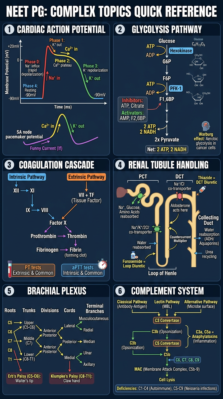
| Section | What's shown |
|---|---|
| Cardiac Action Potential | Phases 0-4, ion channels, SA node pacemaker curve |
| Glycolysis Pathway | Glucose → Pyruvate, rate-limiting PFK-1, regulators, Warburg effect |
| Coagulation Cascade | Intrinsic + Extrinsic pathways, Factor X convergence, PT vs aPTT |
| Renal Tubule Handling | Nephron segments, what's reabsorbed where, drug targets (Furosemide/Thiazide) |
| Brachial Plexus | C5-T1 roots → trunks → cords → terminal nerves, Erb's & Klumpke's palsy |
| Complement System | Classical/Lectin/Alternative pathways, C3b opsonization, MAC, anaphylatoxins |
- Generate a second poster covering other complex topics (e.g. HPA axis, thyroid hormone synthesis, haemostasis, ECG interpretation)?
- Create individual full-page diagrams for any single topic?
- Add this poster into the PDF from before?
Image based question or topic with explanation in a pdf
Which subject/topic should the image-based questions cover? · How many image-based questions should the PDF contain?
histology pathology slide gross specimen diagnosis
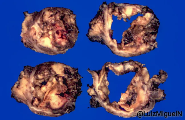
Gross pathology photograph of an angiomatoid fibrous histiocytoma showing a soft tissue tumor with solid nodules and hemorrhagic cystic spaces on cross-section. The four-panel montage reveals variegated tan-brown to red tissue, with hemorrhagic cavities and degenerative areas. This macroscopic appearance aligns with AFH, a mesenchymal soft tissue neoplasm that may present with hemorrhagic, cystic, and pseudoangiomatous features. The specimen type is a resection/excisional soft tissue tumor. No histologic staining or imaging contrast was used in these gross images. Clinically, AFH is more common in children and young adults and often arises in the extremities or trunk; however, location is not specified here. Diagnosis cannot be confirmed on gross morphology alone; histopathology demonstrating nodular growth with fibrous capsule, perivascular cuffing, and immunohistochemical positivity for markers such as CD68, desmin, or EMA can support diagnosis alongside molecular tests (EWSR1-CREB1, EWSR1-ATF1). This image is valuable for education, pattern recognition, and differential diagnosis training in soft tissue tumors and pathology. The image supports radiologic-pathologic correlation by highlighting how gross features may reflect underlying histology, such as hemorrhagic cystic change and fibrous encapsulation, which guide sampling strategy, surgical planning, and diagnostic sequencing in pediatric and young-adult soft tissue oncology.
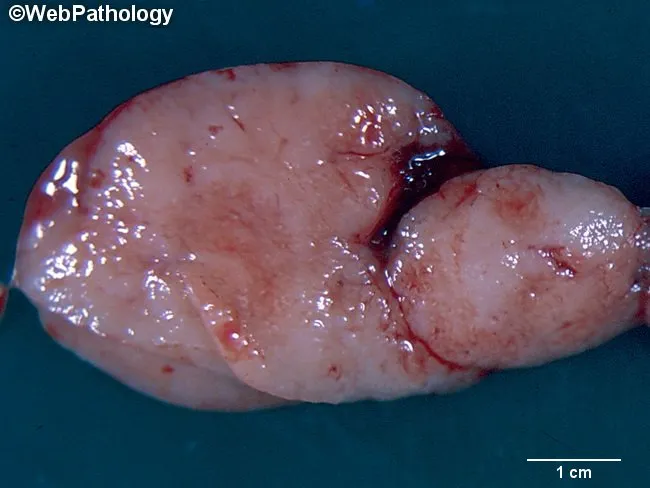
Gross pathology photograph of a surgically excised lymph node demonstrating a pale, glistening, lobulated soft tissue mass approximately 2–3 cm in maximal dimension, fixed in formalin. The specimen exhibits smooth external surface with minor irregularities; no obvious necrosis or hemorrhage on the visible outer contour; the cut surface (if provided) would be expected to reveal tan-white homogeneous to mottled nodal tissue. Context: Hodgkin lymphoma diagnosis typically requires histologic confirmation of Hodgkin and Reed–Sternberg (HRS) cells within a fibrous stroma; cytogenetics and comparative genomic hybridization may reveal chromosomal gains in 2p, 9p, 11q, and losses in 4q and 11q, with amplification of REL (2p16) and 9p24 (JAK2, PD-L1/PD-L2), contributing to NF-κB and JAK/STAT signaling. From a gross image alone, the diagnosis cannot be confirmed; this image is best used for educational demonstration of nodal gross morphology and biopsy guidance. Clinically relevant to staging workup, differential diagnosis includes reactive nodes, other lymphomas, and metastatic cancers. This specimen supports pathologist correlation with microscopic histology, immunophenotyping (CD15, CD30), EBV status, and cytogenetic results to establish classic Hodgkin lymphoma. Potential educational uses include gross pathology atlases, tumor biology teaching, and multidisciplinary case discussions. These cues guide biopsy sampling and correlate with prognosis.
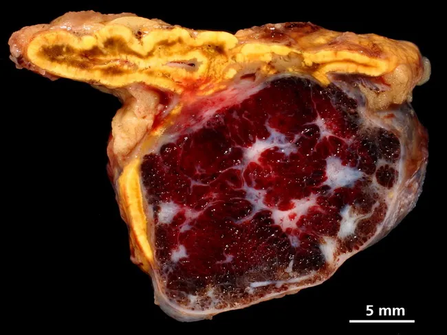
Gross pathology image of an adrenal gland specimen showing a sharply demarcated, rounded, spongy lesion arising within the suprarenal gland. The cortex appears yellow and finely lobulated, surrounding a central, reddish-brown, multi-lobulated mass with conspicuous vascular spaces and areas of hemorrhagic slippage. The surface cut reveals a well-circumscribed lesion with a spongy, sponge-like consistency and a heterogeneous color pattern from bright crimson to dark purplish red, interspersed with white fibrous septa. The margin between the lesion and adjoining adrenal cortex is distinct, consistent with a benign vascular neoplasm. The lesion corresponds to cavernous hemangioma, a vascular malformation characterized by large, dilated blood-filled vascular channels separated by scant stroma. The pathogenesis is related to dilatation of venous channels and thin endothelial linings; histology would demonstrate cavernous, irregular vascular spaces lined by flat endothelium. Final diagnosis: cavernous hemangioma of the adrenal gland. Notable features include well-demarcated borders, spongy gross morphology, and a reddish interior reflecting high vascular content. Clinically, such adrenal vascular tumors are rare and may be incidentalomas; definitive diagnosis relies on histopathology. This specimen is documented for educational reference in gross pathology and for correlation with radiologic and histologic findings. Potential clinical uses include teaching, differential diagnosis of adrenal masses, and study of adrenal pathology.
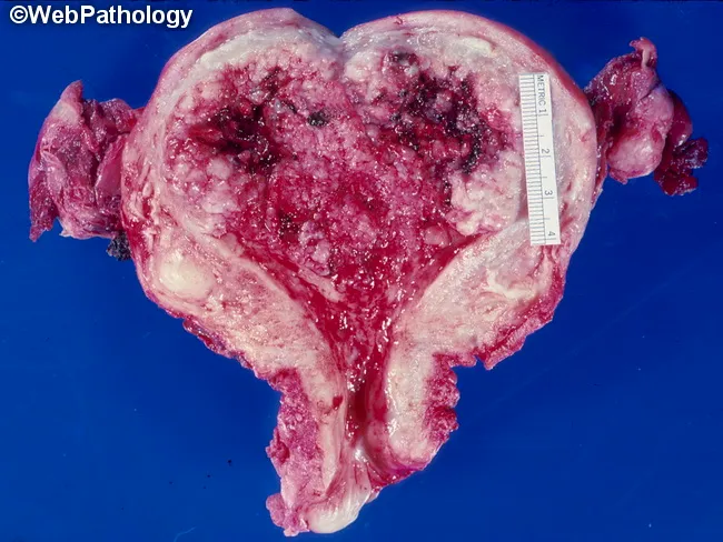
Gross pathology photograph of a hysterectomy specimen showing a hemorrhagic, necrotic endometrial tumor replacing the entire endometrium. The uterus appears enlarged with a bulky, friable mass occupying the endometrial cavity and extending to the myometrium; surfaces show irregular papillary fronds and necrotic red-black areas. A measuring scale is present for gross dimension. The lesion is grossly extensive, with heterogeneous color and soft, oozing tissue; cervical portion may be involved; invasion into surrounding tissues suggested in the cut surfaces but not proven histologically in this view. Diagnosis confirmed on histology as papillary serous carcinoma of the endometrium, a high-grade serous carcinoma characterized by complex papillary architecture, marked nuclear atypia, frequent mitoses, and often psammoma bodies; typically TP53 mutated. Clinically significant as an aggressive endometrial cancer with propensity for early lymphovascular invasion and extrauterine spread; prognosis is poorer than endometrioid adenocarcinoma. Relevance to clinical practice includes accurate histopathologic classification impacting treatment planning (surgery, adjuvant chemotherapy, possible radiotherapy) and staging (FIGO). This image is useful for educational demonstrations of gross endometrial carcinomas, differential diagnosis with other high-grade endometrial neoplasms, and radiology-pathology correlation. Potential applications include medical education, pathology review, and tumor biology research. Biopsy specimens and staging data refine prognosis and therapeutic decision-making in multidisciplinary care.
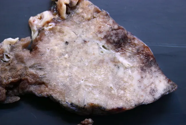
Gross pathology photograph of a lung tissue resection specimen illustrating an invasive mucinous adenocarcinoma with a prominent lepidic component. The tumor demonstrates ill-defined, infiltrative borders at the periphery, with a mucin-rich, gelatinous appearance and patchy grey-white parenchymal involvement. The gross pattern resembles infectious processes such as bronchopneumonia or even tuberculosis, posing a diagnostic pitfall on radiologic and clinical assessment prior to histology. The lesion is located in the peripheral lung (subpleural involvement), with predominant lepidic spread along alveolar septa contributing to the indistinct margins. On macroscopic inspection there is absence of discrete, well-circumscribed nodules; instead, broad-based infiltration disrupts normal parenchyma. Corresponding microscopic diagnosis would require correlation with histopathology to confirm adenocarcinoma with mucin production and lepidic growth, often associated with mucin pools and gland-forming cells. Clinically, recognizing this entity is essential for accurate staging (T modifiers reflecting lepidic invasion) and for planning surgical resection and adjuvant therapy. This image supports education on differential diagnosis for radiologic consolidation, emphasizes the mimicry of infection, and serves as a visual reference for pathologists and surgeons assessing suspicious lung lesions. The gross features underscore the need for tissue confirmation and multidisciplinary discussion to optimize prognosis and therapeutic strategy, correlating radiology, pathology, and clinical data together.
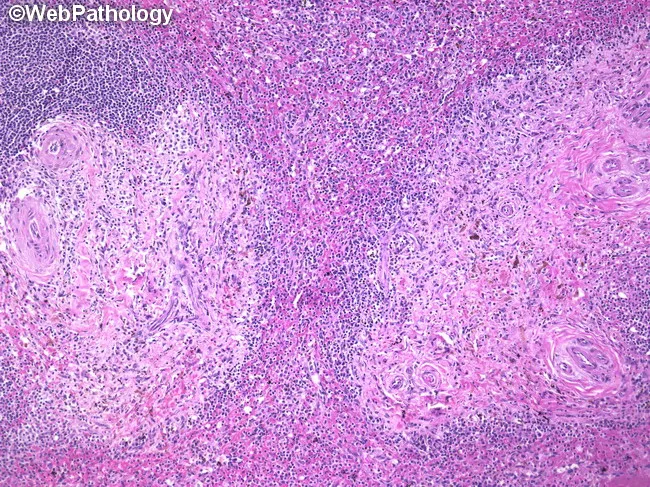
Systemic pathology panel: Histology of a splenectomy specimen from a patient with systemic mastocytosis. Modality: light microscopy on hematoxylin and eosin (H&E) stained sections. Gross features include a thickened splenic capsule with prominent fibrosis and a cut surface showing multiple 1–2 mm nodular foci scattered throughout the parenchyma. Microscopically, these nodules comprise mast cell infiltrates that are spindle-shaped and round, dispersed through both red pulp and white pulp, with a granulomatous appearance. Eosinophils and plasma cells accompany the infiltrate. Immunophenotype aligns with systemic mastocytosis: flow cytometry positive for CD2, CD25, CD117 and tryptase; negative for CD3, CD4, CD20, and CD34. The architectural disruption reflects involvement of the splenic microenvironment, often with hypersplenism and cytopenias as clinical correlates. In this case, patient history includes flushing, dermatologic lesions, diarrhea, hepatosplenomegaly, anemia, and thrombocytopenia; the final diagnosis is systemic mastocytosis with splenic involvement. Radiology is not applicable in this histology image. Clinically, such splenic involvement can be massive in aggressive systemic mastocytosis or systemic mastocytosis with associated hematologic neoplasm. This image illustrates diagnostic clues such as mast cells in fibrotic granulomas within both splenic pulp compartments and provides a reference for differential considerations and therapeutic implications. Useful for education and research instruction purposes.
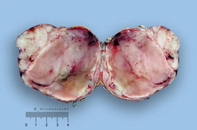
Gross pathology: A thigh soft tissue mass from a 43-year-old male displays a well-circumscribed, encapsulated lesion measuring 8 cm by 8 cm by 5 cm. The external surface is pink-tan with a lobulated, gelatinous cut surface containing focal hemorrhage. The specimen is consistent with a deep-seated lipomatous neoplasm. On microscopic examination, the tumor is diagnosed as myxoid liposarcoma. The peripheral whitish nodules correspond to hypercellular foci, whereas the central myxoid stroma remains relatively hypocellular. There is no definite round cell component identified in this specimen. The myxoid matrix is characterized by abundant mucopolysaccharide-rich ground substance with a delicate capillary network; lipogenic cells show variable adipocytic differentiation. A circumscribed capsule and well-preserved tissue architecture support a localized lesion. Additional ancillary testing (e.g., molecular cytogenetics for FUS-DDIT3 rearrangement) may confirm diagnosis and assist in grading, prognosis, and therapeutic planning. Clinically, myxoid liposarcoma evokes intermediate to low-grade aggressiveness, with potential for local recurrence and distant metastasis, underscoring the importance of complete excision with negative margins. This gross description provides essential context for correlating histology, staging, and treatment strategies in soft tissue sarcoma management. Correlative imaging, margin status, and molecular profile will guide adjuvant therapy decisions and surveillance to optimize outcomes in this case.
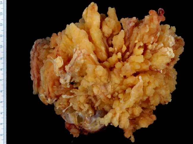
Imaging modality: Gross pathology photograph (macroscopic) of an excised ovarian mass. The specimen displays a cystic lesion with prominent serous papillary architecture. Macroscopic features include a tan-yellow to orange mass with numerous complex, variably sized papillary excrescences projecting from the cyst wall; surfaces are friable and irregular, with some translucent, lobulated fragments. The external contour is distorted by papillary fronds; a portion of ovarian stroma remains visible. Size context is provided by a ruler for scale, indicating a multi-centimeter greatest dimension. This appearance is typical for a serous borderline ovarian tumor (epithelial tumor of serous type) at gross examination, where papillary projections into the lumen are conspicuous but gross invasion is not evident. Histologic confirmation is required to assess borderline malignant potential, microinvasion, and implants. The gross pattern supports the diagnosis of SBOT, guiding surgical management, staging, and prognosis, and helps distinguish it from benign cystadenoma or malignant serous carcinoma on final pathology. Clinically, SBOTs occur in middle-aged to older women and generally have favorable outcomes after complete excision. Educational use includes gross-pathology references, radiologic correlation, differential diagnosis, and surgeon-pathologist communication. Documentation of the gross features complements histology and imaging, enabling accurate grading, prognosis, and multidisciplinary case discussion and planning.
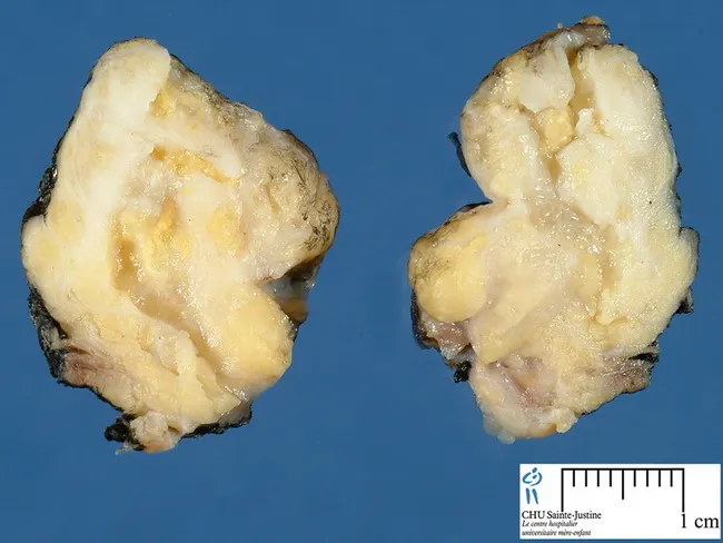
Gross pathology photograph of a pediatric soft tissue lipoblastoma. The lesion is circumscribed, soft, and lobulated, measuring 1-15 cm. On the cut surface tan-white fibrous trabeculae traverse pale yellow adipose tissue, reflecting adipocytic differentiation within a myxoid matrix. Myxoid or gelatinous foci and occasional cystic areas are common. The specimen represents a resected soft tissue mass with lobulated architecture and variable consistency. Image credits: Dr. Jean-Christophe Fournet, Paris, France; humpath.com; Used with permission. Lipoblastoma is a benign pediatric adipocytic neoplasm that typically arises in infancy or early childhood. Gross features described here—well-circumscribed margins, adipose-dominant tissue with fibrous septa and myxoid change—aid differentiation from lipomas and from liposarcomas, which can resemble lipoblastoma histologically but occur less commonly in young patients. Definitive diagnosis requires histopathology showing lipoblasts with scalloped nuclei in a myxoid stroma, with supportive immunohistochemical/molecular studies as needed. Surgical excision with clear margins is curative in most cases, though recurrence can occur with incomplete resection or infiltrative margins. Clinically, gross morphology guides differential diagnosis and surgical planning, and should be correlated with imaging and histology. Educational use includes teaching gross features of pediatric adipocytic tumors, differential with lipoma vs liposarcoma, and prognostic implications. This image highlights lobulation, adipose composition, myxoid variation, and occasional cystic change, informing diagnosis, management, and prognosis.
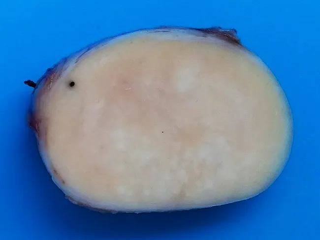
Gross pathology photograph of a dissected lymph node presented as a cross-sectional specimen. The nodal mass is markedly enlarged, with a white-tan to pale cream color and a uniform, homogenous cut surface that is solid in firmness. The capsule appears intact and there is no obvious central necrosis, hemorrhage, or calcification on the visible face. This macroscopic appearance is compatible with massive nodal enlargement often described in Rosai-Dorfman disease, a benign, self-limiting histiocytic disorder, but definitive diagnosis requires microscopic histology and immunohistochemistry. The image emphasizes gross features rather than tissue architecture: a large, well-demarcated lymph node with an even texture and a lack of gross necrotic change. Clinically, Rosai-Dorfman disease commonly presents with fever and weight loss and can involve multiple lymph node groups as well as extranodal sites such as skin or central nervous system; however, the current image depicts only a nodal specimen. The gross image is valuable for educational purposes, illustrating differential considerations in lymphadenopathy, including infectious, inflammatory, and malignant etiologies. In practice, gross assessment guides sampling strategy for histopathology, guiding subsequent techniques such as light microscopy, immunostaining for S-100 and CD68, and emperipolesis demonstration. This image supports gross pathology teaching, case-based discussion, and correlation with clinical presentation.
chest X-ray radiology findings pneumonia pneumothorax
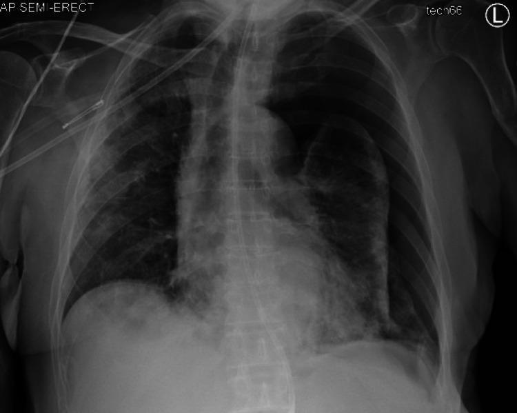
This anteroposterior (AP) semi-erect chest X-ray illustrates a significant secondary spontaneous pneumothorax in the left hemithorax. The image reveals a large, radiolucent space in the left pleural cavity with a complete absence of bronchovascular lung markings, characteristic of a pneumothorax. The left lung is partially collapsed and appears denser as it is displaced medially toward the hilum. Key diagnostic findings include a subtle tracheal deviation to the right and a corresponding mediastinal shift, indicating a degree of tension effect. The right lung field demonstrates multifocal opacities, which in the clinical context of COVID-19, are suggestive of underlying pneumonia or pulmonary edema. The costophrenic angles are visible, and medical monitoring lines are present. This diagnostic image is an educational example of acute pleural pathology and its mechanical effects on thoracic midline structures, suitable for medical students and radiology residents studying respiratory emergencies.
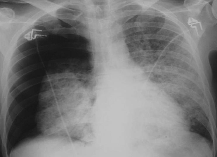
This diagnostic image is a posterior-anterior (PA) chest X-ray demonstrating a large spontaneous right-sided pneumothorax and bilateral interstitial disease. The right hemithorax shows a clear visceral pleural line with a significant peripheral area devoid of lung markings, particularly in the apical and lateral regions, representing approximately 50% lung collapse. Medially, the collapsed right lung and the entire left lung exhibit diffuse, symmetric ground-glass opacities and reticular interstitial markings. These findings are highly characteristic of Pneumocystis jirovecii (formerly carinii) pneumonia (PCP) in the setting of immunocompromise. No significant pleural effusion or mediastinal shift is visualized. Radiopaque medical monitoring electrodes and leads are visible on the upper chest. The radiographic presentation highlights a common complication of PCP, where cystic changes or subpleural blebs lead to spontaneous pneumothorax. Target audience: medical students and radiology residents studying opportunistic infections in HIV/AIDS.
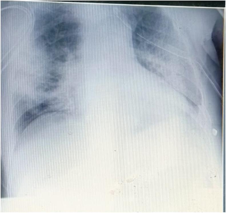
This diagnostic image is an anteroposterior chest X-ray taken following the drainage of a pneumothorax in a patient with COVID-19 pneumonia. The radiograph demonstrates bilateral, diffuse hazy opacities and ground-glass patterns, particularly pronounced in the left lung field, which obscure normal bronchovascular markings and signify extensive parenchymal damage. A chest drainage tube is visible on the right side, consistent with the clinical history of pneumothorax management. The cardiac silhouette is centrally positioned with no evidence of mediastinal shift. The pulmonary findings are characteristic of severe viral pneumonia complications, illustrating typical post-inflammatory changes and the iatrogenic management of gas effusions in a critical care setting.
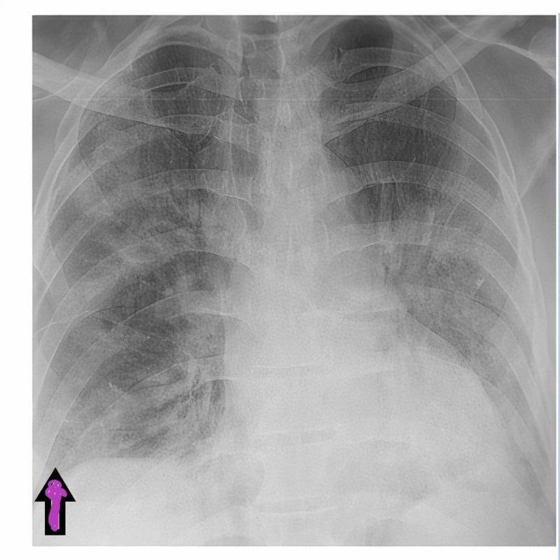
A posterior-anterior (PA) view chest X-ray illustrating characteristic findings in a patient with viral pneumonia, such as COVID-19. The image demonstrates bilateral hazy lung opacities and infiltrates, primarily distributed across both the middle and lower lung zones. These findings represent increased density compared to healthy lung parenchyma, indicative of inflammatory processes. A purple arrow at the right base highlights a clear costophrenic angle, confirming the absence of pleural effusion or pneumothorax at the time of admission. The cardiac silhouette and mediastinal contours appear within normal limits. The clavicles, ribs, and vertebrae are visible and intact. This diagnostic image is used to teach students how to identify multifocal ground-glass opacities and infiltrates on plain radiography, as well as how to differentiate them from secondary complications like pneumomediastinum or fluid accumulation in the costophrenic recesses.
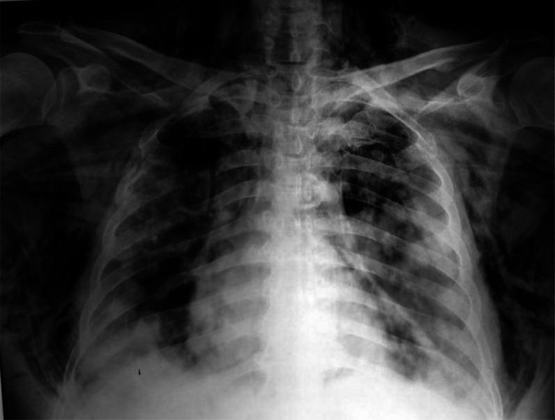
This diagnostic image is an anteroposterior (AP) chest x-ray radiography illustrating severe thoracic pathology. The primary findings include bilateral, predominantly peripheral patchy opacities and ground-glass densities throughout the lung fields, characteristic of atypical or viral pneumonia, such as COVID-19. Critical complications are evident: bilateral pneumothorax is indicated by the visualization of pleural lines with distal lucency lacking lung markings. Additionally, extensive subcutaneous emphysema is visible as characteristic streaky radiolucencies tracking through the soft tissues of the lateral chest walls and supraclavicular regions. There is also evidence of pneumomediastinum, seen as air outlining the mediastinal structures and tracking superiorly into the neck. This image serves as a significant educational example of multi-system thoracic air-leak syndromes and severe parenchymal lung disease, highlighting the radiographic features of barotrauma or spontaneous air dissection in the context of acute respiratory distress.
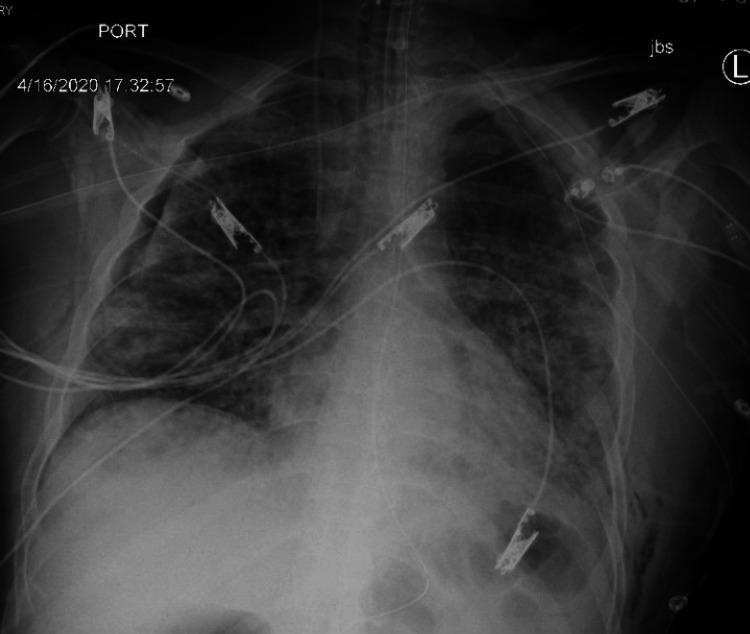
This diagnostic chest X-ray (portable, anteroposterior view) demonstrates post-procedural findings following tube thoracostomy for a pneumothorax. A right-sided chest tube is visible, entering through the axillary region and extending superiorly toward the right lung apex. Despite the tube, a residual small right apical pneumothorax is present, evidenced by a thin pleural line and absence of peripheral lung markings in the apical region. The right lung volume is slightly reduced compared to the left. The left lung remains well-inflated with visible vascular markings. Notable extracorporeal findings include multiple medical monitor leads, various catheters secured with radiopaque clips across the upper thorax and neck, and an endotracheal tube in situ, reflecting intensive care management. Bilateral, diffuse, patchy opacities are present throughout both lung fields, consistent with the patient's primary diagnosis of COVID-19 pneumonia and acute respiratory distress syndrome (ARDS).
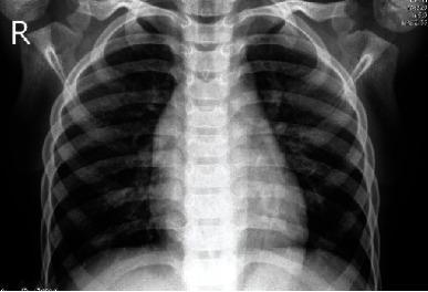
Anteroposterior (AP) chest X-ray of a patient demonstrating classic radiographic signs of pneumonia. The diagnostic image reveals significant bilateral increased pulmonary opacity, particularly prominent in the lower lung zones. These findings are consistent with consolidation or alveolar infiltrates, which obscure the normal branching pulmonary vascular markings. The cardiac silhouette and the diaphragmatic borders appear blurred and poorly defined—an example of the silhouette sign—suggesting the presence of adjacent lung consolidation or pleural fluid accumulation. The bony structures of the rib cage and clavicles are visible, with the patient's right side clearly marked with an 'R' indicator. The central mediastinal shadow and vertebral column are midline, providing anatomical orientation. This clinical imaging is used for medical education to teach the visual identification of infectious lung processes and their impact on normal anatomical silhouettes in diagnostic radiology.
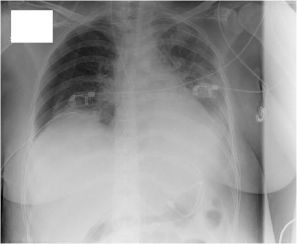
Anteroposterior (AP) chest X-ray demonstrating several critical acute findings. The right hemithorax shows a 'mantle' pneumothorax, evidenced by a thin, peripheral lucency along the apical and lateral lung margins where pulmonary markings are absent, indicating a partial lung collapse. The left lung field displays diffuse, hazy interstitial infiltrates, particularly prominent in the mid-to-lower zones, suggesting interstitial lung disease or inflammatory processes like acute eosinophilic pneumonia. Additionally, there is a left-sided pleural effusion, characterized by the blunting of the left costophrenic angle and an increased basilar density. Monitoring and support equipment are visible, including ECG electrodes and cabling superimposed on the thorax. The radiograph is essential for identifying secondary pulmonary complications, such as spontaneous pneumothorax, in critically ill patients. No obvious cysts, bullae, or cavitary lesions are identified in this view.
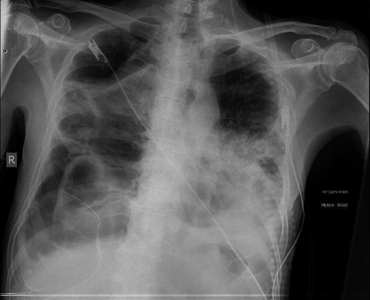
An anteroposterior (AP) chest X-ray demonstrates severe pulmonary parenchymal destruction and complex complications. Key findings include extensive bilateral ground-glass opacities and dense consolidations throughout both lung fields. Multiple large, thin-walled radiolucent structures consistent with cavitations or pneumatoceles are visible, particularly in the right mid-to-lower zones. A right-sided pneumothorax is present, evidenced by a visible pleural line and an absence of lung markings peripherally. A radiopaque intercostal chest drain is in situ on the right side, positioned to manage the pneumothorax. A large right-sided pleural effusion is noted at the base, obscuring the right costophrenic angle and hemidiaphragm. The left lung also shows significant consolidation and reticular opacities. Externally, ECG electrodes are visible on the chest wall. The imaging is consistent with severe pulmonary disease, such as advanced COVID-19 pneumonia complicated by cavitation, pneumothorax, and effusion.
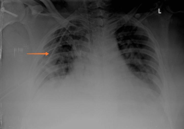
This diagnostic image is an anteroposterior (AP) chest X-ray demonstrating a right-sided pneumothorax and associated medical intervention. Key visual findings include a visible visceral pleural line in the right upper and mid-lung zones, with an absence of peripheral lung markings indicating air in the pleural space. The right lung exhibits partial collapse, particularly visible in the lower lobe area. An orange arrow highlights an intercostal drain (ICD) or chest tube in situ, correctly positioned within the right pleural space to facilitate lung re-expansion. Both lung fields show bilateral patchy opacities, consistent with the patient's underlying viral pneumonia (SARS-CoV-2). The cardiac silhouette is visible, and the tracheal midline appears stable. This image serves as an educational example of managing secondary spontaneous pneumothorax in the context of acute respiratory infection and highlights the radiological appearance of pleural air and therapeutic instrumentation.
skin lesion dermatology rash melanoma psoriasis
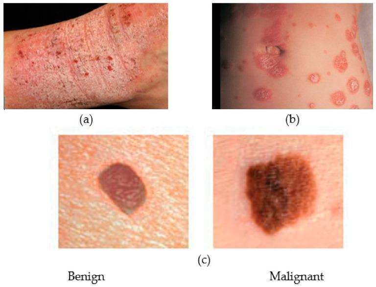
This composite clinical photograph illustrates common dermatological conditions for comparative medical education. Panel (a) shows atopic dermatitis (eczema) presenting as a diffuse, erythematous, and pruritic-appearing rash with visible excoriation, lichenification, and scaling on an extremity. Panel (b) depicts psoriasis, characterized by well-demarcated, erythematous papules and plaques with a silvery-white micaceous scale, distributed in a nummular (coin-shaped) pattern. Panel (c) provides a side-by-side comparison of melanocytic lesions: the 'Benign' lesion (likely a melanocytic nevus) exhibits a symmetrical oval shape, regular borders, and uniform reddish-brown pigmentation; the 'Malignant' lesion (malignant melanoma) demonstrates classic ABCDE warning signs, including asymmetry, irregular or notched borders, and variegated, dark brown-to-black pigmentation. The image serves as a visual guide for distinguishing between inflammatory skin diseases and neoplastic lesions, emphasizing morphology, border definition, and pigmentation patterns in clinical dermatology.
This clinical photograph, presented within a dermatological software interface, depicts a suspicious skin lesion, likely a melanoma, being analyzed for diagnostic segmentation. The primary lesion is a dark red, irregularly shaped macule with jagged, poorly defined borders and a heterogeneous surface texture. It is set against a background of lighter grayish-white skin. The image demonstrates a post-annotation inspection phase in a deep learning workflow. Two distinct red transparent overlays (annotations) are visible: a large, central segment conforming to the primary lesion and a secondary, small circular artifact in the upper right quadrant. A system notification at the top of the GUI displays a 'Warning: Annotation is potentially noisy!', triggered by the detection of multiple segmented objects when only one clinical entity (the lesion) is expected. This visual illustrates the quality control and data cleaning process essential for medical image processing and the training of diagnostic algorithms in dermatology.
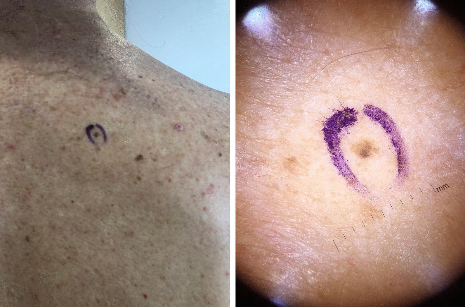
This dual-panel image features a clinical photograph (left) and a dermatoscopic image (right) of a pigmented skin lesion identified as melanoma in situ (MIS). The clinical photograph shows the lesion located on the right scapular region, marked with a purple surgical ink circle. The surrounding skin exhibits actinic damage, characterized by solar lentigines and uneven pigmentation. The dermatoscopic view on the right provides a high-magnification assessment of the same lesion, revealing irregular hyperpigmented areas, varying shades of brown, and poorly defined, asymmetric borders. A millimeter scale is visible at the bottom right of the dermatoscopic frame for size reference, indicating the lesion is approximately 2mm in diameter. The educational focus is on the diagnostic utility of dermatoscopy in identifying early-stage, small-diameter melanomas that may appear as non-specific macules to the naked eye. This content is relevant for dermatology training, specifically in the clinical detection and management of cutaneous malignancies.
This Comparison Chart and Clinical Imaging infographic demonstrates a dermatological diagnostic interface for skin lesion classification. The top left 'Image to explain' displays a malignant melanoma characterized by the ABCDE criteria: asymmetry, highly irregular borders, and color variegation including shades of dark brown, black, and pink-violet. Beside it, a 'Counter example image' shows a melanocytic nevus, which exhibits more uniform pigmentation and relatively smoother, regular boundaries. The interface includes a 'Neighborhood' data panel listing distribution frequencies of similar latent representations among categories such as basal cell carcinoma and dermatofibroma. The lower section features four 'Prototype images'—synthetic exemplars generated by an Adversarial Autoencoder (AAE) to visually represent the core features of the melanoma class. This visual material is used in medical informatics to explain 'black box' machine learning decisions in dermatology, assisting students and clinicians in identifying high-risk morphological features that distinguish melanoma from benign melanocytic nevi.
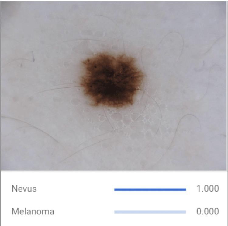
This clinical photograph displays a dermatoscopic-style view of a benign melanocytic nevus, accompanied by artificial intelligence (AI) classification data. The primary lesion is a centrally located, pigmentary skin lesion characterized by relatively regular and well-demarcated borders, a symmetrical oval shape, and a predominantly uniform brown coloration. Fine, light-colored terminal hairs are seen crossing over the lesion surface. The surrounding skin exhibits a faint, net-like pigment pattern. Below the clinical image, an educational graphical interface shows the output of a convolutional neural network (CNN) model, providing a classification score of 1.000 for 'Nevus' and 0.000 for 'Melanoma'. This visual demonstrates the distinguishing morphological features of benign nevi—symmetry, border regularity, and color uniformity—used in clinical dermatology and computer-aided diagnostic systems to differentiate them from malignant melanoma.
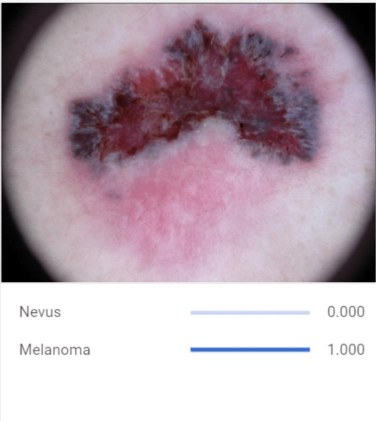
This clinical diagnostic image is a dermoscopic photograph of a malignant melanoma skin lesion, presented alongside a convolutional neural network (CNN) classification output. The lesion demonstrates the classic ABCDE criteria of melanoma: it is highly asymmetrical in shape with irregular, notched, and poorly defined borders. The lesion exhibits significant color variegation, featuring a complex distribution of dark brown, reddish-pink, and focal blue-gray areas. The blue-gray hues are concentrated primarily at the periphery, while the interior shows mixed erythematous and brown pigmentation. Surrounding the primary lesion is a faint pinkish macular area, possibly representing inflammation or regression. The diagnostic significance of this image is to highlight visual biomarkers used in automated dermatological assessment, specifically emphasizing the irregular morphology and multi-chromatic nature of malignant pigmented lesions compared to benign nevi. Below the clinical image, an AI diagnostic tool indicates a probability score of 1.000 for melanoma and 0.000 for nevus, illustrating the application of machine learning in computer-aided diagnosis for dermatology.
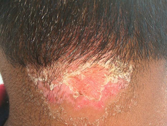
This clinical photograph displays a well-demarcated skin lesion characteristic of scalp psoriasis, located at the posterior hairline and extending onto the neck. The affected area is a prominent erythematous plaque with a vivid reddish-pink hue, indicating underlying inflammation. The lesion is characterized by thick, micaceous (silvery-white to yellowish) scales that are most densely concentrated along the irregular borders, partially adhering to the hair shafts and the scalp surface. The central portion of the plaque shows visible skin thickening and hyperkeratotic texture with less dense scaling. The surrounding skin appears relatively normal, though the plaque exhibits sharp demarcation from the healthy tissue. This image serves as a classic educational example of plaque psoriasis affecting the scalp and hairline, demonstrating key diagnostic features such as well-defined erythema and characteristic scaling patterns relevant for dermatology and primary care education.
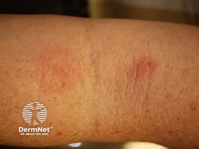
This is a high‑resolution clinical photograph of in vivo human forearm skin documenting an erythematous, patchy rash. Modality: Clinical photography, non‑dermoscopic; close‑up view of the antebrachial skin. Anatomical location: Forearm, integumentary system, with involvement of the superficial epidermis and dermis. Visual features: diffuse erythema with ill‑defined, slightly raised patches, mild desquamation along margins, subtle perifollicular accentuation, and no obvious vesicles or crusts. Skin texture shows mild edema and minimal hyperpigmentation beyond the red areas. The lesion spans a proximal to distal segment of the forearm and exhibits a non‑focal distribution on the extensor surface. Possible etiologies include inflammatory dermatitis such as irritant or allergic contact dermatitis, atopic dermatitis (eczema), or other eczematous processes. Differential diagnosis also includes early psoriasis or drug eruption; however, absence of well‑formed plaques or silvery scale argues against classic plaque psoriasis, and lack of vesicular lesions makes bullous diseases unlikely. Notable significance: image documents a cutaneous inflammatory process and serves as educational dermatology material, triage aid in primary care, and baseline for monitoring therapy response. Clinical utility: baseline documentation, serial assessment of lesion evolution, correlation with environmental exposures, and guiding patch testing and patient counseling. Further documentation may include dermoscopy or biopsy if persistence occurs unexpectedly.
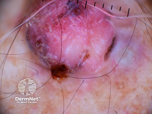
Dermoscopic imaging of a cutaneous melanoma lesion. Modality: dermoscopy (dermoscopy) using contact, non‑invasive imaging of the skin surface. The image shows a solitary pink to flesh‑colored lesion with an irregular vascular pattern, including atypical serpentine and dotted vessels, and peripheral speckling of pigment. The lesion appears with ill‑defined borders, subtle translucency, and a pale to pink background, consistent with malignant melanoma in situ progressing to invasion in some areas. The clinical appearance correlates with histopathologic Breslow depth of 8.3 millimeters, indicating a thick melanoma with high risk for regional spread and poorer prognosis. Dermoscopic clues such as atypical vascularization and peripheral pigment aggregation support a diagnosis of malignant melanoma rather than benign mimics (e.g., benign nevi, seborrheic keratosis). The image highlights key features used in triage and management: lesion asymmetry, color variability, irregular vascular architecture, and peppering pigment. Diagnostic significance: high likelihood of invasive melanoma requiring urgent excisional biopsy, staging workup, sentinel lymph node evaluation, and potential wide local excision with adequate margins. Potential clinical use cases: dermatology education, dermoscopy training, melanoma surveillance, image-based decision support, and research on color/vascular patterns in thick melanomas. Clinically, this lesion warrants histopathologic confirmation and aggressive treatment planning.
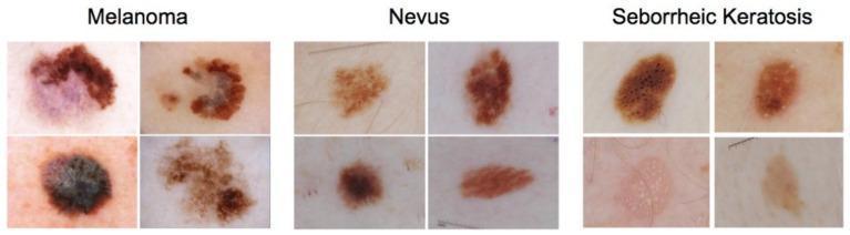
This Comparison Chart presents twelve dermoscopic images categorized into three distinct skin lesion types: Melanoma, Nevus, and Seborrheic Keratosis. The visual aid serves to highlight key diagnostic morphological features used in dermatology and oncology. The Melanoma images demonstrate high clinical concern, characterized by marked asymmetry, irregular or notched borders, and significant color variegation including shades of black, dark brown, and areas of blue-white veil or regression (purple-gray hues). In contrast, the Nevus (common mole) examples display more benign features: symmetry, regular borders, and homogeneous pigment distribution, typically in a single shade of brown. The Seborrheic Keratosis images illustrate classic diagnostic clues for benign epithelial tumors, such as milia-like cysts (small white/yellow spots), comedo-like openings (dark keratin plugs), and a characteristic waxy or 'stuck-on' appearance with a verrucous, scaly texture. This comparative display is essential for medical training in differential diagnosis, particularly in distinguishing between malignant melanoma and common benign mimics. The images originate from the ISIC 2017 dataset, often utilized in benchmarking computer-aided diagnostic algorithms for skin cancer detection.
anatomy cross section diagram brain MRI CT scan
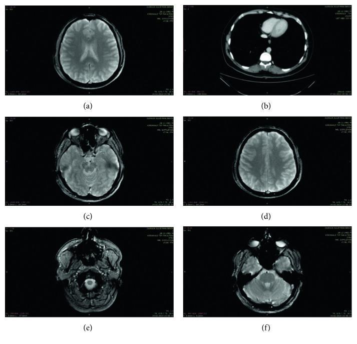
This composite figure presents six diagnostic images illustrating cross-sectional anatomy of the head and thorax. The majority of the images (a, c, d, e, f) are axial Magnetic Resonance Imaging (MRI) scans of the brain at various neuroanatomical levels. (a) and (d) depict the superior cerebrum, highlighting the cerebral cortex, gyri, sulci, and the longitudinal fissure. (c) shows a mid-level axial section including the orbits and temporal lobes. (e) and (f) represent the posterior fossa and skull base, detailing the cerebellum and brainstem. In contrast, image (b) is an axial section of the thorax, likely a Computed Tomography (CT) scan, showing the cardiac silhouette, the ascending and descending aorta, the vertebral body, and the pulmonary parenchyma. These images serve as examples of clinical neuroimaging and thoracic radiology, demonstrating the visualization of soft tissue structures, bony landmarks, and fluid-filled cavities (such as ventricles and orbits) across different imaging modalities and anatomical planes for educational and diagnostic purposes.
This diagnostic image is an axial cross-section of a human brain, likely from a computerized tomography (CT) or magnetic resonance imaging (MRI) scan, focusing on the basal ganglia and internal capsule region. The image illustrates a quantitative method for assessing the orientation of an intracranial infarction relative to the posterior limb of the internal capsule (PLIC). A blue rectangle outlines the anatomical boundaries of the PLIC. A smaller, diagonally oriented red rectangle highlights a localized infarction lesion within this region. The diagram uses orange lines to define specific axes: line segment 'AO' represents the perpendicular bisector of the short axis of the PLIC, while line segment 'BO' represents the long axis of the infarction lesion. The vertex 'O' serves as the common point of intersection, defining angle '∠AOB'. This measurement provides a standardized way to determine the lesion's spatial relationship with surrounding structures like the thalamus and putamen. This content is intended for advanced medical education in neurology or radiology to illustrate clinical research methodology for correlating stroke localization with motor deficits.

This diagnostic image demonstrates a multimodal image fusion used in stereotactic radiosurgery (SRS) preplanning. The visual is a sagittal cross-section featuring a coregistered dataset: an anthropomorphic head phantom CT scan (color-coded in blue) fused with a real patient brain MRI (color-coded in amber). The blue CT component shows the external contour and uniform density of a thermoplastic head phantom, which lacks internal anatomical detail. In contrast, the amber MRI overlay provides high-resolution visualization of internal soft tissue structures, including the cerebral cortex, brainstem, and ventricles. Vertical lines and crosshairs indicate the stereotactic coordinate system and localization framework. This fusion technique is utilized for treatment simulation and dosimetric evaluation, allowing clinicians to perform feasibility studies and initial beam configuration on a surrogate geometry before the final patient-specific CT is acquired on the day of treatment. The image illustrates the coarse manual alignment of external surfaces to match phantom and patient anatomy for clinical workflow optimization.
This diagnostic image is an axial cross-section of the human head, likely from a computerized tomography (CT) scan or magnetic resonance imaging (MRI), emphasizing craniofacial and intracranial anatomy. Visible structures include the orbits, nasal cavity, ethmoid and sphenoid sinuses, and the brain parenchyma within the skull vault. A yellow vertical reference line is superimposed along the midline, specifically aligned to coincide with the falx cerebri, a dural fold that separates the two cerebral hemispheres. A green horizontal line intersects the yellow line, passing through the posterior aspect of the orbits and the sphenoid sinus region. The image illustrates the 'anterior cerebral falx plane method' for establishing a craniofacial midline, a technique used in clinical imaging to assess facial symmetry, plan reconstructive surgery, or conduct cephalometric analysis. This anatomical alignment serves as a reliable internal reference point for measuring deviations in craniofacial morphology.
This figure presents a comparative study of digital watermarking robustness in clinical imaging. On the left is a diagnostic image showing a transverse (axial) cross-section of a human brain, likely from a computerized tomography (CT) or MRI scan. The image displays cerebral anatomy, including the ventricular system and cortical folds. Text above this image indicates a 'clipping(X Direction) 8%' attack, a digital manipulation technique used to test the security of embedded data. On the right, the 'extracted watermarking image' is displayed as a pixelated binary image containing the characters 'HN'. Below this, the value 'NC=1' is shown, representing a Normalized Correlation of 1.0. This indicates a perfect match between the original and extracted watermark despite the 8% horizontal clipping attack. This visual illustrates concepts in medical informatics and multimedia security, specifically demonstrating the resilience of watermarking algorithms in protecting the copyright and integrity of medical diagnostic data against geometric distortions.
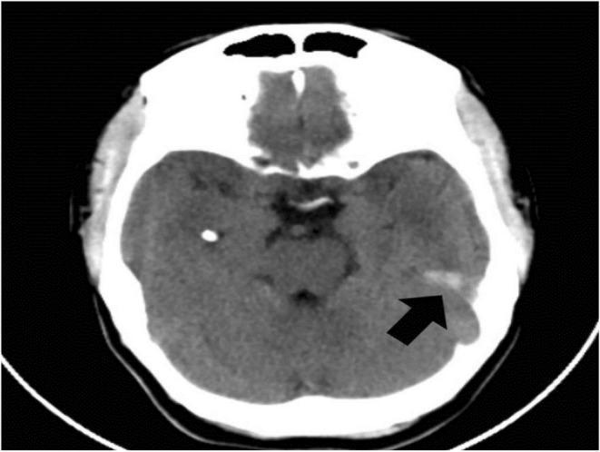
This diagnostic image is an axial non-contrast computed tomography (CT) scan of the brain, a key modality for evaluating neuroanatomical pathology. The scan shows a cross-section of the cerebral hemispheres, including the brain parenchyma, ventricular spaces, and the hyperdense skull vault. A prominent black arrow points to a specific abnormality in the posterior cerebral white matter: a linear, slightly hypoattenuating feature identified as a radial migration line. This finding is a characteristic neuroradiological sign of Tuberous Sclerosis Complex (TSC), representing disordered neuronal migration from the periventricular germinal matrix to the cortex. Additionally, small hyperdense (calcified) foci may be observed elsewhere in the parenchyma, consistent with subependymal nodules or cortical tubers. This visual is intended for medical education regarding the CNS manifestations of phakomatoses, specifically highlighting how CT can identify white matter dysplasias and migration defects when MRI is unavailable.
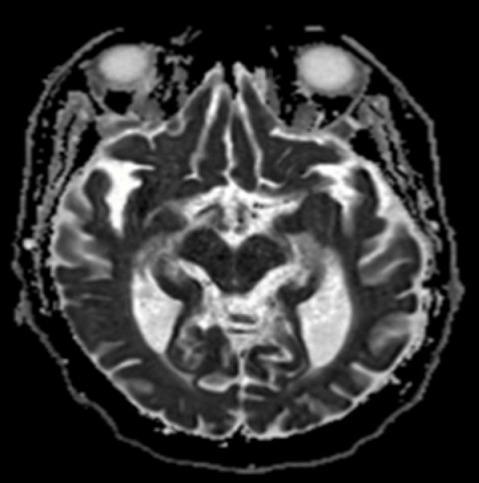
This diagnostic image is an axial magnetic resonance imaging (MRI) scan of the human brain utilizing an apparent diffusion coefficient (ADC) map sequence. The cross-section is taken at the level of the midbrain and temporal lobes, with the orbits visible anteriorly. The primary pathology is a focal area of marked hypointensity (low signal) located in the left medial temporal lobe, specifically involving the hippocampal region. This hypointense signal on the ADC map corresponds to restricted diffusion, which is a classic radiographic sign of acute cytotoxic edema. In this clinical context, the finding represents a metabolic insult likely secondary to severe hypoglycemia. The surrounding brain parenchyma, including the contralateral hippocampus and the cortical ribbons of the temporal and occipital lobes, maintains relatively normal signal intensity. This visual is used to demonstrate the neurological consequences of severe metabolic disturbances and the utility of diffusion-weighted imaging (DWI/ADC) in identifying acute brain lesions that may not be apparent on standard CT scans.
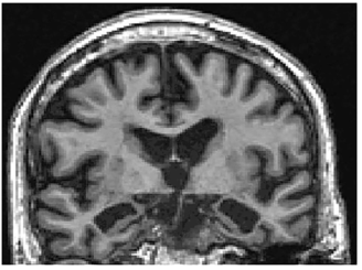
Summary : This image shows a coronal MRI scan of a human brain, displaying anatomical structures in cross-section. photo: Scene Overview : • Main subject is a coronal section of a human brain, as visualized by magnetic resonance imaging (MRI). • The scan is grayscale, with varying intensities highlighting different tissue types. • The image is centered on the brain, showing both hemispheres and midline structures. • The perspective is head-on, with the top of the head at the top of the image. Technical Details : • No scale bar or magnification indicator is visible. • MRI modality, likely T1-weighted due to the contrast between gray and white matter. • No visible text, UI elements, or annotations on the image. Spatial Relationships : • The cerebral cortex is visible around the periphery, with characteristic gyri and sulci. • The lateral ventricles and third ventricle are visible near the center, appearing as dark spaces. • The image shows symmetry between the left and right hemispheres. • The basal ganglia and other deep brain structures are visible below the ventricles. Analysis : • The scan provides a clear view of normal brain anatomy, with well-defined cortical and subcortical structures. • No obvious abnormalities, lesions, or asymmetries are apparent in this image. • The image is suitable for anatomical study or clinical assessment of brain structure.
ECG electrocardiogram findings arrhythmia heart block
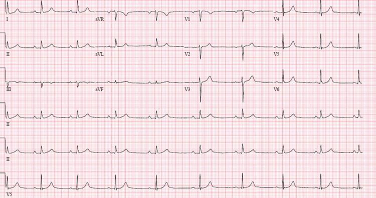
This diagnostic image is a 12-lead electrocardiogram (ECG/EKG) with rhythm strips (II, II, and V5) displayed at the bottom, illustrating a complex cardiac arrhythmia. The key finding is third-degree (complete) atrioventricular (AV) block, characterized by total AV dissociation. P waves are visible and regular (atrial rate), but they bear no consistent temporal relationship to the QRS complexes (ventricular rate), which occur independently. The QRS complexes are widened, measuring >120 ms, and demonstrate a morphology suggestive of an infra-nodal escape rhythm. Notable findings in the precordial leads include absent R-wave progression in V1, V2, and V3, with dominant negative deflections (QS complexes) in these leads. Lead V1 specifically shows a wide, predominantly negative QRS. The underlying ventricular rate is significantly bradycardic compared to the atrial rate. This ECG is a critical educational resource for cardiology, demonstrating the classic findings of complete heart block and the requirement for clinical correlation with hemodynamic stability and potential pacing intervention.
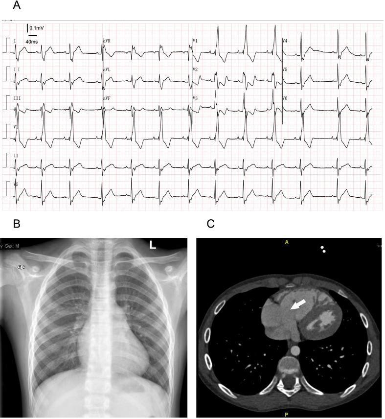
A multi-panel medical diagnostic image set illustrating cardiac findings in a patient with Ebstein's anomaly. Panel A displays a 12-lead electrocardiogram (ECG) demonstrating signs of arrhythmia and a complete right bundle branch block (RBBB), characterized by widened QRS complexes and classic RSR' patterns in precordial leads. Panel B is a posterior-anterior chest radiograph (X-ray) showing prominent enlargement of the right heart border, indicative of right atrial dilation, while the lung fields remain clear and the cardiothoracic ratio is measured at 0.5. Panel C shows an axial contrast-enhanced CT scan of the thorax at the level of the four-chamber view. A white arrow points to the significant enlargement of the tricuspid annulus, a key diagnostic feature of the valvular pathology. This composite figure serves as a diagnostic educational tool for identifying the multi-modality presentation of congenital tricuspid valve malformations and associated right-sided cardiac remodeling.
A 12-lead resting electrocardiogram (ECG) displayed on a digital grid background. The tracing shows a patient in sinus rhythm with a heart rate of 95 bpm and a QRS axis of approximately +70 degrees. Key findings include narrow QRS complexes in the majority of leads, with a transition characterized by an r/S pattern in the precordial leads V2 and V3, and an R/S relationship in the inferior leads II, III, and aVF. Notably, the tracing demonstrates a unifocal premature ventricular contraction (PVC). This ectopic beat exhibits a wide QRS duration, slurred morphology, and a left bundle branch block (LBBB) pattern, indicating an origin in the right ventricle. The image serves as an educational example of arrhythmia detection in the clinical context of post-COVID-19 myocardial assessment, highlighting the distinction between normal conduction and ventricular ectopy.
A 12-lead electrocardiogram (ECG) demonstrating Complete Heart Block (CHB) with significant electrophysiological findings. The tracing reveals a classic atrioventricular (AV) dissociation where P waves and QRS complexes occur independently of each other. The atrial rate is approximately 80-90 beats per minute, while the ventricular rate is slower at approximately 40-45 beats per minute. The escape rhythm is characterized by narrow QRS complexes, suggesting a junctional or intra-Hisian level of block. Notably, the tracing exhibits isorhythmic AV dissociation, where the atrial and ventricular rates exist in a near 2:1 ratio, superficially mimicking a second-degree AV block. Additionally, ventriculophasic sinus arrhythmia is present, evidenced by shorter P-P intervals when a QRS complex is interspersed between them compared to P-P intervals without an intervening QRS. The ST segments and T waves are otherwise unremarkable across the precordial and limb leads.
A 12-lead electrocardiogram (ECG) demonstrating a complex arrhythmia characterized by third-degree (complete) heart block. The tracing reveals distinct P waves occurring at a rapid, regular rate indicative of sinus tachycardia, but with total atrioventricular (AV) dissociation. A junctional escape rhythm is present, evidenced by narrow QRS complexes appearing at a significantly slower, regular rate of approximately 42 beats per minute. The precordial leads (V1-V6) show poor R-wave progression. Specific morphology includes upright T-waves in most leads and discernible P waves most prominent in leads II, III, aVF, and V1. The recording is presented on standard grid paper with a 25 mm/s sweep speed and 10 mm/mV sensitivity. This visual exemplifies the clinical presentation of AV nodal or infra-nodal conduction failure where the atrial and ventricular activities are independent of each other.
A standard 12-lead electrocardiogram (ECG) demonstrating a complex mixed arrhythmia in a patient with dilated cardiomyopathy and cardiogenic shock. The rhythm is irregularly irregular with the absence of discrete P waves, replaced by fine fibrillatory waves, characteristic of atrial fibrillation. Notable features include frequent premature ventricular complexes (PVCs) and a widened QRS duration (>120ms). Lead V1 shows a tall R wave followed by a deep S wave, and Lead V6 exhibits broad, slurred S waves, diagnostic of a complete right bundle branch block (RBBB). Significant ST-segment and T-wave abnormalities are visible across the precordial leads (V1-V6), likely representing secondary repolarization changes from the RBBB or underlying myocardial ischemia. The ECG indicates severe cardiac conduction system involvement and ventricular irritability in the context of acute decompensated heart failure.
This diagnostic image is a 12-lead electrocardiogram (ECG) demonstrating a complex cardiac arrhythmia in a 71-year-old male. The tracing shows complete atrioventricular (AV) dissociation, a hallmark of third-degree heart block. A sinus rhythm (atrial rate approximately 60 bpm) is present, indicated by P-waves that bear no constant relationship to the QRS complexes. A junctional escape rhythm (ventricular rate approximately 45 bpm) is noted, marked by blue arrows in the rhythm strip. The QRS morphology indicates a nonspecific interventricular conduction delay. Additionally, the ECG captures frequent ventricular ectopy; green arrows highlight premature ventricular complexes (PVCs) occurring with an 'R-on-T' phenomenon, where the ectopic beat falls very close to the preceding T-wave. This pattern is clinically significant as a precursor to malignant ventricular arrhythmias such as Torsades de Pointes. The baseline undulations and varying morphologies of the ectopic beats further suggest electrical instability. This ECG is essential for teaching advanced cardiovascular life support (ACLS) concepts, specifically the recognition and management of complete heart block and high-risk ventricular ectopy.
Diagnostic electrocardiogram (ECG) rhythm strips showing an acute cardiac arrhythmia on standard grid paper. The image consists of four consecutive rhythm leads displaying polymorphic ventricular tachycardia (PVT). The tracing is characterized by a rapid, chaotic rhythm with continuously changing QRS complex morphology, amplitude, and axis. There is a complete absence of identifiable P waves and distinct T waves, indicating severe ventricular dysrhythmia. The waveforms show a characteristic 'twisting' or undulating appearance around the isoelectric line, consistent with Torsades de Pointes or similar polymorphic ventricular patterns. This diagnostic image illustrates a critical clinical state that can occur post-adrenaline administration in patients with underlying conduction issues, such as heart block. It serves as an educational example of high-grade ventricular irritability and unstable cardiac rhythms that require immediate medical intervention.
Give me pdf