
What is plantar fasciitis
plantar fasciitis anatomy diagram heel pain
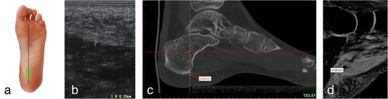
This multicomponent educational figure demonstrates a comparative multimodal imaging approach for measuring the plantar fascia thickness at its calcaneal origin. Panel A is an anatomical diagram of the plantar foot surface showing a longitudinal probe placement (green rectangle) over the medial tubercle of the calcaneus. Panel B is a longitudinal musculoskeletal ultrasound image (sonogram) displaying the hyperechoic, fibrillar plantar fascia with a caliper measurement of 0.28 cm. Panel C shows a sagittal reconstruction CT scan in a bone window, highlighting the skeletal anatomy of the calcaneus and midfoot with a software-based measurement of the plantar fascia origin at 2.69 mm. Panel D presents a sagittal T1-weighted or proton-density MRI sequence, providing superior soft tissue contrast of the plantar aponeurosis and heel pad, also displaying a measurement of 2.69 mm. This comparison illustrates the consistency between imaging modalities (ultrasound, CT, and MRI) for the clinical assessment of plantar fasciitis and anatomical morphometry.
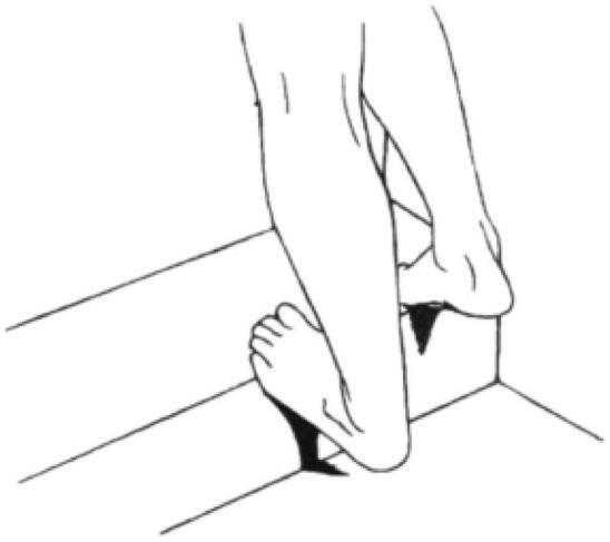
This medical illustration depicts a calf and Achilles tendon stretching exercise, a common physical therapy intervention for plantar fasciitis and Achilles tendinopathy. The anatomical diagram shows a posterior-oblique view of the lower legs and feet. One foot is positioned with the forefoot and metatarsal heads on the edge of a raised step, allowing the heel to drop below the level of the step. This position induces passive dorsiflexion at the ankle joint, creating a lengthening force on the gastrocnemius and soleus muscles (calf complex) and the Achilles tendon. The contralateral foot is placed flat on the floor behind the step for stability. The illustration demonstrates the structural mechanics of an eccentric or static stretch used in orthopedics and sports medicine to improve ankle range of motion and reduce tension in the posterior leg compartment. The clinical relevance includes rehabilitation protocols for heel pain and tendon flexibility.
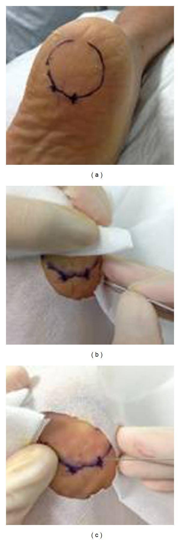
This composite of clinical photographs illustrates the surgical procedure for miniscalpel-needle (MSN) release treatment for plantar fasciitis. Panel (a) shows the plantar aspect of a human heel with a circular surface landmark and 'X' markings used to identify the most painful tender point over the medial tubercle of the calcaneum. Panel (b) depicts the intraoperative phase where a 0.80 mm diameter MSN is inserted vertically into the marked tender point while the patient is in a prone position. The clinician's gloved hands are visible managing the surgical field. Panel (c) shows the immediate post-procedure result, demonstrating a small, linear puncture wound at the insertion site with minimal local skin trauma. The series highlights a minimally invasive approach to musculoskeletal pain management, specifically targeting the release of thickened plantar fascia under local anesthesia. The educational focus is on the localization of anatomical landmarks and the technique for needle-based surgical intervention in chronic heel pain.
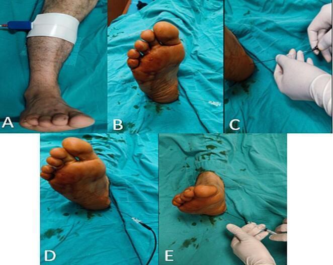
A multi-panel clinical photograph series depicting the sequential stages of pulsed radiofrequency ablation (PRFA) for the treatment of plantar fasciitis or heel pain. Panel A illustrates the preparatory phase with a return electrode (grounding pad) secured to the patient's posterior calf using white adhesive tape. Panels B through E demonstrate the procedural intervention at the anteromedial calcaneus of the foot, which is positioned on a sterile blue surgical drape. The sequence shows the initial application of a hypodermic cannula with a stylet (B), the removal of the stylet and subsequent insertion of the radiofrequency electrode by a gloved clinician (C), the delivery of radiofrequency energy with the electrode connected to a black cable (D), and the post-procedure administration of a local anesthetic substance (e.g., bupivacaine) via syringe (E). This educational visual demonstrates the precise anatomical targeting, instrument handling, and sterile technique required for minimally invasive nerve ablation in podiatry and pain management.
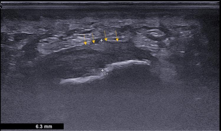
This diagnostic image is a sagittal B-mode ultrasound of the plantar heel in a 38-year-old female patient, demonstrating sonographic features characteristic of plantar fasciitis. The primary focus is the plantar fascia, which appears as a significantly thickened, hypoechoic band-like structure indicated by yellow arrows in the mid-upper region. Calipers (marked 'D1') measure the fascial thickness at 6.3 mm, which is pathological compared to normal physiological limits. There is a notable loss of the normal organized fibrillar pattern within the fascial body. Deep to the plantar fascia, the hyperechoic cortical surface of the calcaneus is visible, characterized by a bright, dense acoustic reflection. This image serves as a clinical example of musculoskeletal imaging for diagnosing chronic heel pain, highlighting key diagnostic criteria: thickening >4 mm, reduced echogenicity, and loss of internal architectural detail.
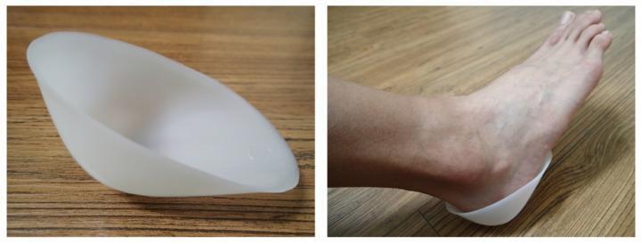
This clinical photograph consists of two panels demonstrating a plastic heel cup, a common orthopedic device used in the management of plantar fasciitis and heel pain. The left panel shows the standalone device: a semi-rigid, translucent white plastic orthotic with a contoured, bowl-like shape designed to anatomically cradle the calcaneal region. The right panel displays the device in situ on a human foot. It illustrates the clinical application where the cup encloses the posterior and plantar aspects of the heel, providing confinement for the soft tissue heel pad. The device is designed to prevent the lateral expansion (collapse) of the heel pad under weight-bearing loads, thereby enhancing shock absorption and reducing plantar pressure. This visual is relevant for orthopedic and podiatric education regarding conservative treatment options for plantar heel pain and mechanical stabilization of the calcaneal fat pad.
"plantar fasciitis"[MeSH Terms] AND treatment
Plantar Fasciitis
What Is It?
Risk Factors
-
BMI > 30 kg/m² (obesity) - the single biggest risk factor
-
Pes planus (flat feet) or pes cavus (high arches)
-
Limited ankle dorsiflexion
-
Tight gastrocnemius-soleus complex (calf muscles)
-
Prolonged standing or walking on hard surfaces
-
Running (especially a sudden increase in mileage or a change to harder surfaces)
-
Faulty or unsupportive footwear
-
Harrison's Principles of Internal Medicine 22E; Miller's Review of Orthopaedics 9th Ed
Pathology
Symptoms
- Sharp, stabbing pain at the bottom of the heel, worst with the first steps in the morning or after prolonged sitting (post-static dyskinesia)
- Pain typically lessens after a few steps but worsens again with continued activity or prolonged standing
- Pain increases when walking barefoot or climbing stairs
- Bilateral symptoms are not uncommon
- Swelling, numbness, and tingling are uncommon
Diagnosis
- Point tenderness over the medial plantar surface of the calcaneus at the proximal insertion of the plantar fascia
- Imaging is not usually needed, but:
- X-ray (weight-bearing): may show heel spur; more importantly rules out stress fractures, arthritis, or tumor
- Ultrasound: shows thickening of the fascia (>4 mm is pathological) and hypoechogenicity
- MRI: most sensitive - shows fascia thickening and surrounding edema; rarely required
Treatment
Conservative (First-Line)
- Plantar fascia-specific stretching and Achilles tendon stretching - the most important interventions
- Ice/heat and massage
- NSAIDs - primarily for analgesic effect (not anti-inflammatory, since the condition is largely degenerative)
- Cushioned heel inserts and orthotics (medial arch support)
- Night splints (to keep the ankle in a neutral position overnight)
- Physical therapy
- Temporary activity modification
Injection Therapy
- Corticosteroid injections: provide short-term pain relief (~1 month), but carry risks of plantar fascia rupture and fat pad atrophy. Studies suggest the rupture risk rises significantly after an average of ~2.67 injections.
- Platelet-rich plasma (PRP): has shown promise in limited studies
- Extracorporeal shock wave therapy (ESWT): has demonstrated success in some studies, especially for chronic cases
Surgery (Last Resort)
- Indicated in fewer than 5% of patients
- Only considered after at least 6-12 months of failed conservative treatment
- Procedure: limited plantar fasciotomy (release of the medial half only)
- Complete release risks destabilizing the arch and causing metatarsal stress fractures
- Gastrocnemius recession may be considered in patients with a confirmed tight calf complex