
Leuconychia
Reading File
Leuconychia refers to white discoloration of the nails. It can be true (involving the nail plate itself) or apparent (involving the nail bed, visible through the plate). Let me search the medical library for detailed coverage.
Searching sources...
Finding Sources
Finding Sources
Searching PubMed
leuconychia nail white discoloration
Finding Sources
Finding Sources
Reading File
Reading File
Finding Sources
Reading File
Reading File
Reading File
Reading File
Loading Image
I now have comprehensive information from multiple authoritative dermatology textbooks. Here is the full structured answer:
Leuconychia (White Nail Discoloration)
Leuconychia (also spelled leukonychia) is the most common nail color change. It refers to white discoloration of the nail and is broadly divided into two fundamentally different types based on the anatomical source of the whiteness.
Classification: True vs. Apparent
| Feature | True Leuconychia | Apparent Leuconychia |
|---|---|---|
| Origin | Nail plate (parakeratotic cells in ventral portion) | Nail bed (pallor/edema beneath transparent plate) |
| Nail plate transparency | Lost - opaque | Maintained - plate stays transparent |
| Fades with pressure? | No | Yes (blanches) |
| Moves distally with nail growth? | Yes | No |
| Anatomical site of damage | Distal nail matrix | Nail bed |
True Leuconychia
The nail plate looks white because parakeratotic (nuclei-retaining) cells are present in its ventral portion - a result of disturbed distal nail matrix keratinization. It does not fade with pressure and moves distally as the nail grows.
Morphologic Variants
1. Punctate Leuconychia
- Small, discrete opaque white spots in the nail plate
- Migrate distally with nail growth; may disappear before reaching the free edge
- Most common form - due to trauma (repetitive minor injury to the distal matrix)
- Most often seen in the fingernails of children
2. Striate (Transverse) Leuconychia
- One or more transverse opaque white parallel lines across the nail
- Also migrates distally with nail growth
- Common in fingernails of women after manicures (matrix trauma) or toenails from tight shoes
- Clinically important: Mees' lines - transverse white bands seen in arsenic and thallium poisoning; also associated with HIV, Kawasaki disease, acitretin, and anticancer drugs
3. Diffuse Leuconychia (Leukonychia Totalis / "Porcelain Nails")
- Nail plate completely or almost completely opaque and white
- Rare; genetic forms due to mutations in:
- PLCD1 - encodes phospholipase C delta 1 (expressed in nail matrix)
- GJA1 - encodes connexin 43; associated with keratoderma and hypotrichosis
- GJB2 - encodes connexin 26; causes Bart-Pumphrey syndrome (deafness + knuckle pads + palmoplantar keratoderma + leukonychia)
- CAST loss-of-function mutations - associated with peeling skin, acral punctate keratoses, cheilitis, and knuckle pads
- Also associated with: typhoid fever, Hansen disease (leprosy), cirrhosis, ulcerative colitis, HIV, nail biting, trichinosis, emetine or vorinostat use, complex regional pain syndrome, anticancer therapies, electron beam therapy
4. Longitudinal Leuconychia
- White bands running lengthwise; seen in Hailey-Hailey disease and with onychopapillomas
5. Partial (Leukonychia Partialis)
- Associated with tuberculosis, nephritis, selenium deficiency, Hodgkin disease, chilblains, metastatic carcinoma, complex regional pain syndrome, Hansen disease, or idiopathic
Apparent Leuconychia
White color arises from nail bed pallor or edema, not the nail plate. The nail plate stays transparent. Whiteness fades with pressure (compressing the nail bed vessels/fluid causes blanching) and does not move with nail growth.
Three classic clinical patterns:
1. Muehrcke's Nails
- Multiple transverse whitish bands parallel to the lunula
- First described in hypoalbuminemia (nephrotic syndrome, 1956)
- Very common in patients receiving combination chemotherapy
- The white bands represent areas where the nail bed has become edematous
2. Half-and-Half Nails (Lindsay's Nails)
- Proximal half of the nail is white (apparent leukonychia); distal half is pink-red or brown
- Found in up to 25% of patients with chronic renal disease on hemodialysis
- First reported by Bean (1963)
- Also occasionally seen in normal individuals
3. Terry's Nails
- Entire nail white except for a 1-2 mm distal pink/brown band
- Classic sign of liver cirrhosis - present in up to 80% of patients (Terry, 1954)
- Also seen in congestive heart failure, type 2 diabetes, and normal aging
- Frequently observed in normal individuals as well
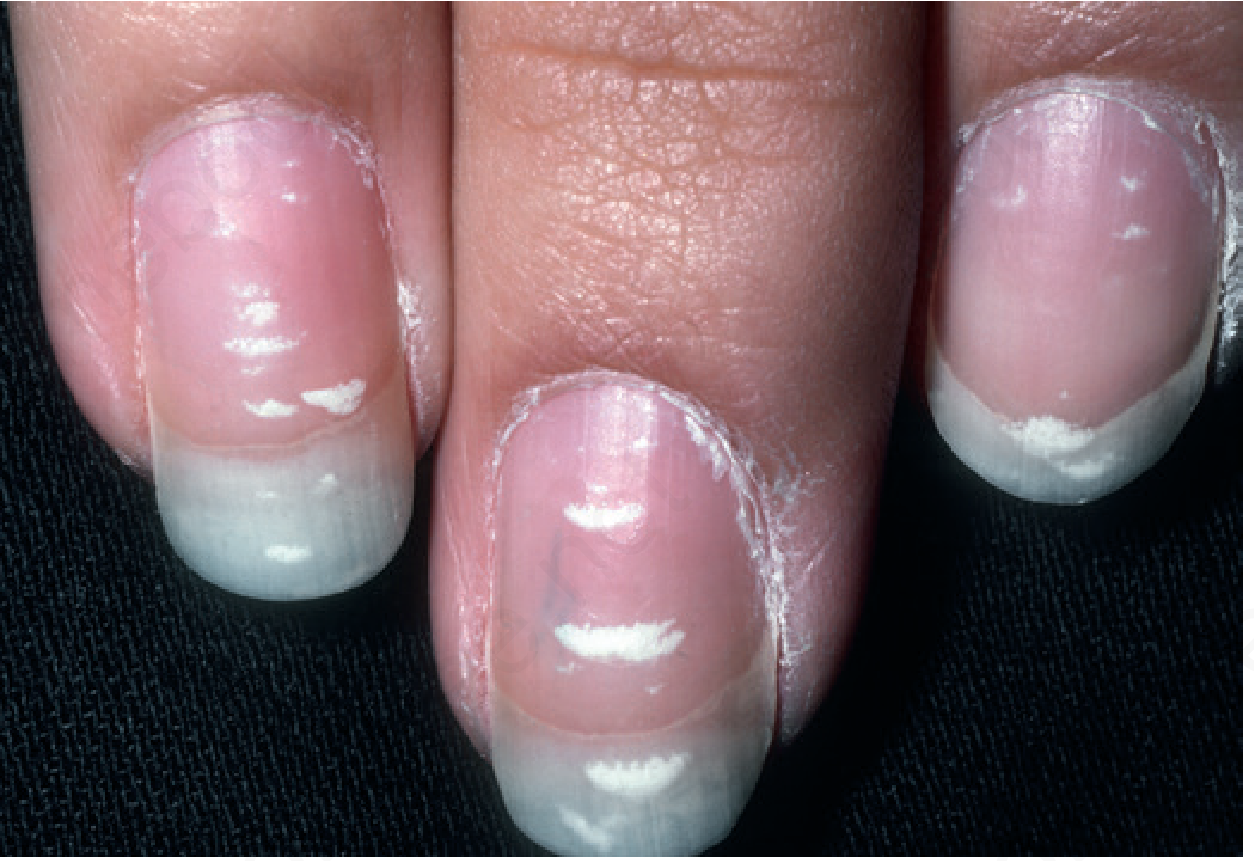
Transverse (striate) leuconychia - Andrews' Diseases of the Skin
Differential Diagnosis - Key Mimics
- Superficial white onychomycosis (pseudo-leuconychia): Friable, chalky white spots on the nail plate surface that can be scraped off; caused by T. mentagrophytes, occasionally molds. Distinguish by KOH and culture.
- Onycholysis: Distal nail plate detachment - looks white due to air under the nail; NOT true leukonychia
- Proximal subungual onychomycosis: White opaque proximal nail plate in immunosuppressed patients (HIV, transplant); due to T. rubrum invading the ventral nail plate - a marker of immunosuppression
Alopecia Areata Association
Punctate leuconychia is a recognized nail manifestation of alopecia areata, along with geometric pitting, erythema of the lunula, and onychomadesis.
Summary of Systemic Causes
| Pattern | Associated Condition |
|---|---|
| Mees' lines (striate) | Arsenic/thallium poisoning |
| Muehrcke's bands (apparent) | Hypoalbuminemia, nephrotic syndrome, chemotherapy |
| Half-and-half nails (apparent) | Chronic renal failure / hemodialysis |
| Terry's nails (apparent) | Liver cirrhosis, cardiac failure, diabetes |
| Leukonychia totalis (true) | PLCD1/GJA1/GJB2 mutations, Bart-Pumphrey syndrome, systemic infections |
| Punctate/striate (true) | Trauma (most common), alopecia areata |
Sources: Dermatology 2-Volume Set 5e (Bolognia et al.), Andrews' Diseases of the Skin 2e, Goldman-Cecil Medicine International Edition
This is a shared conversation. Sign in to Orris to start your own chat.