
History and examination of patient of sarcoidosis
sarcoidosis clinical features skin lung examination
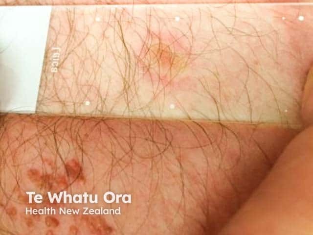
This is a clinical photography of a cutaneous sarcoidosis lesion on the trunk, evaluated with diascopy to elicit an apple-jelly appearance. The imaging modality is a straightforward skin examination with a diascopic test, capturing surface and subcutaneous texture of a trunk lesion. The anatomical location is the torso, with involvement of the dermal and subcutaneous layers beneath erythematous, nodular plaques. Visual features include firm, well-circumscribed nodules and plaques with brown to orange-brown coloration that become more conspicuous under pressure, producing the characteristic apple-jelly nodules. The distribution appears on the central trunk, without obvious mucosal involvement, and there is minimal epidermal change overlying the lesion. Pathophysiology involves granulomatous inflammation with epithelioid histiocytes and multinucleated giant cells within the dermis, consistent with sarcoid granulomas. Clinically, diascopy-positive apple-jelly nodules support a granulomatous process and help distinguish sarcoidosis from other differential diagnoses such as lupus vulgaris and granulomatous dermatitis. Diagnostic significance lies in correlating with systemic sarcoidosis workup, including chest imaging and serum ACE levels. Potential clinical use cases include dermatology educational resources, differential diagnosis practice, biopsy decision-making, and monitoring response to therapy. This image thus serves as a representative dermatologic sign and provides visual context for cutaneous sarcoidosis.
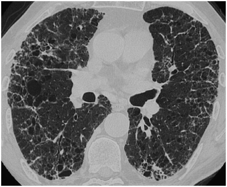
This diagnostic image is an axial high-resolution computerized tomography (HRCT) scan of the thorax, specifically focusing on the lung parenchyma at the level of the lower lobes. The scan demonstrates a Usual Interstitial Pneumonia (UIP)-like pattern in a patient with clinical sarcoidosis. Key visual features include extensive reticulations and traction bronchiectasis (abnormal bronchial widening) within areas of fibrotic change. Characteristic honeycombing is present, appearing as subpleural, clustered cystic airspaces with well-defined, thickened walls. These findings exhibit a distinct basal and peripheral (subpleural) predominance, involving both lungs. This pattern of architectural distortion is representative of advanced pulmonary fibrosis. The educational focus is on identifying atypical presentations of sarcoidosis that mimic UIP, illustrating the transition from granulomatous inflammation to chronic fibrotic disease. This resource is intended for medical students and radiology residents studying interstitial lung disease (ILD) classification and diagnostic imaging.
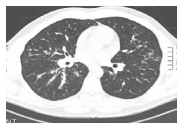
This diagnostic image is an axial chest CT scan using a lung window, demonstrating findings characteristic of Stage 2 sarcoidosis. The primary pathological features include bilateral hilar adenopathy, visualized as symmetric soft tissue masses at the lung roots that obscure normal vascular and bronchial margins. Additionally, there is evidence of diffuse parenchymatous infiltration throughout both lung fields. These infiltrates present as ill-defined nodular opacities with a perilymphatic distribution pattern, typical of granulomatous inflammation. The image serves as a clinical example of pulmonary sarcoidosis involvement, illustrating both lymphatic enlargement and parenchymal disease. It is suitable for medical education regarding thoracic imaging and interstitial lung diseases.
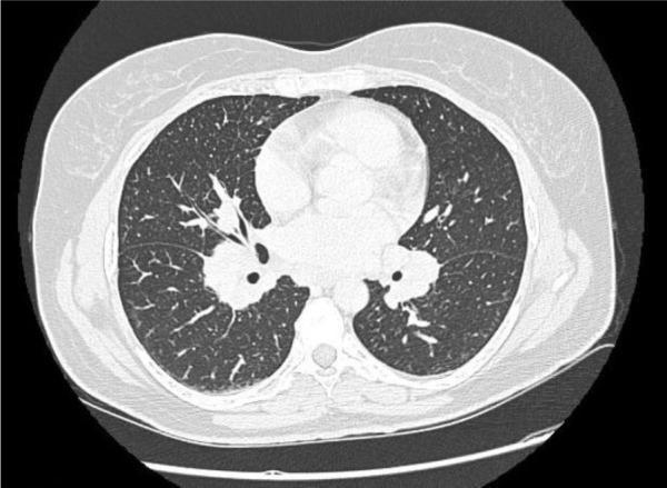
This diagnostic axial CT image of the thorax, presented in a lung window, demonstrates classic radiographic features of pulmonary sarcoidosis. Key findings include prominent, systemic bilateral hilar and mediastinal lymphadenopathy, characterized by enlarged, soft-tissue density nodes at the root of both lungs. The lung parenchyma exhibits a subtle but widespread reticulonodular infiltration pattern, most notably in the mid-to-lower lung zones, consisting of fine linear opacities and small nodular densities scattered throughout the interstitial space. These findings lack significant consolidation or pleural effusion. In the clinical context of Löfgren’s syndrome—an acute presentation of sarcoidosis—this imaging captures the characteristic triad involving lymph node enlargement and pulmonary parenchymal involvement. This case serves as an educational example for radiology and pulmonology students to recognize the distribution of sarcoid-related adenopathy and the characteristic 'ground-glass' like texture of interstitial lung disease.
lupus pernio sarcoidosis facial skin lesion
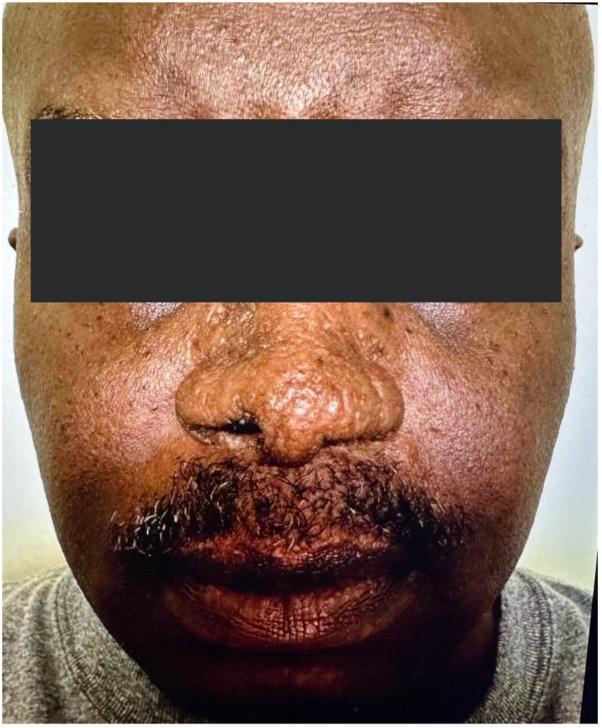
This clinical photograph shows the frontal view of a patient with cutaneous sarcoidosis, specifically lupus pernio, following systemic treatment. The image focuses on the mid-face region, including the nose, cheeks, and upper lip. While there is evidence of post-inflammatory texture changes and some subtle residual thickening, there is a marked absence of the highly characteristic indurated, violaceous, or reddish-brown shiny nodules and plaques typically seen in active lupus pernio. The nasal bridge and alae appear relatively smooth and the previous bulbous distortion has largely resolved. The skin tone on the cheeks and perioral area is more uniform, though some mild follicular prominence and chronic scarring may persist. This image serves as an educational example of the therapeutic response of cutaneous sarcoidosis to Janus kinase (JAK) inhibitor therapy, illustrating the significant reduction in granulomatous infiltration and the restoration of normal facial contours.
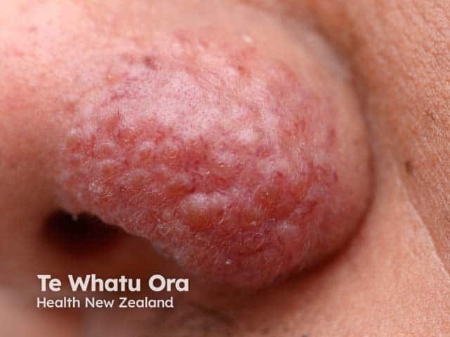
Imaging modality: Clinical dermatologic photography of a localized cutaneous plaque on the left nasal ala. The lesion is an indurated, erythematous-to-violaceous plaque with a rough surface and subtle nodularity, coalescing into a conspicuous patch along the ala nasi. The appearance is classic for lupus pernio-like cutaneous sarcoidosis, though biopsy is often required for definitive confirmation. The image emphasizes surface texture with increased dermal density and faintly infiltrated margins against surrounding normal facial skin. Involvement is confined to the external nose region; no adjacent mucosal ulceration or necrosis is evident in this view. The lesion color ranges from reddish to purplish pink, reflecting granulomatous inflammation beneath the epidermis. Clinically, such plaques may precede systemic sarcoidosis or accompany pulmonary, ocular, or multiorgan involvement; thus, this photo is valuable for differential diagnosis and educational case discussion. When compared with other granulomatous dermatoses, this presentation helps distinguish sarcoid skin lesions from infectious etiologies or rosacea variants. Potential clinical applications include guiding biopsy planning, monitoring therapeutic response to corticosteroids or steroid-sparing agents, and prompting systemic evaluation for sarcoidosis. This image serves as a teaching example for dermatologists, radiologists, and medical trainees studying cutaneous markers of systemic granulomatous disease. Clinical relevance includes monitoring progression and response.
History and Examination in Sarcoidosis
Overview
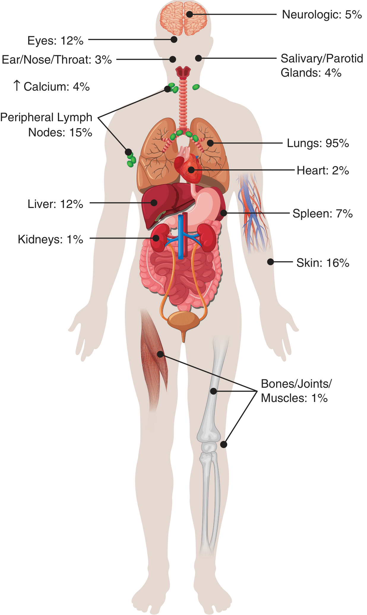
HISTORY
1. Demographic Profile
- Age: Peak incidence 35-45 years; a second peak occurs after age 55
- Sex: Women more commonly affected (ratio usually <2:1)
- Race/Ethnicity: Highest incidence in African Americans (35.5/100,000) and Northern Europeans (24/100,000). Severity is higher in Black patients, who present with more extrapulmonary disease
- Occupation/Exposure: Ask about dust, silica, beryllium, organic antigens (risk factors)
- Season: Spring predominance of new diagnoses
- Family history: Elevated familial risk - three times higher in Black patients
2. Mode of Presentation
| Presentation | Features |
|---|---|
| Asymptomatic | Incidental finding on chest X-ray (most common in some populations) |
| Acute/Subacute | Löfgren syndrome, Heerfordt syndrome (see below) |
| Chronic/Insidious | Gradual onset of respiratory, systemic, or organ-specific symptoms |
Löfgren Syndrome (Acute Presentation - Good Prognosis)
- Bilateral hilar adenopathy + fever + polyarthritis + erythema nodosum
- Common in Europeans and women; typically self-limiting
Heerfordt-Waldström Syndrome
- Parotitis + fever + facial nerve palsy + uveitis
- Associated with sarcoidosis
3. Respiratory Symptoms (>90% have lung involvement)
- Dyspnea (most common, often exertional)
- Chronic nonproductive cough
- Chest tightness or discomfort
- Some patients have normal lung function despite active radiographic disease
- Severe cases: progressive respiratory failure
4. Constitutional/Systemic Symptoms
- Fatigue (very common, often disproportionate)
- Fever (low-grade, especially in acute presentations)
- Night sweats
- Weight loss
- Malaise
5. Skin Symptoms (~16-25%)
- Red-brown, non-scaly papules and plaques - especially around nose, mouth, and face
- Lupus pernio - indurated violaceous plaques on nose, cheeks, lips, ears; ask specifically about this (strongly associated with chronic disease, pulmonary fibrosis, upper airway involvement)
- Infiltration of old scars or tattoos (pathognomonic clue in history)
- Subcutaneous nodules
- Erythema nodosum - tender nodules on anterior shins (common in Europeans and in Löfgren syndrome)
- Lesions are usually not painful or pruritic
6. Ocular Symptoms (~12%)
- Blurred vision, photophobia, ocular pain, redness (anterior uveitis most common)
- Posterior uveitis - may be asymptomatic; ask about scotomas, visual field defects
- Lacrimal gland swelling (dry eyes, sicca symptoms)
7. Neurological Symptoms (~5%)
- Facial nerve palsy (most common cranial nerve involved - good prognosis)
- Headache, altered mentation (basal meningitis)
- Visual disturbances (optic nerve involvement)
- Peripheral numbness, pain (small-fiber neuropathy - common cause of pain)
- Autonomic dysfunction
- Seizures (rare)
- Neuroendocrine symptoms - diabetes insipidus, amenorrhea (hypothalamic/pituitary involvement)
8. Cardiac Symptoms (~2% clinically, higher at autopsy)
- Palpitations, syncope, presyncope (arrhythmias, conduction blocks)
- Dyspnea, peripheral edema (heart failure from cardiomyopathy)
- Sudden cardiac death (rare - can be first manifestation)
- Note: Japanese patients have higher rates of cardiac involvement
9. Musculoskeletal Symptoms (~1%)
- Arthritis/polyarthralgia (especially ankles in Löfgren syndrome)
- Bone pain (bone cysts, particularly hands/feet)
- Muscle weakness (granulomatous myopathy - rare)
10. Abdominal/Other Symptoms
- Hepatomegaly symptoms: RUQ discomfort, jaundice (rare)
- Splenomegaly: early satiety, LUQ fullness
- Parotid swelling (Heerfordt syndrome)
- Renal symptoms: kidney stones (hypercalciuria from granuloma-produced calcitriol)
- Hypercalcemia symptoms: polyuria, polydipsia, constipation, nausea
11. Drug and Medication History
- Immune checkpoint inhibitors, antiretroviral therapy, interferon, TNF-α antagonists can cause sarcoid-like reactions - must be excluded
PHYSICAL EXAMINATION
General Appearance
- May appear well (subclinical disease) or chronically ill
- Fever (acute presentations)
- Signs of weight loss
Respiratory Examination
- Inspection: Tachypnea in advanced disease; bilateral symmetrical chest expansion (usually normal early)
- Palpation: Trachea central; vocal fremitus often normal
- Percussion: Usually resonant; dullness if pleural effusion (rare)
- Auscultation:
- Often normal lung sounds even with extensive radiographic disease (important distinguishing feature)
- Bilateral fine end-inspiratory crackles in fibrotic/advanced disease
- Wheeze if airflow obstruction (seen in ~1/3 of cases)
- Note: Crackles are less common in sarcoidosis than in IPF, despite similar imaging findings
Lymph Node Examination
- Bilateral hilar adenopathy - the most common intrathoracic finding (not directly palpable but note neck/peripheral nodes)
- Peripheral lymphadenopathy (~15%): palpable cervical, axillary, inguinal nodes - non-tender, rubbery, mobile
- Superficial nodes may be biopsied for diagnosis
Skin Examination
- Specific sarcoid granulomatous lesions:
- Red-brown to violaceous smooth papules and plaques on face (especially peri-nasal, perioral, eyelids)
- Lupus pernio: indurated violaceous plaques on nose, cheeks, lips, ears - ask for diascopy (press with glass slide) to elicit "apple-jelly" appearance (yellowish-orange color under pressure), characteristic of granulomatous disease
- Infiltration of scars and tattoos (pathognomonic)
- Subcutaneous nodules (Darier-Roussy nodules)
- Annular, ulcerative, or verrucous plaques
- Hypopigmented or hyperpigmented macules
- Non-specific lesions:
- Erythema nodosum (tender raised nodules on anterior shins - especially in Löfgren)
- Alopecia (scalp sarcoidosis)
Ocular Examination
- Conjunctival injection, corneal band keratopathy
- Slit-lamp findings: anterior uveitis (cells and flare), posterior synechiae
- Fundoscopy: choroidal granulomas, optic disc swelling, retinal vasculitis
- Lacrimal gland enlargement
- An ophthalmologic evaluation is mandatory in all newly diagnosed patients (silent uveitis is common)
Cardiovascular Examination
- Pulse: irregular (arrhythmia, conduction block)
- S3/S4 gallop (cardiomyopathy)
- Murmurs (valvular involvement - rare)
- Signs of right heart failure: raised JVP, peripheral edema, hepatomegaly (cor pulmonale from pulmonary fibrosis)
Abdominal Examination
- Hepatomegaly (~12%) - usually mild, non-tender
- Splenomegaly (~7%) - smooth, non-tender
- Ascites (rare, portal hypertension from hepatic sarcoidosis)
Neurological Examination
- Cranial nerves: Especially VII (facial palsy), II (optic), VIII (hearing loss - Heerfordt)
- Peripheral nervous system: reduced sensation in a stocking-glove distribution (small-fiber neuropathy)
- Motor: weakness (myopathy or mononeuritis multiplex)
- Cerebellar signs, long tract signs (spinal cord involvement - rare)
Musculoskeletal Examination
- Ankle periarthritis (in Löfgren syndrome - periarticular swelling of ankles)
- Joint swelling in polyarthritis (knees, wrists, small joints)
- Bone tenderness (cystic bone lesions in hands/feet - dactylitis)
Other
- Parotid gland enlargement (Heerfordt syndrome; bilateral sausage-shaped swelling)
- Salivary gland enlargement (~4%)
- Nasal mucosal examination: Granulomatous infiltration (lupus pernio of nasal mucosa), associated with upper respiratory tract involvement
BASELINE INVESTIGATIONS (Always Ordered After History and Exam)
| Investigation | Purpose |
|---|---|
| Pulmonary function tests | Restrictive pattern or obstruction, DLCO reduction |
| Chest X-ray / CT | Scadding staging (I-IV); perilymphatic nodules on CT |
| Eye exam (slit-lamp) | Detect silent uveitis |
| ECG | Conduction blocks, arrhythmias |
| Serum calcium, creatinine | Hypercalcemia, renal involvement |
| Alkaline phosphatase, LFTs | Hepatic granulomas |
| Full blood count | Lymphopenia, anaemia, thrombocytopenia |
| Serum ACE level | Elevated in ~60%; useful for monitoring activity |
| 25-OH and 1,25-OH Vitamin D | Assessing vitamin D/calcium metabolism |
| Biopsy (affected organ) | Noncaseating granulomas; exclude infection/malignancy |
KEY CLINICAL CLUES ON HISTORY AND EXAMINATION
| Clue | Significance |
|---|---|
| Bilateral hilar adenopathy on CXR + young adult | Classic; always suspect sarcoidosis |
| Lesion developing in old scar or tattoo | Pathognomonic of sarcoidosis |
| Lupus pernio | Chronic sarcoidosis; associated with pulmonary fibrosis and upper airway disease |
| Erythema nodosum + ankle periarthritis + bilateral hilar adenopathy | Löfgren syndrome - favorable prognosis |
| Parotitis + fever + uveitis + facial palsy | Heerfordt-Waldström syndrome |
| Facial nerve palsy in sarcoidosis | Good prognosis among neurologic findings |
| Normal chest auscultation with abnormal CXR | Classic dissociation in sarcoidosis |
| Hypercalcemia | Granulomas convert 25-OH-D to 1,25-OH-D (calcitriol) autonomously |
| Small-fiber neuropathy | Common underdiagnosed cause of pain in sarcoidosis |