
Mucoepidermoid carcinoma of parotid gland
Reading File
Now let me search the medical library and PubMed in parallel for mucoepidermoid carcinoma of the parotid gland.
Finding Sources
Finding Sources
Searching PubMed
mucoepidermoid carcinoma parotid gland
Good results. Now let me read the key sections from the most relevant textbooks in parallel.
Reading File
Reading File
Reading File
Finding Sources
Excellent - very rich content. Let me fetch the histology image and read more from the Scott-Brown section and Cummings grading/treatment section.
Loading Image
Reading File
Reading File
Now let me read the treatment/management section from Cummings and also the molecular genetics portion.
Reading File
Finding Sources
I now have comprehensive information. Let me also get the treatment/prognosis details from Scott-Brown and any staging data.
Reading File
Finding Sources
I now have comprehensive data from multiple authoritative textbooks to write a thorough answer. Let me compile everything.
Mucoepidermoid Carcinoma of the Parotid Gland
Overview
Mucoepidermoid carcinoma (MEC) is the most common malignant salivary gland neoplasm, accounting for 12-29% of all salivary gland malignancies. The parotid gland is its most frequent site. It is also the most common salivary gland carcinoma in children and young adults, with a peak incidence in the second decade in pediatric populations and around the fifth decade overall. It affects women more frequently than men.
- Cummings Otolaryngology, p. 1531 | Scott-Brown's Otorhinolaryngology, p. 1149 | Bailey and Love's Surgery, p. 862
Etiology / Risk Factors
- Radiation exposure is the major known risk factor. Prior therapeutic radiation carries a latent period of 7-32 years before MEC development.
- MECs are known to occur following radiation or chemotherapy in childhood.
- Scott-Brown's Otorhinolaryngology
Clinical Presentation
- Typically a painless, slow-growing mass in the parotid region.
- Low-grade tumors are soft and often cystic, sometimes with a bluish hue resembling a mucocele (especially intraoral minor gland tumors).
- High-grade tumors may present with skin or bone involvement, pain, or facial nerve involvement.
- Distant metastases (mainly to lungs) are seen predominantly with high-grade disease.
Gross Pathology
- Tumors are circumscribed or infiltrative, predominantly solid with tan-white to pink cut surface.
- Often have cystic components filled with viscous mucinous (brown) fluid.
- Low-grade: macrocystic, well-circumscribed.
- High-grade: solid, infiltrative.
Microscopic Pathology (Histology)
The hallmark is the three cell types:
| Cell Type | Appearance |
|---|---|
| Mucous cells (mucocytes) | Abundant light-blue mucin in cytoplasm; peripherally displaced nuclei; line cystic spaces |
| Squamoid (epidermoid) cells | Large, abundant pink cytoplasm; look squamoid but lack true keratinization |
| Intermediate cells | Small-medium, modest pink/clear cytoplasm; usually predominate |
The architecture is a mixture of cystic and solid elements - sheets, nests, or duct-like structures.
True keratinization is rare in MEC; its presence should raise the alternative diagnosis of adenosquamous carcinoma.
Special stains (PAS, mucicarmine, Alcian blue) can highlight mucin in cases where it is scant.
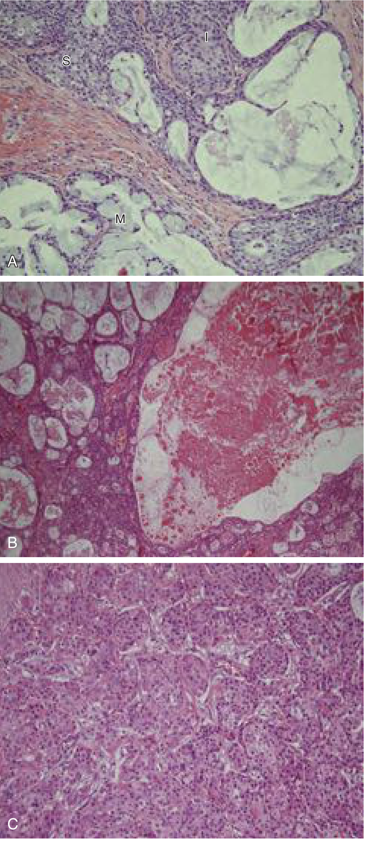
Fig. Mucoepidermoid carcinoma. (A) Three cell types - intermediate (I), mucous (M), and squamoid (S) at x200. (B) Tumor with extensive cystic change at x100. (C) High-grade MEC with solid sheets of intermediate cells, minimal mucous differentiation, and no cystic change at x200.
- Cummings Otolaryngology, p. 1531
Histologic Grading
Grading is critical and strongly correlates with clinical behavior. Two widely used systems exist:
Auclair Grading System:
| Parameter | Points |
|---|---|
| Cystic component <20% | +2 |
| Neural invasion | +2 |
| ≥4 mitoses/10 hpf | +3 |
| Necrosis | +3 |
| Anaplasia | +4 |
| Grade | Score |
|---|---|
| Low (Grade 1) | 0-4 |
| Intermediate (Grade 2) | 5-6 |
| High (Grade 3) | ≥7 |
Brandwein Grading System (modified, more strict - designed to avoid undergrading):
| Parameter | Points |
|---|---|
| Cystic component <25% | +2 |
| Tumor front invades in small nests/islands | +2 |
| Pronounced nuclear atypia | +2 |
| Lymphatic/vascular invasion | +3 |
| Neural invasion | +3 |
| Necrosis | +3 |
| ≥4 mitoses/10 hpf | +3 |
| Bony invasion | +3 |
| Grade | Score |
|---|---|
| Low (Grade 1) | 0 |
| Intermediate (Grade 2) | 2-3 |
| High (Grade 3) | ≥4 |
- Cummings Otolaryngology, p. 1531 (Table 85.6)
Grade-specific histology summary:
- Low-grade: Predominantly cystic, well-circumscribed, rich in mucous cells, minimal atypia.
- Intermediate-grade: Less circumscribed, more solid, predominant intermediate cells; most challenging for pathologists.
- High-grade: Solid, infiltrative, nuclear atypia, necrosis, perineural invasion, lymphovascular emboli; resembles SCC - diagnosis requires demonstration of at least focal intracellular mucin.
Molecular Genetics
MEC has a characteristic chromosomal translocation:
| Fusion Oncogene | Frequency | Notes |
|---|---|---|
| CRTC1-MAML2 | >80% | Present in low and intermediate grade tumors |
| CRTC3-MAML2 | <10% | Also present in lower grades |
These translocations arise from t(11;19)(q21;p13). Detection is by FISH, PCR, or DNA sequencing and has growing diagnostic utility.
- Scott-Brown's Otorhinolaryngology, Table 26.9
Differential Diagnosis
- Necrotizing sialometaplasia - nonneoplastic reactive change in minor salivary glands (hard palate); can mimic MEC.
- Squamous cell carcinoma - especially for high-grade MEC; mucin stains are key to differentiate.
- Mucocele - for low-grade intraoral MEC with bluish hue.
- Adenosquamous carcinoma - when true keratinization is present.
FNA Cytology
FNA yields a characteristic mixture - both glandular and squamoid components must be present for diagnosis:
- Mucous cells: abundant vacuolated cytoplasm
- Intermediate cells: small, round, scant cytoplasm, open chromatin
- Squamoid cells: moderate dense homogeneous cytoplasm; appear orange on Papanicolaou staining
Treatment
-
Complete surgical excision with wide margins is the treatment of choice for all grades.
-
Parotidectomy (superficial or total depending on tumor location and extent) with facial nerve preservation when possible.
-
Adjuvant radiotherapy is indicated for intermediate- to high-grade MEC.
-
Neck dissection is performed when there is clinical or radiographic evidence of nodal metastasis; elective neck dissection is considered for high-grade tumors.
-
Distant metastases (predominantly to lungs) are treated with systemic therapy.
-
Bailey and Love's Surgery, p. 861
Prognosis
| Grade | Behavior |
|---|---|
| Low | Rarely metastasizes; essentially never results in death if strictly defined by Brandwein criteria |
| Intermediate | Variable; least pathologist agreement; therapeutic decisions rely on additional clinicopathologic features |
| High | Locally aggressive; bone/skin involvement; nodal and distant (lung) metastases common |
- Clinical stage is as important as histologic grade and the two should always be considered together.
- MEC of the submandibular gland has a more aggressive course than parotid MEC.
- Cummings Otolaryngology, p. 1531
Key Summary Points
- Most common malignant salivary gland tumor; most common site = parotid
- Most common salivary gland carcinoma in children and adults
- Triphasic: mucous + squamoid + intermediate cells
- Graded low/intermediate/high (Auclair or Brandwein systems)
- Molecular signature: CRTC1-MAML2 translocation in >80%
- Treatment: surgery ± adjuvant radiotherapy for intermediate/high grade
- Prognosis directly correlates with histologic grade and clinical stage
This is a shared conversation. Sign in to Orris to start your own chat.