
Anemia, classification of anemia
classification of anemia by mean corpuscular volume microcytic normocytic macrocytic diagram
red blood cell morphology anemia peripheral smear microcytic hypochromic
anemia classification diagnosis
Anemia: Definition
1. Morphologic Classification (by Mean Corpuscular Volume, MCV)
- Iron deficiency anemia
- Thalassemia syndromes
- Anemia of inflammation/chronic disease (some cases)
- Sideroblastic anemia (congenital)
- Lead poisoning
- Early iron deficiency
- Anemia of inflammation/chronic disease (most cases)
- Anemia of chronic kidney disease
- Sickle cell anemia
- Aplastic anemia (many cases)
- Endocrine deficiency (hypothyroidism, adrenal insufficiency)
- Marrow replacement/infiltration, myelodysplastic syndrome
- Physiologic anemia of pregnancy
- Oval macrocytes (impaired DNA synthesis): vitamin B12 deficiency, folate deficiency, myelodysplasia, chemotherapy/antiseizure drugs
- Round macrocytes (membrane defects/other): alcohol use, liver disease, hypothyroidism, reticulocytosis, smoking
2. Mechanistic (Kinetic) Classification
- Blood loss: acute (trauma, hemorrhage) or chronic (GI lesions, menorrhagia)
- Hemolysis
- Inherited: membrane disorders (hereditary spherocytosis, elliptocytosis), enzyme deficiencies (G6PD, pyruvate kinase), hemoglobinopathies (sickle cell disease, thalassemia)
- Acquired: autoimmune hemolytic anemia, drug-induced, transfusion reactions, paroxysmal nocturnal hemoglobinuria
- Mechanical: microangiopathic hemolytic anemia (TTP, HUS, DIC), defective cardiac valves
- Infections: malaria, babesiosis
- Nutritional deficiency: iron, vitamin B12, folate, copper, vitamin C
- Marrow failure/stem cell defects: aplastic anemia, pure red cell aplasia, Fanconi anemia, acute/chronic leukemia, myelodysplastic syndrome
- Marrow infiltration/replacement: metastatic cancer, granulomatous disease, myeloma
- Erythropoietin deficiency: chronic kidney disease, anemia of inflammation, anemia of aging
Practical approach
Deep dive into iron deficiency anemia: causes, diagnosis, and treatment
"iron deficiency anemia" AND treatment
iron deficiency anemia diagnosis treatment guideline 2025
iron deficiency anemia peripheral blood smear microcytic hypochromic red cells
Overview
- Isolated/latent iron deficiency: stores are low/absent (ferritin <15-30 μg/L) but erythropoiesis is still adequately supplied - red cells remain normocytic/normochromic.
- Iron-deficiency anemia: transferrin saturation falls below 15-20%, iron supply to erythropoiesis becomes insufficient, and red cells become hypochromic and microcytic - the classic hematologic picture (Harrison's, p. 808).
- Absolute vs. functional deficiency: absolute deficiency means stores are truly exhausted; functional deficiency means stores are adequate (or even increased) but iron is sequestered and unavailable to erythroid precursors (e.g., due to high hepcidin in inflammation) - Harrison's Principles of Internal Medicine, p. 806.
Causes
- Menstrual blood loss - menstruating women lose ~30 mg iron per cycle; heavy menstrual bleeding causes much greater loss (Katzung's Basic and Clinical Pharmacology, p. 932)
- Gastrointestinal bleeding - the most common cause in men and postmenopausal women (ulcers, colorectal cancer, angiodysplasia, NSAID/aspirin-related lesions, hookworm infection in endemic areas). Unexplained IDA in these groups mandates a GI evaluation (Katzung's; Goldman-Cecil Medicine)
- Celiac disease
- Helicobacter pylori infection
- Inflammatory bowel disease
- Post-bariatric surgery (gastric bypass), post-gastrectomy states
- Achlorhydria; chronic use of proton pump inhibitors or H2-blockers (reduced gastric acid impairs ferric-to-ferrous conversion needed for absorption)
- Common in resource-poor settings or restrictive diets (vegetarian/vegan diets with lower iron bioavailability - heme iron from meat is absorbed far more efficiently, ~20%, than nonheme iron from plants, ~1-2%)
- Pregnancy (especially 2nd/3rd trimester), infancy, rapid growth in adolescence, erythropoiesis-stimulating agent (ESA) therapy
- Rare: TMPRSS6 mutations cause iron-refractory iron-deficiency anemia (IRIDA), with reduced absorption and refractoriness to oral iron
- GWAS studies show variants in TMPRSS6 and TF (transferrin gene) confer susceptibility (Harrison's, p. 807)
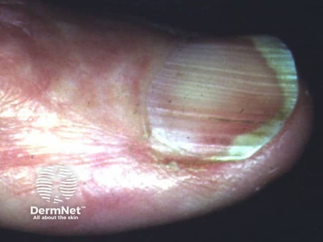
Diagnosis
- CBC: as deficiency progresses, Hb falls and cells become progressively more hypochromic and microcytic (low MCV, low MCHC). Very early iron deficiency can have a normal CBC despite falling ferritin. Thrombocytosis may accompany severe IDA (shared erythroid-megakaryocyte progenitor commitment).
- Serum ferritin is the single best test in patients without chronic inflammatory disease - Symptom to Diagnosis: An Evidence-Based Guide. A ferritin <15 ng/mL carries a likelihood ratio (LR+) of ~51 for iron deficiency; ferritin <30 ng/mL has LR+ ~46. A ferritin >100 ng/mL essentially rules out iron deficiency (LR- 0.08) in general populations.
- Limitation: ferritin is an acute-phase reactant and rises with inflammation, so a "normal" ferritin can mask coexisting iron deficiency in patients with chronic illness (IBD, CKD, malignancy, infection). In these cases, use transferrin saturation (TSAT = serum iron/TIBC) alongside ferritin:
- Absolute iron deficiency: TSAT <20%, ferritin <100 ng/mL
- Functional iron deficiency (inflammation): TSAT <20%, ferritin ≥100 ng/mL
- Transferrin saturation ≤5% has the best single LR+ (~10.5) among the "other" tests
- Additional biomarkers (less sensitive/specific than ferritin alone): serum iron, TIBC, red cell distribution width (RDW, typically elevated), red cell protoporphyrin, reticulocyte hemoglobin content, soluble transferrin receptor (sTfR - useful in inflammation since it is not an acute-phase reactant).
- Gold standard: absence of stainable iron on bone marrow aspirate - rarely needed given the accuracy of noninvasive markers.
- Therapeutic trial: a reticulocyte response within 1 week or a hemoglobin rise of >1 g/dL within 2-4 weeks of iron supplementation supports the diagnosis in ambiguous cases; failure to respond should prompt reassessment (poor adherence, ongoing bleeding, malabsorption, or wrong diagnosis).
- Men and postmenopausal women with unexplained IDA: bidirectional endoscopy (upper endoscopy + colonoscopy) to exclude GI malignancy or bleeding source (AGA guidance, reflected in Katzung's and Goldman-Cecil Medicine)
- Premenopausal women: a trial of oral iron is reasonable first if no alarm symptoms; investigate menstrual history; consider GI workup if bleeding is unexplained or iron therapy fails
- Screen for celiac disease and H. pylori in adults with unexplained IDA
Treatment
Oral iron (first-line for most patients)
- Ferrous salts are preferred (best absorbed): ferrous sulfate (65 mg elemental iron/325 mg tablet), ferrous gluconate (36 mg/325 mg), ferrous fumarate (106 mg/325 mg)
- Traditional dosing: 200-400 mg elemental iron/day in divided doses (about 50-100 mg/day incorporated into hemoglobin; ~25% of oral ferrous iron is absorbed)
- Emerging evidence favors alternate-day dosing: stable-isotope studies show alternate-day administration maximizes fractional absorption (avoids the hepcidin peak triggered by daily dosing) and is better tolerated than daily dosing - consistent with a 2024 systematic review comparing daily vs. alternate-day oral iron (PMID: 37979057)
- Take on an empty stomach when tolerated (food reduces absorption), with ~80 mL orange juice or natural vitamin C to enhance absorption; avoid dairy/calcium and PPIs/antacids around dosing time
- Duration: continue for 3-6 months after the underlying cause is corrected, to replenish stores and prevent early recurrence, not just to normalize hemoglobin
- Response monitoring: reticulocytosis within ~1 week, Hb rise ≥1 g/dL by 2-4 weeks; if inadequate, reassess for adherence, ongoing blood loss, malabsorption, or wrong diagnosis
- Side effects (30-60% of patients): nausea, epigastric discomfort, cramping, constipation or diarrhea, black stools (benign but can mask ongoing GI bleeding on stool guaiac). Managed by lowering dose, taking with food, or switching salts.
- Newer oral option: ferric maltol (Accrufer/Feraccru), FDA-approved for adults and (as of 2025) children ≥10 years, useful in IBD-associated IDA with better GI tolerability profile in some patients.
Intravenous (parenteral) iron
- Modern formulations (ferric carboxymaltose, ferumoxytol, iron isomaltoside) allow full-dose replacement in one or two infusions and no longer require a test dose; infusion reactions occur in <0.1% of cases and are usually mild.
- Ferric carboxymaltose is associated with a recognized risk of hypophosphatemia (via FGF23 elevation) - monitor phosphate with repeated dosing.
- Ferumoxytol can interfere with MRI imaging (obtain MRI before dosing if imaging is anticipated) and carries an FDA black-box warning for rare severe allergic reactions.
- A 2025 systematic review found IV iron generally preferable to blood transfusion for correcting IDA when rapid but non-emergent repletion is needed (PMID: 40577932), and IV iron has been shown cost-effective compared with alternatives in women with heavy menstrual bleeding-related IDA per recent ASH reporting.
Transfusion
Special populations
- Pregnancy: oral iron is first-line; IV iron is used for more severe deficiency, especially in the 2nd/3rd trimester, when oral repletion cannot keep pace with demand. A 2025 systematic review on recombinant erythropoietin for IDA in pregnancy found it used adjunctively in specific refractory scenarios (PMID: 39087437), but iron repletion remains the backbone of therapy.
- CKD/dialysis: parenteral iron is preferred because oral absorption is often inadequate for the elevated requirements imposed by ESA therapy.
Note on evolving guidance
Define hemolytic anemia its causes, symptoms and lab diagnosis
"hemolytic anemia" diagnosis
hemolytic anemia classification causes lab diagnosis 2025
Definition
- Extravascular hemolysis - red cells are removed by splenic (and hepatic) macrophages, usually because reduced deformability prevents them from traversing splenic sinusoids. Produces jaundice/hyperbilirubinemia, splenomegaly, and (if chronic) pigment gallstones.
- Intravascular hemolysis - red cells rupture directly within the circulation from mechanical trauma, complement fixation, or toxins. Produces hemoglobinemia, hemoglobinuria, and hemosiderinuria, since free hemoglobin is small enough to be filtered by the kidney.
- Intrinsic (intracorpuscular) - defect within the red cell itself (usually inherited)
- Extrinsic (extracorpuscular) - defect outside the red cell (usually acquired)
Causes
| Category | Examples |
|---|---|
| Membrane disorders | Hereditary spherocytosis, hereditary elliptocytosis, hereditary stomatocytosis/xerocytosis |
| Enzyme defects | G6PD deficiency, pyruvate kinase deficiency, hexokinase deficiency |
| Hemoglobin abnormalities | Sickle cell disease, thalassemia syndromes, unstable hemoglobins |
- Immune-mediated: Autoimmune hemolytic anemia (warm-antibody IgG-mediated or cold-antibody IgM-mediated/cold agglutinin disease), alloimmune hemolysis (hemolytic transfusion reactions, hemolytic disease of the newborn), drug-induced immune hemolysis
- Mechanical/traumatic: Microangiopathic hemolytic anemia (TTP, HUS, DIC, HELLP syndrome), defective prosthetic heart valves, march hemoglobinuria
- Infectious: Malaria, babesiosis, Clostridium perfringens sepsis, infectious mononucleosis/EBV
- Toxic/chemical: Certain drugs and chemicals, snake venom, severe burns
- Membrane lipid abnormalities: Liver disease, abetalipoproteinemia (spur cell anemia)
- Paroxysmal nocturnal hemoglobinuria (PNH): acquired clonal deficiency of GPI-anchored complement-regulatory proteins
- Hypersplenism/sequestration
Symptoms and Clinical Features
- Fatigue, loss of stamina, exertional dyspnea
- Tachycardia, wide pulse pressure, forceful heartbeat, strong peripheral pulses, systolic flow murmur
- Pallor of mucous membranes, skin, palmar creases, and nail beds
- Jaundice and scleral icterus (from unconjugated hyperbilirubinemia) - more prominent in extravascular hemolysis
- Splenomegaly - reflects "work hyperplasia" of splenic macrophages in extravascular hemolysis
- Dark "cola-colored" urine - from hemoglobinuria/methemoglobinuria, seen in intravascular or severe extravascular hemolysis
- Red-colored urine - seen with very rapid intravascular hemolysis (fresh, non-oxidized hemoglobin)
- Flank pain - can occur with abrupt-onset hemolysis
- Fever in some acute hemolytic episodes
- Long-term complications of chronic hemolysis: pigment (bilirubin) gallstones, iron deficiency (from urinary iron loss in chronic intravascular hemolysis), and extramedullary hematopoiesis in severe cases
Laboratory Diagnosis
Step 1 - Confirm hemolysis (general markers)
| Test | Finding in hemolysis |
|---|---|
| Reticulocyte count (corrected) | Increased (marrow compensation) |
| Serum LDH | Increased - can be up to 10x normal in intravascular hemolysis |
| Serum haptoglobin | Decreased/absent (binds and clears free hemoglobin); <25 mg/dL is ~95% specific for hemolysis |
| Indirect (unconjugated) bilirubin | Increased, especially in extravascular hemolysis |
| Peripheral blood smear | Essential - look for spherocytes, schistocytes, bite cells, sickle cells, agglutination |
| Plasma free hemoglobin / urinary hemoglobin | Positive in intravascular hemolysis (plasma free Hb >40-50 mg/dL) |
| Urinary/fecal urobilinogen | Increased |
| MCV/MCH | Usually increased (reflecting reticulocytosis) |
Step 2 - Determine the cause
- Direct antiglobulin test (DAT/Coombs test): the pivotal test to separate immune from non-immune causes.
- Positive DAT → autoimmune hemolytic anemia (further typed as warm IgG-mediated vs. cold IgM/complement-mediated) or alloimmune hemolysis
- Negative DAT → look for hereditary defects, mechanical/microangiopathic causes, enzyme deficiencies, infections, or toxins
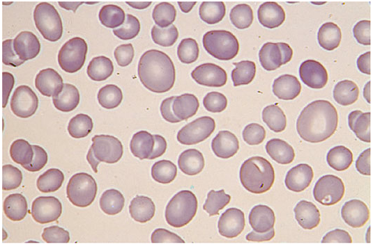
- Peripheral smear morphology narrows the differential substantially:
- Spherocytes (small, no central pallor) → hereditary spherocytosis or warm AIHA
- Schistocytes (fragmented cells) → microangiopathic hemolytic anemia (TTP, HUS, DIC, HELLP, mechanical heart valve)
- Bite cells/Heinz bodies → G6PD deficiency
- Sickle cells → sickle cell disease
- Elliptocytes → hereditary elliptocytosis
- Osmotic fragility test / molecular testing - for hereditary spherocytosis (osmotic fragility is neither sensitive nor specific alone; molecular testing is definitive)
- G6PD enzyme assay - for suspected enzymopathy (best checked after the acute hemolytic episode resolves, as levels can transiently normalize during reticulocytosis)
- Hemoglobin electrophoresis - for suspected hemoglobinopathies (sickle cell, thalassemia)
- Flow cytometry (CD55/CD59) - for suspected PNH
Summary table (adapted from Rosen's Emergency Medicine, Table 109.6)
| Cause | LDH | Haptoglobin | Reticulocytes | Coombs (DAT) | Smear |
|---|---|---|---|---|---|
| G6PD deficiency | ↑ | ↓ | ↑ | Negative | Bite cells |
| Hereditary spherocytosis | ↑ | ↓ | ↑ | Negative | Spherocytes |
| Autoimmune hemolytic anemia | ↑ | ↓ | ↑ | Positive | Spherocytes/agglutination |
| Microangiopathic hemolysis (TTP/HUS/DIC) | ↑↑ | ↓↓ | ↑ | Negative | Schistocytes |
Recent evidence notes
Define Megaloblastic anemia, it's causes , symptoms and lab diagnosis
"megaloblastic anemia" OR "vitamin B12 deficiency" diagnosis
megaloblastic anemia causes symptoms lab diagnosis 2025 guideline
hypersegmented neutrophil peripheral blood smear megaloblastic anemia macro-ovalocytes
Definition
Causes
A. Vitamin B12 (Cobalamin) Deficiency
| Mechanism | Examples |
|---|---|
| Decreased intake | Strict vegan/vegetarian diet, breastfed infant of a B12-deficient mother |
| Lack of intrinsic factor | Pernicious anemia (autoimmune destruction of gastric parietal cells/type A gastritis) - the most common cause overall; gastrectomy/gastric bypass; congenital intrinsic factor defect |
| Impaired absorption at terminal ileum | Ileal resection, Crohn disease/ileitis, congenital cubam receptor defect (Imerslund-Grasbeck syndrome) |
| Competitive/parasitic uptake | Bacterial overgrowth (blind loops, diverticula), fish tapeworm (Diphyllobothrium latum), tropical sprue |
| Drug-induced | Metformin, H2-blockers/proton pump inhibitors, nitrous oxide abuse or anesthesia |
| Inborn errors | Transcobalamin deficiency, CblC-J mutations |
B. Folate Deficiency
| Mechanism | Examples |
|---|---|
| Decreased intake | Poor diet lacking fresh fruit/green leafy vegetables, alcoholism (most common cause in developed countries), anorexia/eating disorders, infancy |
| Malabsorption | Celiac disease, tropical sprue, Crohn disease |
| Increased requirement/loss | Pregnancy, hemolytic anemias, disseminated cancer, hemodialysis |
| Drug-induced (antifolates) | Methotrexate, pyrimethamine, trimethoprim, sulfasalazine, phenytoin (rare), oral contraceptives |
C. Other/rarer causes
- Copper deficiency (e.g., excess zinc or penicillamine use, as in Wilson disease treatment) - can mimic B12 deficiency with anemia plus ataxia
- Nitrous oxide abuse ("whippets") - oxidizes cobalamin, functionally inactivating it
- Congenital enzyme defects of folate/DNA metabolism
- Drugs unresponsive to B12/folate replacement: methotrexate and other direct inhibitors of DNA synthesis
Symptoms and Clinical Features
- Fatigue, weakness, pallor, dyspnea, palpitations
- Glossitis (sore, smooth, "beefy" red tongue), anorexia, weight loss
- Mild jaundice (from intramedullary hemolysis of defective precursors)
- GI symptoms - diarrhea, nausea (from megaloblastic change in rapidly dividing gut epithelium)
- Leukopenia and thrombocytopenia may cause increased infection risk or easy bruising in severe cases
- Subacute combined degeneration of the spinal cord - demyelination of the dorsal and lateral columns
- Symmetric paresthesias and numbness of hands/feet (often the earliest sign, can precede anemia)
- Loss of vibration and proprioceptive sense, sensory ataxia, positive Romberg sign
- Progressive spastic paraparesis, combined hyperreflexia (brisk knee jerk) with hyporeflexia (absent ankle jerk) - a classic combined upper+lower motor neuron pattern
- Cognitive changes, irritability, depression, and in severe cases psychosis ("megaloblastic madness")
- Important: neurologic disease can occur with or without anemia, and giving folate alone in a B12-deficient patient can correct the blood picture while the neurologic damage silently progresses or worsens - so B12 status must always be excluded before treating with folate alone.
Laboratory Diagnosis
Step 1 - Confirm megaloblastic morphology
| Test | Finding |
|---|---|
| CBC | Macrocytic anemia, MCV usually markedly elevated (often >110 fL) |
| Peripheral smear | Macro-ovalocytes (large oval red cells), anisocytosis, poikilocytosis, hypersegmented neutrophils (nuclei with ≥5-6 lobes) - highly characteristic |
| Reticulocyte count | Low/inappropriately normal despite anemia (reticulocytopenia) - reflects ineffective erythropoiesis |
| WBC/platelets | Leukopenia and thrombocytopenia may accompany anemia in more severe cases (pancytopenia) |
| Bone marrow (if done) | Hypercellular marrow with megaloblastic erythroid precursors - nuclear-cytoplasmic asynchrony; giant metamyelocytes |
| Indirect bilirubin, LDH | Mildly elevated (from intramedullary hemolysis of defective precursors) |
Step 2 - Distinguish B12 vs. folate deficiency
| Test | Vitamin B12 deficiency | Folate deficiency |
|---|---|---|
| Serum vitamin B12 | Low | Normal or high |
| Serum/RBC folate | Normal or high | Low |
| Serum homocysteine | Elevated | Elevated |
| Serum methylmalonic acid (MMA) | Elevated (most sensitive/specific marker of B12 deficiency) | Normal |
| Neurologic findings | May be present | Absent |
- Methylmalonic acid is the single most sensitive and specific biochemical marker for cobalamin deficiency, because the B12-dependent enzyme methylmalonyl-CoA mutase requires B12 as cofactor; MMA accumulates only in B12 (not folate) deficiency (Goldman-Cecil Medicine).
- Serum homocysteine rises in both deficiencies because both vitamins participate in the homocysteine-to-methionine conversion pathway - it does not distinguish between the two.
- Intrinsic factor antibodies and parietal cell antibodies: highly specific for pernicious anemia when B12 deficiency is confirmed.
- Schilling test: historically used to determine the mechanism of B12 malabsorption (distinguishing pernicious anemia from ileal disease), but is now largely obsolete due to reliance on radioactive tracers; replaced by intrinsic factor antibody testing and MMA/homocysteine measurement.
- Therapeutic response: reticulocytosis and rising hematocrit beginning about 5 days after parenteral B12 (or oral folate) administration confirms the diagnosis and adequacy of treatment.
- Endoscopy/gastric biopsy: may show atrophic gastritis in pernicious anemia; patients with pernicious anemia carry increased long-term risk of gastric carcinoma and should be monitored.