


Please make flow chart of the types of necrosis in the above photo
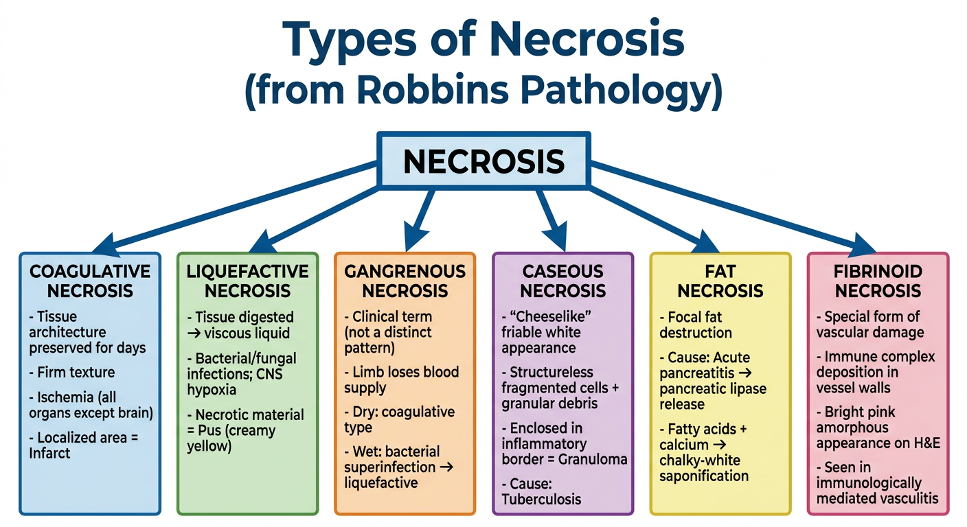
A clean, professional medical flowchart titled "Types of Necrosis" (from Robbins Pathology). The central node at the top reads "NECROSIS" in bold. Six branches radiate downward to six labeled boxes, each with a subtitle and bullet points: 1. COAGULATIVE NECROSIS - Tissue architecture preserved for days - Firm texture - Ischemia (all organs except brain) - Localized area = Infarct 2. LIQUEFACTIVE NECROSIS - Tissue digested → viscous liquid - Bacterial/fungal infections; CNS hypoxia - Necrotic material = Pus (creamy yellow) 3. GANGRENOUS NECROSIS - Clinical term (not a distinct pattern) - Limb loses blood supply - Dry: coagulative type - Wet: bacterial superinfection → liquefactive 4. CASEOUS NECROSIS - "Cheeselike" friable white appearance - Structureless fragmented cells + granular debris - Enclosed in inflammatory border = Granuloma - Cause: Tuberculosis 5. FAT NECROSIS - Focal fat destruction - Cause: Acute pancreatitis → pancreatic lipase release - Fatty acids + calcium → chalky-white saponification 6. FIBRINOID NECROSIS - Special form of vascular damage - Immune complex deposition in vessel walls - Bright pink amorphous appearance on H&E - Seen in immunologically mediated vasculitis Use a white background, color-coded boxes (light blue for coagulative, light green for liquefactive, light orange for gangrenous, light purple for caseous, light yellow for fat necrosis, light pink for fibrinoid), dark readable text, and clear connecting arrows from the central NECROSIS node to each type.
| Type | Key Cause | Hallmark Feature |
|---|---|---|
| Coagulative | Ischemia (all organs except brain) | Tissue architecture preserved; firm texture; = Infarct |
| Liquefactive | Bacterial/fungal infection; CNS hypoxia | Tissue digests into viscous liquid; pus (creamy yellow) |
| Gangrenous | Limb loses blood supply | Dry = coagulative; Wet = bacterial superinfection |
| Caseous | Tuberculosis | Cheeselike white debris; granuloma border |
| Fat | Acute pancreatitis (lipase release) | Chalky-white saponification; fatty acids + calcium |
| Fibrinoid | Immune complex deposition in vessels | Bright pink amorphous deposits on H&E; vasculitis |
Note: Gangrenous necrosis is a clinical term, not a distinct histological pattern - it overlaps with coagulative (dry) and liquefactive (wet) types.