
Sciatic nerve surface marking
The sciatic nerve is the largest nerve in the body. Its surface marking runs from the midpoint between the posterior superior iliac spine (PSIS) and the ischial tuberosity, down to the midpoint of the back of the thigh, then to the apex of the popliteal fossa.
Searching sources...
Reading File
Finding Sources
Finding Sources
S Das has a dedicated section on sciatic nerve surface marking (a clinical surgery text). Let me read that alongside Gray's Anatomy.
Reading File
Reading File
The S Das section started mid-content. Let me read the actual surface marking section from S Das and also find the specific surface marking paragraph.
Reading File
Finding Sources
Excellent - found strong sources. Let me read the key sections: Gray's Anatomy detail on surface marking and the THIEME Atlas reference lines.
Reading File
Reading File
Reading File
I now have excellent detail from multiple sources. Let me also fetch the key anatomical diagram.
Loading Image
Loading Image
I now have comprehensive information from multiple authoritative sources. Here is the complete answer:
Sciatic Nerve - Surface Marking
The sciatic nerve (L4, L5, S1, S2, S3) is the largest nerve in the body. Its surface marking is drawn as a line with three key reference points.
Reference Points (Bony Landmarks)
| Landmark | How to locate |
|---|---|
| Posterior superior iliac spine (PSIS) | Felt as the lateral point of the Michaelis rhomboid |
| Ischial tuberosity | Palpable just above the gluteal fold |
| Greater trochanter | Hard bony protuberance ~one hand's width below the midpoint of the iliac crest |
The Surface Line
Point A (gluteal exit point): The nerve emerges from the infrapiriform foramen at the midpoint of a line drawn from the PSIS to the ischial tuberosity (spine-tuberosity line). This is where the sciatic nerve, inferior gluteal nerve, pudendal nerve, and inferior gluteal vessels all emerge together.
Point B (gluteal region): The nerve then passes downward and lies between the medial and middle thirds of the line drawn from the ischial tuberosity to the greater trochanter (tuberosity-trochanter line). At this point it is deep to gluteus maximus, at the midpoint between the ischial tuberosity and greater trochanter.
Point C (posterior thigh): From Point B, the nerve descends along the midline of the posterior thigh (roughly the midpoint of the back of the thigh), heading toward the apex of the popliteal fossa, where it divides into the common fibular (peroneal) nerve and tibial nerve.
Summary of the Line
PSIS → midpoint → Ischial tuberosity
         ↓
    (exits infrapiriform foramen here)
         ↓
Midpoint between ischial tuberosity and greater trochanter
    (deep to gluteus maximus)
         ↓
Midline of posterior thigh
         ↓
Apex of popliteal fossa
    (divides into tibial + common fibular nerves)
Gluteal Quadrant Method (for injection safety)
The gluteal region is divided into four quadrants using:
- A vertical line descending from the highest point of the iliac crest
- A horizontal line passing midway between the iliac crest and ischial tuberosity plane
The sciatic nerve curves through the upper lateral corner of the lower medial quadrant and descends along the lateral margin of that quadrant. Safe intramuscular injections are given in the upper lateral (anterolateral) quadrant to avoid the nerve.
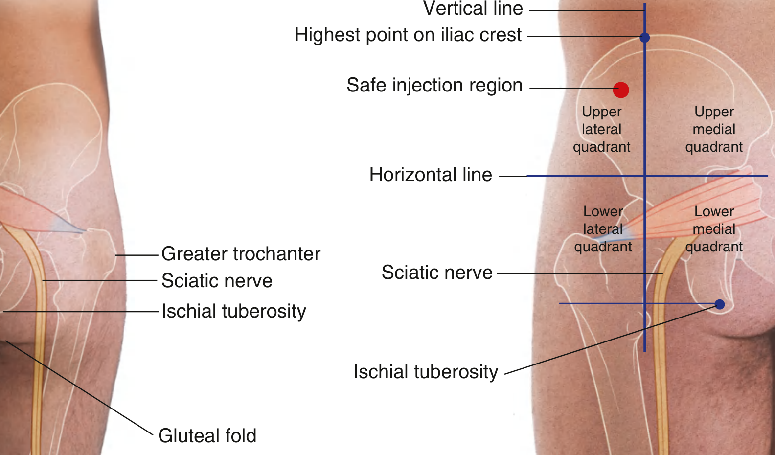
Anatomical Diagram
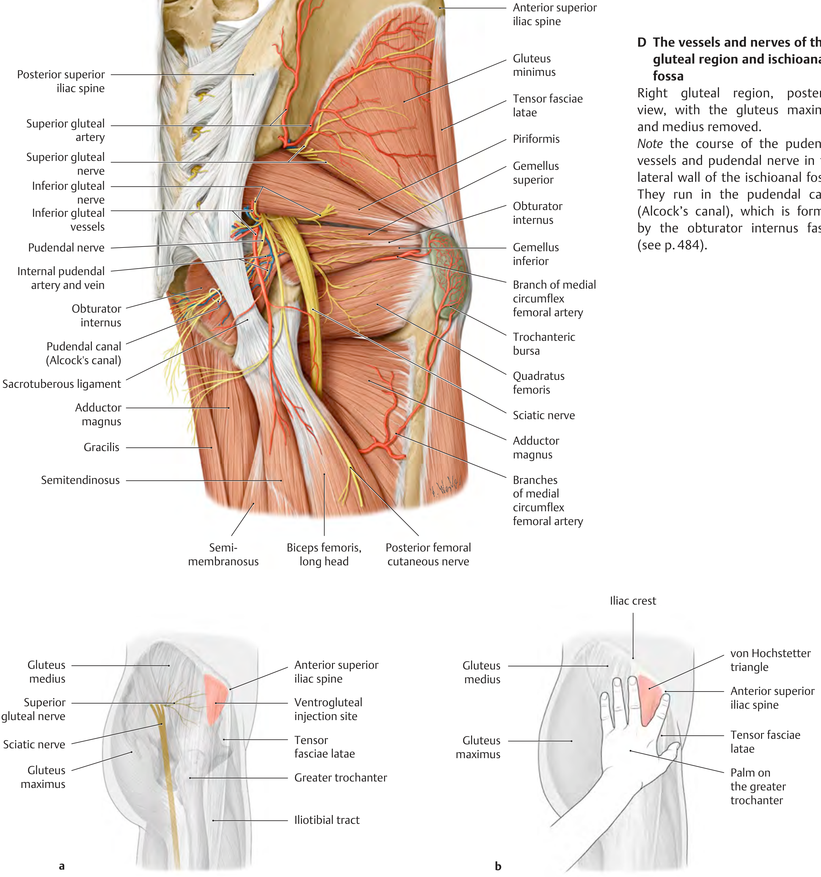
Key Anatomical Relations in the Gluteal Region
- Exits pelvis through the greater sciatic foramen, below piriformis
- Crosses posterior surfaces of: obturator internus + gemelli, then quadratus femoris
- Lies between the superficial (gluteus maximus) and deep muscle groups (piriformis, obturator internus, gemelli, quadratus femoris)
- Enters posterior thigh at the lower margin of quadratus femoris
- In the thigh, it lies on adductor magnus, covered by the long head of biceps femoris
- Divides at the apex of the popliteal fossa (or sometimes higher up in the thigh) into:
- Common fibular nerve (carries L4-S2 posterior divisions)
- Tibial nerve (carries L4-S3 anterior divisions)
Sources: Gray's Anatomy for Students, p. 682 & p. 770; THIEME Atlas of Anatomy - General Anatomy and Musculoskeletal System, p. 579
This is a shared conversation. Sign in to Orris to start your own chat.