
Branches of radial artery
radial artery branches forearm hand anatomy diagram
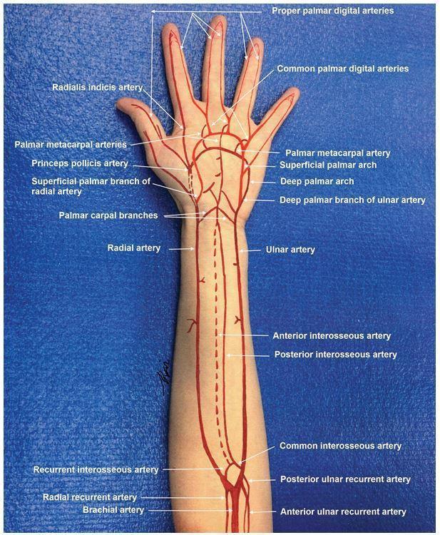
Anatomical diagram superimposed on a photograph of a human arm, illustrating the arterial circulation from the distal humerus to the fingertips. The brachial artery is shown bifurcating into the radial and ulnar arteries. Key forearm vessels are labeled, including the radial and ulnar recurrent arteries, common interosseous artery, and the anterior and posterior interosseous arteries. In the hand, the illustration details the complex vascular anastomoses: the superficial palmar arch (primarily from the ulnar artery) and the deep palmar arch (primarily from the radial artery). Specific terminal branches are identified, such as the princeps pollicis artery, radialis indicis artery, palmar metacarpal arteries, common palmar digital arteries, and proper palmar digital arteries supplying each digit. This educational graphic serves as a guide for understanding upper extremity vascular anatomy, essential for surgical planning, trauma assessment, and performing procedures like the Allen's test.
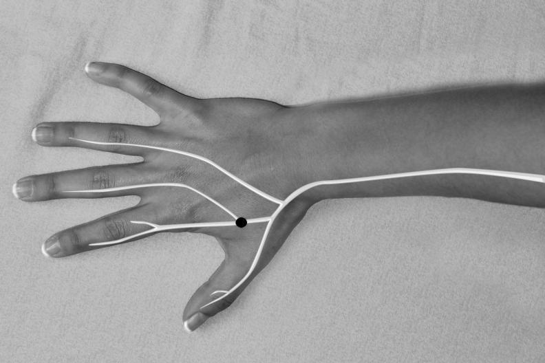
This clinical photograph/anatomical diagram hybrid depicts the dorsal aspect of a human hand and distal forearm in a grayscale format. Superimposed on the anatomy is a schematic representation of the superficial branch of the radial nerve. A thick white line originates from the radial side of the forearm, traveling distally across the wrist. Upon reaching the dorsum of the hand, the line branches into multiple digital nerves that extend toward the thumb, index, middle, and ring fingers. A prominent black circular marker is placed on the first dorsal interosseous space (the web space between the thumb and index finger), identifying the Superficial Radial Neuropuncture Point (SRNP). The image illustrates the neuroanatomical basis for specific acupuncture or electroacupuncture sites, emphasizing the relationship between traditional medicine points and peripheral nerve distribution. This visual is intended for healthcare professionals or students studying neuroanatomy, physical medicine, or integrative pain management protocols.
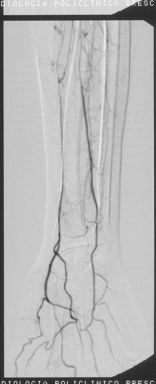
This diagnostic image is a digital subtraction angiogram (DSA) of the right forearm and wrist, captured following a thrombolysis procedure. The image depicts the arterial anatomy of the distal upper extremity, showing contrast opacification of the radial artery and its progression toward the hand. The radial artery exhibits continuous flow with a regular caliber, indicating a successful restoration of patency. In the wrist region, the image demonstrates clear vascularization with branching patterns and anastomoses forming the palmar arches. Smaller collateral vessels and tertiary branches are visible around the radio-carpal joint. The clinical significance of this imaging is to confirm the resolution of previous vasospasm or thromboembolic occlusion and to ensure adequate distal perfusion to the hand. This visual serves as an educational example of normal post-interventional vascular anatomy in the context of interventional radiology and vascular surgery.
Branches of the Radial Artery
1. In the Forearm
| Branch | Details |
|---|---|
| Radial recurrent artery | First and most important branch; ascends to anastomose with the radial collateral branch of the profunda brachii, contributing to the periarticular anastomosis around the elbow |
| Muscular branches | Multiple small branches supplying muscles on the lateral (radial) side of the forearm |
| Palmar carpal branch | A small branch given off near the wrist; joins the palmar carpal branch of the ulnar artery to form the palmar carpal arch, supplying the carpal bones and joints |
| Superficial palmar branch | Given off just before the radial artery leaves the wrist; passes through (or superficial to) the thenar muscles and anastomoses with the superficial palmar arch of the ulnar artery |
2. At the Wrist / Dorsum of Hand
| Branch | Details |
|---|---|
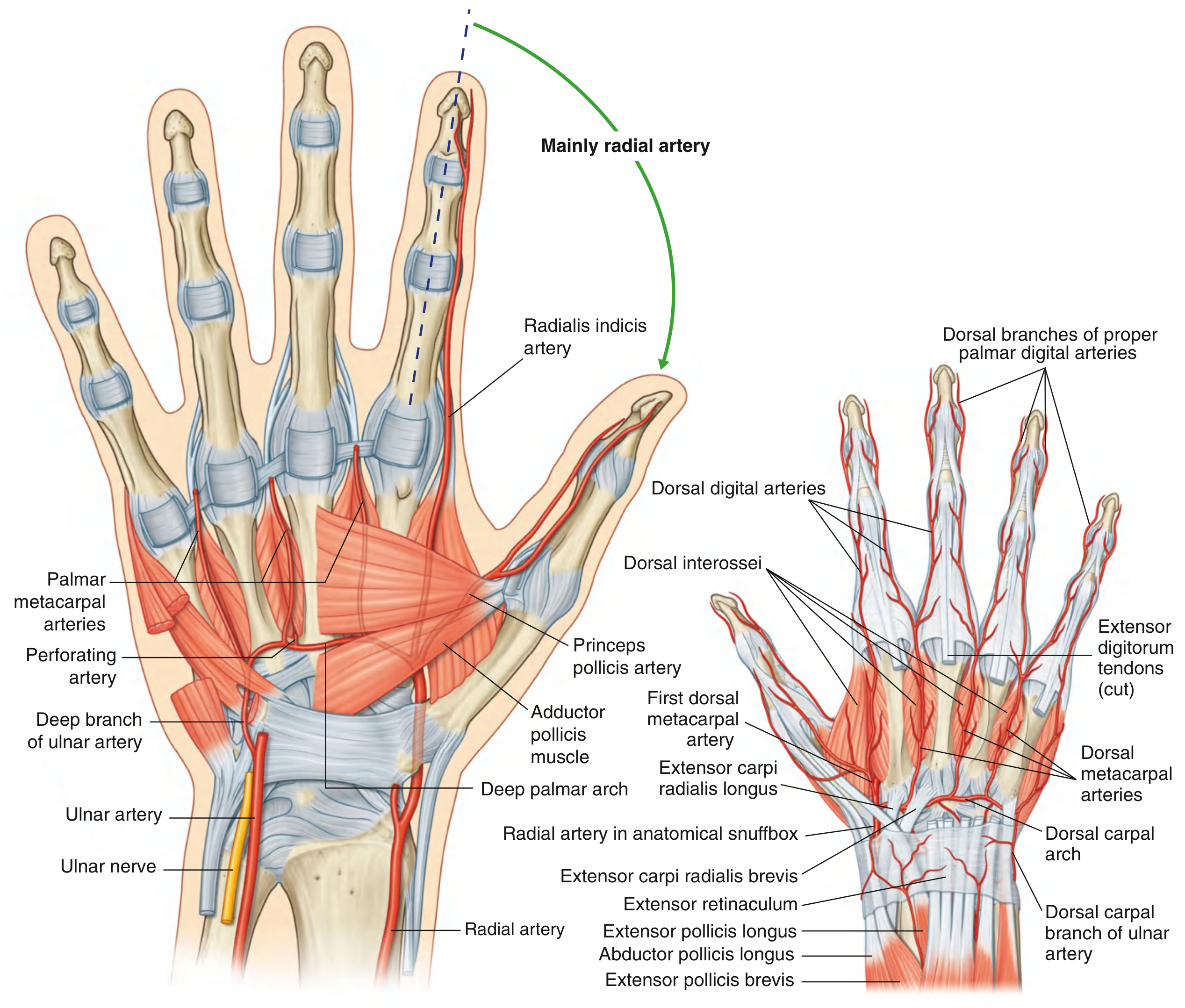
| Dorsal carpal branch | Passes medially to form the dorsal carpal arch (with the dorsal carpal branch of the ulnar artery); gives rise to 3 dorsal metacarpal arteries, which divide into small dorsal digital arteries for the fingers |
| First dorsal metacarpal artery | Arises just before the radial artery dips into the palm; supplies the adjacent sides of the thumb and index finger on their dorsal aspects |
3. In the Hand (Deep Palm)
| Branch | Details |
|---|---|
| Princeps pollicis artery | "Prince of the thumb" - the major blood supply to the thumb; divides into two palmar digital arteries for the medial and lateral sides of the thumb |
| Radialis indicis artery | Arises from the radial artery (or from the princeps pollicis); supplies the lateral (radial) side of the index finger |
| Deep palmar arch | The terminal continuation of the radial artery; curves medially between the metacarpals and the long flexor tendons, completing the arch by anastomosing with the deep palmar branch of the ulnar artery |
- 3 palmar metacarpal arteries - pass distally to join the common palmar digital arteries of the superficial arch
- 3 perforating branches - pass posteriorly between the bases of the dorsal interossei to anastomose with the dorsal metacarpal arteries
Summary Diagram (Forearm - Anterior View)
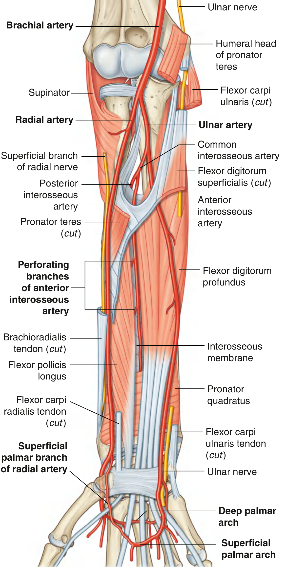
Deep Palmar Arch and Hand Branches
Quick Memory Aid
"R-M-PC-SP | DC-FDM | PP-RI-DPA"
- Forearm: Radial recurrent, Muscular, Palmar Carpal, Superficial Palmar
- Wrist/Dorsum: Dorsal Carpal, First Dorsal Metacarpal
- Hand: Princeps Pollicis, Radialis Indicis, Deep Palmar Arch