I now have comprehensive data. Let me update todos and compile the full detailed response.# Stroke Management - Harrison's Principles of Internal Medicine 22e (2025)
Source: Harrison's Principles of Internal Medicine, 22nd Edition - Chapters 438 & 439
MASTER FLOWCHART: STROKE MANAGEMENT
PATIENT PRESENTS WITH ACUTE NEUROLOGICAL DEFICIT
              |
              v
    ┌─────────────────────────────┐
    │   IMMEDIATE STABILIZATION   │
    │   ABC (Airway/Breathing/    │
    │   Circulation)              │
    │   Finger-stick glucose       │
    │   Treat hypo/hyperglycemia  │
    └─────────┬───────────────────┘
              |
              v
    ┌─────────────────────────────┐
    │  EMERGENCY NON-CONTRAST     │
    │       HEAD CT SCAN          │
    │  (within minutes of arrival)│
    └─────────┬───────────────────┘
              |
       ┌──────┴────────┐
       |               |
       v               v
  ISCHEMIC         HEMORRHAGIC
  STROKE           STROKE
  (No blood        (Blood on CT)
  on CT)               |
       |               └────────────────────────┐
       v                                        v
  [See Section A]                          [See Section B]
SECTION A: ACUTE ISCHEMIC STROKE
A1. IMMEDIATE INVESTIGATIONS
CONFIRMED ISCHEMIC STROKE (CT negative for blood)
              |
  ┌───────────┼───────────────────────┐
  v           v                       v
MANDATORY   CARDIAC               VASCULAR
LABS        WORKUP                IMAGING
  |           |                       |
• CBC        • 12-lead ECG           • CT Angiography
• BMP        • Troponin               (if EVT candidate)
• PT/PTT/    • Echocardiogram        • MRA Brain/Neck
  INR          (TTE or TEE)          • Carotid Doppler
• LFT/RFT   • Holter monitor         (if carotid territory)
• Glucose     (paroxysmal AF)
• HbA1c
• Lipids
• Type & Screen
Additional targeted investigations:
- Hypercoagulability panel (young stroke, no clear cause): Protein C/S, antithrombin III, factor V Leiden, prothrombin gene mutation, antiphospholipid antibodies
- ESR/CRP (vasculitis workup)
- Toxicology screen (cocaine, amphetamines)
- MRI brain with DWI (most sensitive for early ischemia - restricted diffusion within minutes)
- CT Perfusion: identifies ischemic core vs salvageable penumbra for late-window EVT selection
A2. SIX PILLARS OF ISCHEMIC STROKE TREATMENT
(Harrison's Ch. 438: "Treatments designed to reverse or lessen tissue infarction fall within six categories")
                    ACUTE ISCHEMIC STROKE
                          |
        ┌────────┬─────────┼──────────┬─────────┬──────────┐
        v        v         v          v         v          v
    MEDICAL   IV-tPA   ENDOVASCULAR  ANTI-    NEURO-    STROKE
    SUPPORT  THROMBO-  THROMBECTOMY THROMBOT  PROTECTN  CENTER &
             LYSIS     (EVT)        THERAPY           REHAB
A3. MEDICAL SUPPORT (Pillar 1)
| Parameter | Target / Action |
|---|
| Blood Pressure | Do NOT lower unless >220/120 mmHg (no thrombolysis) OR >185/110 mmHg (if tPA planned) |
| BP agent of choice | Labetalol IV, Nicardipine IV, Clevidipine IV |
| Fever | Treat with antipyretics (fever worsens neuronal injury) |
| Glucose | Maintain euglycemia; treat hypo- AND hyperglycemia |
| Airway | Intubate if GCS <8 or unable to protect airway |
| DVT prophylaxis | Subcutaneous heparin (UFH or LMWH) + Pneumatic compression stockings |
| Infections | Active surveillance for pneumonia, UTI, pressure ulcers |
| Nutrition | Early nasogastric feeds if dysphagia present |
| Head position | Flat (0 degrees) initially to maximize cerebral perfusion (unless raised ICP) |
A4. IV THROMBOLYSIS - rt-PA / ALTEPLASE (Pillar 2)
Dosing
Alteplase 0.9 mg/kg IV (maximum 90 mg)
- 10% of dose as IV bolus over 1 minute
- Remaining 90% as IV infusion over 60 minutes
TIME WINDOW FLOWCHART
ONSET OF SYMPTOMS
      |
      |-- ≤3 hours ────> STANDARD WINDOW (all eligible patients)
      |
      |-- 3-4.5 hours ──> EXTENDED WINDOW (most patients, extra exclusions apply)
      |
      |-- >4.5 hours ───> IV tPA NOT indicated
                          (consider EVT if large vessel occlusion)
INDICATIONS FOR IV tPA
| Criterion | Standard (0-3h) | Extended (3-4.5h) |
|---|
| Age | ≥18 years | ≥18 years |
| Clinical diagnosis of ischemic stroke | Yes | Yes |
| Measurable neurological deficit | Yes | Yes |
| Time from symptom onset | <3 hours | 3-4.5 hours |
| BP before treatment | <185/110 mmHg | <185/110 mmHg |
| Blood glucose | 50-400 mg/dL | 50-400 mg/dL |
| CT shows no hemorrhage | Yes | Yes |
| Extra exclusions (3-4.5h only) | N/A | Age >80, NIHSS >25, prior stroke + DM, oral anticoagulant use |
CONTRAINDICATIONS TO IV tPA
ABSOLUTE CONTRAINDICATIONS:
| Contraindication | Rationale |
|---|
| CT evidence of intracranial hemorrhage | Direct bleeding risk |
| Symptoms consistent with subarachnoid hemorrhage | Aneurysm rupture |
| Intracranial/intraspinal surgery in prior 3 months | Surgical wound hemorrhage |
| Serious head trauma in prior 3 months | Wound/contusion hemorrhage |
| History of intracranial hemorrhage | High recurrence risk |
| Uncontrolled hypertension >185/110 mmHg at treatment time | Cannot be corrected |
| Active internal bleeding | Systemic hemorrhage |
| Bleeding diathesis: platelets <100,000/mm³ | Inadequate hemostasis |
| Heparin in prior 48h AND elevated aPTT | Active anticoagulation |
| INR >1.7 (warfarin use) | Active anticoagulation |
| Blood glucose <50 or >400 mg/dL | Metabolic cause (reversible) |
| Current use of direct thrombin inhibitors or factor Xa inhibitors | Active anticoagulation |
| Infective endocarditis | Embolic mycotic aneurysm risk |
| Aortic arch dissection | Catastrophic hemorrhage risk |
RELATIVE CONTRAINDICATIONS:
| Contraindication | Comment |
|---|
| Minor/rapidly improving symptoms | Risk may outweigh benefit |
| Major surgery or serious trauma in prior 14 days | Wound hemorrhage |
| GI/urinary hemorrhage in prior 21 days | Rebleeding risk |
| Post-MI pericarditis | Pericardial tamponade risk |
| Pregnancy | Case-by-case basis |
| Seizure at onset | If deficit from seizure (Todd's palsy), not stroke |
| Lumbar puncture in prior 7 days | Spinal hematoma |
POST-tPA MONITORING PROTOCOL
tPA INFUSION COMPLETE
        |
        v
• No antithrombotics for 24 hours
• No arterial punctures (use existing lines)
• No NG/Foley for 30 min post-infusion
• BP monitoring q15 min x 2h, then q30 min x 6h, then q1h x 16h
• Repeat CT head at 24h BEFORE starting antithrombotics
• Watch for: angioedema, orolingual edema, hemorrhagic transformation
A5. ENDOVASCULAR THROMBECTOMY / EVT (Pillar 3)
Indications for Mechanical Thrombectomy
CANDIDATE SCREENING FOR EVT
            |
    ┌───────┼───────────────────────┐
    v       v                       v
 CLINICAL  IMAGING              VESSEL
 CRITERIA  CRITERIA             CRITERIA
    |           |                   |
• NIHSS ≥6  • CT/MRI no          • LVO confirmed
• Premorbid   large infarct        on CTA/MRA:
  mRS 0-1    (ASPECTS ≥6)          - ICA
• Age ≥18   • Penumbra:            - M1 MCA
            • Core <70mL           - M2 MCA (selected)
            • Mismatch >1.8        - Basilar artery
                                   - Vertebral artery
            ALL THREE CRITERIA MET?
                     |
                ┌────┘
                v
          PROCEED TO EVT
Time Windows for EVT
| Window | Criteria |
|---|
| 0-6 hours | Standard window - all LVO patients meeting criteria |
| 6-16 hours | DAWN/DEFUSE-3 criteria: clinical-imaging mismatch (ischemic penumbra > core) |
| 16-24 hours | DAWN criteria: age ≥80 + NIHSS ≥10, or age <80 + NIHSS ≥10 + core 0-51 mL |
| >24 hours | Not routinely recommended |
EVT Procedure Note
- Preferred: Stent retriever devices (Merci, Solitaire, Trevo) + aspiration
- Access: Transfemoral or transradial approach
- Anticoagulation during procedure: Heparin 70 units/kg IV bolus
- Target: mTICI ≥2b reperfusion (successful in ~80-90% of cases)
Contraindications to EVT
- CT showing large completed infarct (ASPECTS <6 = poor outcome territory)
- No LVO on vascular imaging
- Premorbid severe disability (mRS 3-5)
- Life expectancy <3 months
- Coagulopathy not correctable (INR >3)
A6. ANTITHROMBOTIC THERAPY (Pillar 4)
For Non-Cardioembolic Stroke (Atherothrombotic/Lacunar)
ANTIPLATELET THERAPY - CHOOSE ONE:

1. ASPIRIN 325 mg PO loading, then 81-325 mg/day
   - Give within 24-48h of stroke onset
   - If tPA given: wait 24h, confirm no hemorrhage on CT, THEN start

2. ASPIRIN 25 mg + EXTENDED-RELEASE DIPYRIDAMOLE 200 mg (Aggrenox) BID
   - Superior to aspirin alone for secondary prevention
   - SE: headache (common, usually resolves)

3. CLOPIDOGREL 75 mg/day
   - Alternative if aspirin intolerant
   - Preferred in high atherosclerotic burden

4. ASPIRIN + CLOPIDOGREL (DAPT) - SHORT-TERM (21 days)
   - For high-risk TIA (ABCD2 ≥4) or minor stroke (NIHSS ≤3)
   - CHANCE trial: reduces recurrent stroke without major bleeding increase
   - Then switch to monotherapy
For Cardioembolic Stroke (Atrial Fibrillation)
ANTICOAGULATION - CHA₂DS₂-VASc GUIDED:

Score 0: Aspirin or no antithrombotic
Score 1: Aspirin OR OAC (individualize)
Score ≥2: ORAL ANTICOAGULANT (OAC) - MANDATORY

PREFERRED OAC (Non-Valvular AF):
┌─────────────────────────────────────────────────────┐
│  DOAC (Direct Oral Anticoagulants) - PREFERRED      │
│                                                     │
│  Apixaban: 5 mg BID (2.5 mg BID if ≥2 of:         │
│           age ≥80, weight ≤60kg, Cr ≥1.5)          │
│                                                     │
│  Rivaroxaban: 20 mg once daily with evening meal    │
│                                                     │
│  Dabigatran: 150 mg BID (75 mg BID if CrCl 15-30) │
│                                                     │
│  Edoxaban: 60 mg once daily (30 mg if CrCl 15-50, │
│           weight ≤60 kg, P-gp inhibitors)          │
└─────────────────────────────────────────────────────┘

WARFARIN (Valvular AF, mechanical valves):
Target INR: 2.0-3.0 (mitral mechanical: 2.5-3.5)

TIMING OF ANTICOAGULATION AFTER CARDIOEMBOLIC STROKE:
• Small infarct: Start OAC at day 3-5
• Moderate infarct: Start OAC at day 6-8
• Large infarct: Start OAC at day 12-14
• TIA: Start OAC immediately
("1-3-6-12 rule" - days after stroke onset)
A7. NEUROPROTECTION (Pillar 5)
Harrison's acknowledges this remains an unproven area in clinical practice:
- Numerous neuroprotective agents tested in trials (NMDA antagonists, calcium channel blockers, free-radical scavengers) have failed to show benefit
- Current "neuroprotection" is indirect: maintain euglycemia, treat fever, maintain BP, optimize oxygenation
- Normobaric oxygen: no proven benefit in unselected patients
- Hypothermia: under investigation; not yet standard
A8. STROKE CENTERS & REHABILITATION (Pillar 6)
ACUTE MANAGEMENT                    REHABILITATION
        |                                  |
• Dedicated Stroke Unit             • Begin within 24-48h
• Continuous monitoring             • Physiotherapy (mobility)
• Stroke protocol activation        • Occupational therapy (ADLs)
• Multidisciplinary team            • Speech therapy (dysphagia/aphasia)
• Telemedicine (tele-stroke)        • Cognitive rehabilitation
  in non-stroke centers             • Psychological support
                                    • Driving assessment
                                    • Vocational rehabilitation
SECTION B: INTRACEREBRAL HEMORRHAGE (ICH)
(Harrison's Ch. 439)
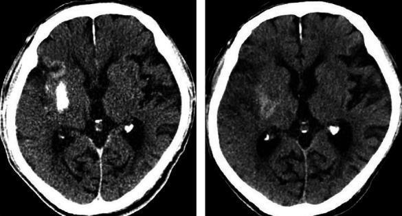
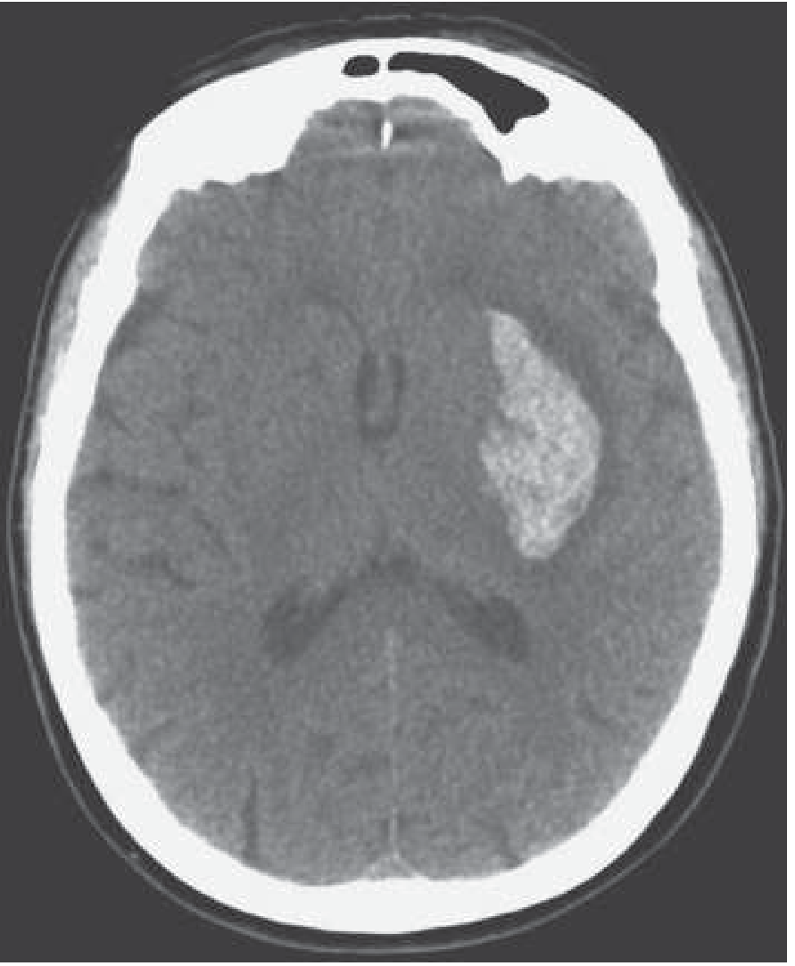
FIGURE 439-1: Hypertensive ICH - left putamen (Harrison's 22e)
B1. EMERGENCY MANAGEMENT OF ICH
HEMORRHAGIC STROKE CONFIRMED ON CT
              |
    ┌─────────┼────────────────────┐
    v         v                    v
AIRWAY &   BLOOD               REVERSAL OF
NEURO      PRESSURE            ANTICOAGULATION
STATUS     CONTROL
    |         |                    |
• Intubate • Target SBP          • Warfarin: Vit K +
  if GCS<8   <140 mmHg             4-factor PCC (Kcentra)
• ICP       (INTERACT2)           • DOAC: Andexanet alfa
  monitoring • Agents:              (anti-Xa) or
• Neurosurg  - Labetalol IV         Idarucizumab
  consult    - Nicardipine IV       (dabigatran)
             - Clevidipine IV     • Heparin: Protamine sulfate
             - Enalaprilat IV
Blood Pressure Management in ICH
- INTERACT2 trial: Target SBP <140 mmHg vs <180 mmHg
- <140 mmHg group: 52% death/major disability at 90 days
- <180 mmHg group: 55.6% death/major disability (p = 0.06, non-significant, but significant ordinal shift)
- Current recommendation: Acutely lower SBP to <140 mmHg if initial SBP was 150-220 mmHg (considered safe)
Common Causes of ICH by Location
| Location | Common Cause |
|---|
| Putamen / Basal ganglia | Hypertension (most common) |
| Thalamus | Hypertension |
| Pons | Hypertension |
| Cerebellum | Hypertension, AVM |
| Lobar (cortical/subcortical) | Cerebral amyloid angiopathy, AVM, tumor, anticoagulation |
| Multiple locations | Amyloid angiopathy, metastases, DIC |
Investigations in ICH
ALL ICH PATIENTS:
• Non-contrast CT (initial - confirms hemorrhage)
• CTA or MRA (if AVM/aneurysm suspected - young patients, lobar location)
• MRI with GRE/SWI (detects microbleeds - amyloid angiopathy)
• CBC, coagulation panel (PT/PTT/INR), platelet count
• BMP, LFT
• Toxicology screen (cocaine, amphetamines - especially young patients)
• 12-lead ECG
• Urinalysis

SELECTIVE:
• Catheter angiography (DSA) - gold standard for AVM/aneurysm
• Lumbar puncture - if subarachnoid extension suspected and imaging equivocal
• Anti-amyloid PET - research setting only
Surgical Intervention in ICH
SURGICAL CONSIDERATIONS:

Indications (STRONGEST):
• Cerebellar hemorrhage >3 cm with neurological deterioration
• Cerebellar hemorrhage compressing brainstem or hydrocephalus
  → External ventricular drain (EVD) + suboccipital craniectomy

Indications (CONDITIONAL):
• Lobar hemorrhage >30 mL within 1 cm of surface
  with neurological deterioration - consider craniotomy
• Progressive hydrocephalus from IVH → EVD

NOT routinely recommended:
• Deep hemorrhages (putamen, thalamus) - surgery not shown to improve outcomes
  (STICH trial: surgical hematoma evacuation = medical management)
• Minimally invasive surgery (MIS) with rt-PA clot lysis (MISTIE III):
  Reduced clot size but no functional benefit

ICP Management if elevated:
• Head of bed 30 degrees
• Hypertonic saline (3% NaCl) or Mannitol 0.25-1 g/kg IV
• Sedation + hyperventilation (temporary: target pCO₂ 30-35 mmHg)
• Decompressive craniectomy (large territory - last resort)
SECTION C: SECONDARY PREVENTION
(Harrison's Ch. 438-439, Block 49)
C1. RISK FACTOR MODIFICATION
| Risk Factor | Target / Intervention |
|---|
| Hypertension (most important) | Target <130/80 mmHg; Thiazides and ACE-I have strongest evidence |
| SPRINT data | SBP <120 mmHg reduced stroke+MI by 43% vs <140 mmHg |
| Dyslipidemia | LDL <70 mg/dL with statin; Atorvastatin 80 mg/d (SPARCL trial) |
| Diabetes | Good glycemic control; Pioglitazone benefits prediabetes/insulin resistance only |
| Smoking | Cessation mandatory (doubles stroke risk) |
| Oral contraceptives | Switch to alternative if possible (particularly with migraine + aura) |
| Sleep apnea | CPAP therapy |
| Atrial fibrillation | Rate control + anticoagulation (see Section A6) |
C2. ANTIPLATELET AGENTS FOR NON-CARDIOEMBOLIC STROKE PREVENTION
FIRST-LINE:
Aspirin 81-325 mg/day OR
Aspirin 25 mg + Dipyridamole ER 200 mg BID (ESPS-2, ESPRIT trials) OR
Clopidogrel 75 mg/day

ASPIRIN + CLOPIDOGREL DAPT (SHORT-TERM - 21 days):
• For high-risk TIA (ABCD₂ score ≥4) or minor stroke (NIHSS ≤3)
• CHANCE trial (Chinese): 60% reduction in recurrent stroke vs aspirin alone
• POINT trial (US): Similar benefit; increased minor bleeding, similar major bleeding

LONG-TERM DAPT (>3 months): NOT recommended
• Increases hemorrhagic risk without additional benefit
• MATCH trial and SPS3 trial: No benefit of long-term DAPT
C3. SURGICAL & ENDOVASCULAR SECONDARY PREVENTION
Carotid Stenosis Management
SYMPTOMATIC CAROTID STENOSIS (ipsilateral TIA or stroke):

>70% stenosis: CEA (Carotid Endarterectomy) STRONGLY INDICATED
               • NNT = 6 to prevent 1 stroke over 2 years
               • Best performed within 2 weeks of TIA/stroke

50-69% stenosis: CEA BENEFICIAL (especially in males)
               • NNT = 22

<50% stenosis: MEDICAL MANAGEMENT ONLY (no surgical benefit)

CAS (Carotid Artery Stenting):
• Alternative to CEA in high surgical risk patients
• CREST trial: CAS = CEA for combined outcomes
• Higher periprocedural stroke risk with CAS in older patients
Intracranial Stenosis
INTRACRANIAL STENOSIS (50-99%) - WARFARIN vs ASPIRIN:
• WASID trial: Warfarin = Aspirin (no benefit, more bleeding)
• Current standard: Aspirin + aggressive risk factor management
• Intracranial stenting (SAMMPRIS): Stenting WORSE than aggressive medical therapy
• Aggressive medical management: Aspirin 325 mg + Clopidogrel x 90 days,
  then aspirin alone + intensive statin + BP control
Patent Foramen Ovale (PFO) Closure
PFO CLOSURE vs MEDICAL THERAPY (cryptogenic stroke):
• CLOSURE I, PC Trial: No benefit
• RESPECT, REDUCE, CLOSE trials (2017-2018):
  PFO closure SUPERIOR to medical therapy in selected patients
  (age <60, large PFO, atrial septal aneurysm, no other cause found)
• Current: PFO closure considered in age <60, cryptogenic stroke,
  no other identifiable cause, after discussion of risks/benefits
COMPLETE STROKE MANAGEMENT SUMMARY FLOWCHART
ACUTE STROKE PRESENTATION
          |
          v
    ABCs + Glucose
          |
          v
    Noncontrast CT HEAD (STAT)
          |
    ┌─────┴──────────────────┐
    |                        |
    v                        v
NO BLOOD = ISCHEMIC      BLOOD = HEMORRHAGIC
    |                        |
    v                        |
 WITHIN 4.5h?                |
  ┌───┴──────┐               |
  YES        NO              |
  |          |               |
  v          v               v
 tPA        CTA +     ICH Management:
(0.9 mg/kg  LVO?     • BP <140 mmHg
 IV, max    |        • Reverse anticoag
 90 mg)    YES       • Airway management
  |         |        • Neurosurgery consult
  |         v        • Cerebellar ICH >3cm
  |       EVT           → Surgery
  |     (0-24h)
  |
  └─────────┐
            v
    Antithrombotics at 24h
    (confirm no hemorrhage on repeat CT)
          |
          v
    CARDIOEMBOLIC?  ──YES──> OAC (DOAC preferred)
          |
          NO
          |
          v
    Antiplatelet therapy
    (Aspirin ± Clopidogrel DAPT x21d
     for high-risk TIA/minor stroke)
          |
          v
    Risk factor modification:
    • Statin (atorvastatin 80 mg)
    • BP control (<130/80 mmHg)
    • Smoking cessation
    • Diabetes management
          |
          v
    STROKE UNIT + REHABILITATION
    (PT/OT/Speech - begin 24-48h)
KEY CLINICAL PEARLS FROM HARRISON'S 22e
-
No reliable clinical signs distinguish ischemic from hemorrhagic stroke - CT is mandatory before any treatment.
-
BP in ischemic stroke: Do NOT routinely lower unless >220/120 mmHg (no tPA) or >185/110 mmHg (pre-tPA) - collateral flow may be BP-dependent.
-
tPA "wake-up stroke" or unknown onset time: MRI DWI/FLAIR mismatch can identify patients who may benefit within extended window.
-
EVT superiority: The five major 2015 trials (MR CLEAN, ESCAPE, EXTEND-IA, SWIFT PRIME, THRACE) established EVT as standard of care for LVO, with NNT of 2.6 for independent outcome.
-
DAPT timing: Start aspirin within 24-48h of ischemic stroke onset for non-cardioembolic stroke; if tPA given - wait 24h.
-
SPARCL trial: Atorvastatin 80 mg/day reduces recurrent stroke by 16% (absolute risk reduction) in patients with recent stroke/TIA.
-
ICH surgical caveat: STICH trial showed no benefit of early surgical evacuation for most ICH - except cerebellar hemorrhage >3 cm with deterioration.
-
CHA₂DS₂-VASc: Score ≥2 = mandatory anticoagulation; DOACs preferred over warfarin for non-valvular AF (lower intracranial hemorrhage risk).
Clinical images for reference:
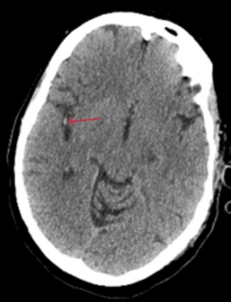
CT brain: Hyperdense MCA sign - early acute ischemic stroke, indicating thromboembolic occlusion of M2 MCA
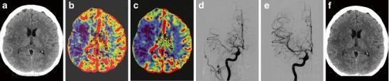
Multimodal imaging in MCA stroke: (a) hyperdense MCA sign, (b-c) CT perfusion deficit, (d) DSA showing occlusion, (e) post-EVT recanalization, (f) 6-week follow-up
Sources: Harrison's Principles of Internal Medicine 22e (2025), Chapter 438 (Acute Ischemic Stroke) and Chapter 439 (Intracerebral Hemorrhage)