
Suprapubic catheterisation md emergency medicine exam
Reading File
Now let me search the medical library and web simultaneously for comprehensive exam content on suprapubic catheterisation.
Finding Sources
Finding Sources
Searching the Web
suprapubic catheterisation emergency medicine exam notes indications contraindications technique complications
I have two excellent primary sources - Roberts and Hedges' Clinical Procedures in Emergency, and Pfenninger and Fowler's Procedures for Primary Care. Let me read both simultaneously.
Reading File
Reading File
Excellent content from both books. Let me read more from Pfenninger (technique continuation) and also check Grainger & Allison for radiology guidance:
Reading File
Reading File
Finding Sources
Now let me read the full Pfenninger technique section, and get any remaining content from Roberts & Hedges:
Reading File
Reading File
I now have comprehensive content from the textbooks. Let me also fetch the Grainger & Allison illustration and read the full Pfenninger technique section:
Reading File
Loading Image
I now have all the information needed for a comprehensive exam-focused answer. Let me compile everything.
Suprapubic Catheterisation - Emergency Medicine Exam Notes
Definition
Suprapubic cystostomy (SPC) is a percutaneous procedure to drain urine directly from the urinary bladder through the anterior abdominal wall, bypassing the urethra entirely. It is used when urethral catheterisation is impossible, dangerous, or contraindicated.
Relevant Anatomy
- The bladder lies in the retropubic space behind the symphysis pubis.
- When distended (>300 mL), it rises above the pubic symphysis as an extraperitoneal structure - the peritoneal reflection is pushed cephalad. This creates a safe "window" for percutaneous access without entering the peritoneum.
- The insertion point is 2-3 cm above the pubic symphysis in the midline, exploiting this extraperitoneal space.
- Key structures at risk: bowel (if bladder not adequately distended), inferior epigastric vessels (stay midline), peritoneum.
Indications
Emergency Indications
| Indication | Notes |
|---|---|
| Acute urinary retention where urethral catheterisation fails or is contraindicated | Most common ED scenario |
| Urethral trauma / pelvic fracture with suspected urethral injury | Absolute indication - urethral catheter risks converting partial to complete rupture |
| Urethral stricture (impassable) | Unable to advance catheter past obstruction |
| Bladder neck contracture / obstruction | |
| Elevated bladder neck or enlarged median prostatic lobe | Unable to pass catheter past obstruction |
| Severe phimosis precluding urethral access | |
| Acute prostatitis / significant urethral infection | Urethral instrumentation risks spreading infection |
| Recent urethral or bladder neck reconstructive surgery | Must avoid disrupting repair |
| Inability to tolerate urethral catheter + refuses self-catheterisation |
Elective Indications
- Long-term bladder drainage in neurogenic bladder (spinal cord injury, MS, diabetic neuropathy)
- Chronic urinary retention when patient cannot void
- Preference over urethral catheter for comfort or nursing reasons
"In general, any patient requiring a urethral catheter, but in whom a catheter cannot be safely passed, is a candidate for a suprapubic cystostomy tube." - Roberts and Hedges' Clinical Procedures in Emergency Medicine, p.1333
Contraindications
Absolute
- Non-distended or non-palpable bladder (< ~300 mL) - most important - risk of bowel/vessel injury is unacceptably high without a clear target. This is the #1 exam answer.
- Suspected bladder cancer - risk of tumour seeding along the tract
- Uncooperative patient (unable to achieve still positioning)
Relative
- Coagulopathy or uncorrected bleeding diathesis (correct first if possible)
- Previous lower abdominal/pelvic surgery (adhesions, bowel may be interposed in the retropubic space)
- Prior abdominal irradiation
- Concurrent antithrombotic therapy
- Pelvic anatomic abnormality from cancer or trauma
Key exam point: "The absence of an easily palpable or ultrasonographically localised distended urinary bladder is an absolute contraindication." (Medscape/eMedicine)
Equipment
- Sterile preparation: povidone-iodine, sterile drape, gloves, mask
- Local anaesthetic: 10 mL lidocaine 1-2%, 10 mL syringe, 22-gauge spinal needle
- No. 11 or No. 15 scalpel blade
- Suprapubic catheter kit - several options:
- Bonnano catheter (Becton-Dickinson) - pigtail self-retaining tip
- Stamey percutaneous set (Cook Medical) - Malecot tip with wings
- SupraFoley introducer (Rusch/Teleflex Medical)
- Cook peel-away sheath (Chiou Suprapubic Kit) - uses Seldinger technique, preferred for ED use
- Guidewire (for Seldinger technique)
- Foley catheter (for Seldinger/peel-away approach)
- Closed urinary drainage bag
- 2-0 nylon suture to secure catheter
- Bedside ultrasound (strongly recommended)
Pre-Procedure
- Informed consent - explain indications, alternatives, risks
- Warn patient: mild-to-moderate suprapubic discomfort for hours to days post-procedure
- Confirm bladder is distended - by palpation and/or ultrasound (>300 mL needed)
- If bladder not palpable due to obesity: ultrasound mandatory; if still unclear, consider cystoscopy or delay procedure
- Review coagulation status; correct if needed
Technique (Step-by-Step)
Method 1: Trocar/Obturator Technique (Traditional - Pfenninger & Fowler)
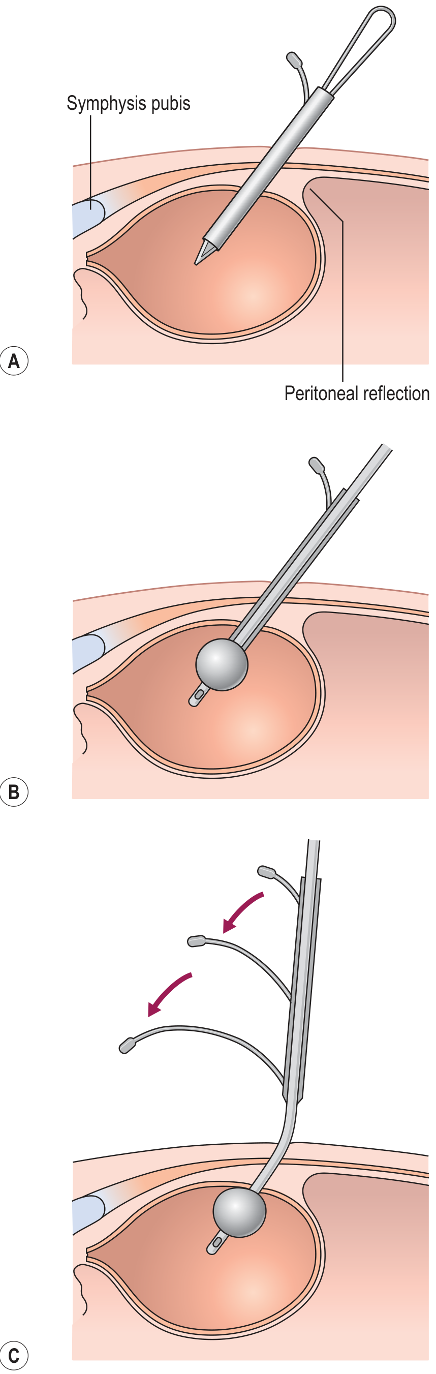
- Position: Patient supine.
- Sterile prep: Clean suprapubic skin with antiseptic; sterile drape.
- Local anaesthesia: Infiltrate skin, subcutaneous tissue, fascia, and down to the bladder dome at a point 5 cm above the pubic symphysis in the midline. Aspirate before each injection.
- Skin incision: Make a 1 cm horizontal skin incision at the anaesthetic site (some also incise the anterior rectus fascia).
- Bladder confirmation (optional but recommended): Pass a 22-gauge spinal needle into the bladder to verify location and confirm urine return before inserting the trocar. If bladder is not distended enough, fill with sterile saline via this needle.
- Assemble catheter: Place the metal obturator through the catheter lumen so the sharp tip extends beyond the catheter tip.
- Insert: Advance the obturator + catheter through the incision at a 60-degree caudal angle (toward the bladder neck / mid-perineum) with firm pressure. Pass through rectus sheath, muscle, and into the bladder dome (~5 cm below skin in adults).
- Advance a further 5 cm to ensure correct positioning.
- Remove obturator: Urine will drain. The Malecot wings expand OR the Bonnano pigtail coils to self-retain. If balloon-type: inflate the balloon with sterile saline.
- Secure catheter: Suture to skin with 2-0 nylon. Connect to closed drainage system.
Method 2: Seldinger (Guidewire) Technique - Preferred for Emergency Department
The Cook peel-away sheath system is the most ED-friendly approach:
- Prep and drape skin - sterile technique.
- Inject local anaesthetic 2-3 cm above the pubic symphysis in the midline.
- Confirm bladder by palpation or ultrasound. Advance a 22-gauge spinal needle with attached syringe, aspirating continuously until urine returns.
- Remove syringe; thread guidewire through the needle into the bladder.
- Remove needle, leaving guidewire in place.
- Make a small stab incision at the skin-guidewire interface.
- Pass the peel-away sheath + fascial dilator over the wire into the bladder.
- Remove the guidewire and fascial dilator, leaving the sheath.
- Pass a Foley balloon catheter through the sheath; inflate the balloon.
- Withdraw (peel away) the sheath, leaving the Foley in the bladder.
"Among the most user-friendly devices for suprapubic bladder access is the Cook peel-away sheath unit. It uses the Seldinger technique to gain bladder access and allows suprapubic placement of a Foley balloon catheter for definitive bladder drainage. This device is readily suitable for ED use." - Roberts and Hedges', p.1332
Emergency shortcut: Any central venous catheter kit can be inserted suprapubically using the Seldinger technique when no dedicated SPC kit is available.
Needle Angulation - Key Exam Point
| Patient | Direction of needle |
|---|---|
| Adults | Direct needle slightly caudad (toward feet) - peritoneum is pushed cephalad by distended bladder |
| Infants | Needle perpendicular (10-20 degrees from vertical) - bladder is an abdominal organ in infants; angling caudad risks missing the bladder |
Complications
Immediate
| Complication | Notes |
|---|---|
| Bowel perforation | Most feared; rate 0.3% with image guidance vs 2.4-2.7% with blind/cystoscopic technique |
| Haematuria (gross) | Common, usually transient - resolves within hours to days |
| Haemorrhage / haematoma | Injury to inferior epigastric vessels (avoid by staying midline) |
| Failed procedure | Catheter not in bladder, no urine return |
| Injury to adjacent structures | Bowel, blood vessels, peritoneum |
Early
| Complication | Notes |
|---|---|
| Infection / UTI | Universal risk with any indwelling catheter |
| Cellulitis / abscess at insertion site | Site should be cleaned with soap and water regularly |
| Catheter displacement or kinking | Can occur in early period before tract matures |
| Urinary leakage around catheter | |
| Bladder spasm |
Delayed
| Complication | Notes |
|---|---|
| Post-obstructive diuresis | Monitor all patients in ED for 2-3 hours; admit if it occurs for IV fluid + electrolyte management |
| Catheter blockage | Simple saline irrigation usually resolves; if malposition suspected, do cystography |
| Non-deflating balloon | Manage by: guidewire to unblock channel; US-guided needle puncture of balloon; transrectal US-guided puncture (with antibiotics) |
| Catheter encrustation | Long-term complication |
| Bladder stones | Long-term |
| Chronic infection | Long-term |
Ultrasound Guidance
- Strongly recommended in all cases, mandatory when:
- Bladder not clinically palpable (obesity, etc.)
- Previous lower abdominal surgery
- Pelvic anatomic abnormalities
- Reduces bowel perforation rate from ~2.4% to 0.3%
- Confirms adequate bladder volume before insertion
- Image-guided insertion at a dedicated centre: bowel perforation rate 0.3% (Grainger & Allison's Diagnostic Radiology, p.2178)
- CT guidance indicated for complex pelvic anatomy (prior surgery, congenital abnormalities)
Catheter Changing / Exchange
- Speed is critical when changing an established suprapubic catheter
- New catheter must be inserted within 5-10 minutes of removing the old one - the tract closes rapidly
- Never remove a suprapubic catheter unless you are ready to replace it immediately
- Use an exchange wire technique if needed; appropriate dilators can upsize to a permanent catheter
Comparison: Suprapubic vs Urethral Catheter
| Feature | Suprapubic | Urethral |
|---|---|---|
| Route | Anterior abdominal wall | Urethra |
| Urethral trauma | None | Possible |
| Patient comfort (long-term) | Better | Worse |
| UTI rate (long-term) | Lower | Higher |
| Trial of void | Easier (clamp and observe) | Requires removal and reinsertion |
| Mobility | Less restricted | More restricted |
| Nursing care | Easier | More complex in disabled patients |
| Contraindications | Non-distended bladder, bladder Ca | Urethral injury, stricture |
Key High-Yield Exam Points Summary
- Absolute contraindication: non-distended / non-palpable bladder - risk of bowel perforation
- Most common emergency indication: urethral trauma (pelvic fracture), acute retention where urethral route fails
- Insertion site: 2-3 cm (Roberts & Hedges) or 5 cm (Pfenninger - traditional trocar) above pubic symphysis, midline
- Angle in adults: caudal (toward bladder neck); in infants: perpendicular
- Seldinger technique preferred in ED (Cook peel-away sheath)
- Always use ultrasound when bladder not palpable
- Bladder cancer = absolute contraindication (tract seeding risk)
- Coagulopathy = relative contraindication (correct first)
- Post-obstructive diuresis: observe 2-3 hours post-procedure; admit if occurs
- Catheter change: never remove without immediate replacement; tract closes in 5-10 minutes
Sources:
- Roberts and Hedges' Clinical Procedures in Emergency Medicine, Chapter 55 (Suprapubic Cystostomy), pp.1332-1334
- Pfenninger and Fowler's Procedures for Primary Care, 3rd ed., Chapter 113, pp.809-812
- Grainger & Allison's Diagnostic Radiology, Chapter 85, p.2178 (image-guided insertion)
- StatPearls: Suprapubic Bladder Catheterization (NCBI)
This is a shared conversation. Sign in to Orris to start your own chat.