
How to diagnose familial hypercholesterolemia?
"familial hypercholesterolemia"[MeSH Terms] AND diagnosis
familial hypercholesterolemia xanthoma xanthelasma diagnosis
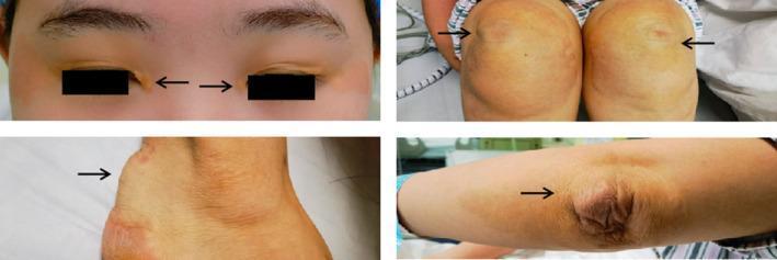
This composite clinical photograph displays multiple cutaneous manifestations of xanthomas in a patient with homozygous familial hypercholesterolemia. The image is divided into four panels, each highlighting a different anatomical region with arrows pointing to the pathology. Top left: Ocular region showing xanthelasma palpebrarum, characterized by yellowish, slightly raised plaques on the upper and lower eyelids. Top right: Bilateral patellar region demonstrating tuberous xanthomas, which appear as firm, yellowish-orange nodules over the knees. Bottom left: Posterior view of the ankle showing a prominent tendon xanthoma of the Achilles tendon, presenting as a large, subcutaneous nodular thickening. Bottom right: Cubital fossa/elbow region showing a tuberoeruptive xanthoma with a wrinkled, raised, yellowish appearance over the extensor surface. Collectively, these findings illustrate the systemic deposition of cholesterol-rich material in skin and tendons, serving as a hallmark diagnostic sign for severe primary hyperlipidemia and associated cardiovascular risk.
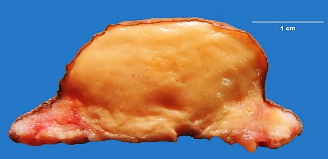
This is a gross pathology photograph of an excised tuberous xanthoma from an 18-year-old female with familial hypercholesterolemia (type IIa). The specimen shows a solitary, well-circumscribed, nodular lesion removed from periarticular soft tissue around a knee (and noted at elbows and toes in the clinical history). On gross inspection, the cut surface is homogeneously pale to yellow-orange, soft, and slightly lobulated, with a glossy, lipid-rich appearance consistent with lipid-laden macrophage infiltration. Histology is diagnostic of xanthoma, characterized by dense aggregates of foamy macrophages (lipid-laden histiocytes) in the dermis/subcutaneous tissue with accompanying chronic inflammatory infiltrate and varying degrees of fibrosis in long-standing lesions. The lipid-rich cytoplasm creates the characteristic yellow hue. Systemic findings include elevated total cholesterol and LDL cholesterol with normal triglycerides, VLDL, and HDL; thyroid function is normal. This phenotype corresponds to familial hypercholesterolemia, type IIa, typically due to LDL receptor or ApoB gene defects. Xanthomas are not neoplasms but reactive lipid deposits that reflect prolonged hyperlipidemia and local tissue remodeling. Clinically relevant implications include increased risk of premature atherosclerotic cardiovascular disease, guiding lipid-lowering therapy and family screening. Correlation with histologic confirmation supports diagnosis and informs genetic counseling, lifestyle modification, and pharmacotherapy, such as statins, PCSK9 inhibitors, or ezetimibe.
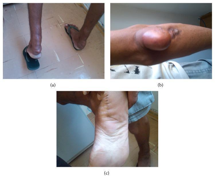
This composite of three clinical photographs displays classic dermatological manifestations of Familial Hypercholesterolemia (FH). Panel (a) shows bilateral Achilles tendon xanthomas, presenting as large, firm, rounded subcutaneous swellings at the posterior aspect of the heels that significantly distort the local anatomy. Panel (b) illustrates a tuberous xanthoma on the elbow, characterized by a large, well-circumscribed, flesh-colored to slightly yellowish nodular protrusion with a smooth overlying surface. Panel (c) shows the plantar aspect of the foot, demonstrating planar xanthomas that cause diffuse thickening and yellowish discoloration of the sole, along with a visible vertical surgical incision line with sutures. These findings are highly characteristic of severe hyperlipidemia, specifically FH, where lipid deposits accumulate in the skin and tendons. The images serve as an educational reference for identifying physical stigmata of lipid metabolism disorders in a clinical setting.
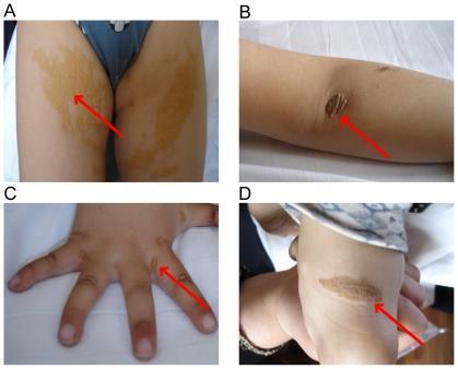
This composite of four clinical photographs (labeled A-D) illustrates the dermatological and musculoskeletal manifestations of Familial Hypercholesterolemia (FH) in a pediatric patient. Panel A displays large, bilateral, yellowish-tan cutaneous xanthomas on the buttocks and upper thighs, characterized by irregular borders and a plaque-like distribution. Panel B shows a distinct, yellowish-tan nodule with a roughened surface over the extensor surface of the elbow, consistent with a tuberous xanthoma. Panel C depicts the dorsal aspect of a hand with several small, raised, yellowish cutaneous xanthomas located on the fingers and interdigital webs. Panel D reveals a prominent, elongated yellowish nodule overlying the Achilles tendon, indicative of a tendinous xanthoma. Red arrows in each panel highlight the specific lesions. These clinical findings are hallmark physical signs of severe lipid metabolic disorders, representing localized accumulations of lipid-laden macrophages (foam cells) in the skin and tendons due to extreme hyperlipidemia.
"familial hypercholesterolemia" AND "Dutch Lipid Clinic" OR "Simon Broome" diagnosis criteria
Diagnosis of Familial Hypercholesterolemia (FH)
1. Clinical Suspicion - When to Consider FH
- Adults (≥20 years): LDL-C > 190 mg/dL (persistently elevated after ruling out secondary causes)
- Children: LDL-C consistently elevated, especially with positive family history
- Family history: First-degree relative with premature ASCVD (men <55 years, women <65 years) or very high LDL-C
- Physical stigmata present at any age
2. Clinical Features (Physical Examination)
| Feature | Description | Notes |
|---|---|---|
| Tendinous xanthomas | Firm, nodular cholesterol deposits in Achilles tendon, extensor tendons of hands/feet | Most specific sign; pathognomonic in right clinical context |
| Tuberous/tuberoeruptive xanthomas | Yellow-orange nodules over knees, elbows, buttocks | More common in homozygous FH |
| Xanthelasma palpebrarum | Yellowish plaques on eyelids | Less specific - can occur in normolipidemia |
| Corneal arcus | White/grey ring around cornea | Significant if present before age 45 |
| Planar xanthomas | Flat yellow deposits, especially interdigital webs | Suggest homozygous FH |
3. Laboratory Investigations
| Parameter | Heterozygous FH | Homozygous FH |
|---|---|---|
| LDL-C | >190 mg/dL (typically >220 mg/dL) | >400 mg/dL |
| Total cholesterol | Markedly elevated | Severely elevated (often >600-700 mg/dL) |
| Triglycerides | Usually normal (key distinguishing feature) | |
| HDL-C | Usually normal |
- Apolipoprotein B-100 can be elevated
- Elevated LDL-C from birth is characteristic; FH does not cause hypertriglyceridemia in isolation
4. Formal Diagnostic Criteria
A. Dutch Lipid Clinic Network (DLCN) - Point-Based System
| Category | Criterion | Points |
|---|---|---|
| Family history | First-degree relative with tendinous xanthoma or premature ASCVD | 1-2 |
| First-degree relative with known FH | 1 | |
| First-degree relative with LDL-C >95th percentile | 1 | |
| Clinical history | Patient has premature ASCVD (<55M / <60F) | 2 |
| Patient has premature cerebrovascular or peripheral artery disease | 1 | |
| Physical examination | Tendinous xanthoma | 6 |
| Corneal arcus before age 45 | 4 | |
| LDL-C level | ≥8.5 mmol/L (≥328 mg/dL) | 8 |
| 6.5-8.4 mmol/L | 5 | |
| 5.0-6.4 mmol/L | 3 | |
| 4.0-4.9 mmol/L | 1 | |
| Genetic testing | Pathogenic LDLR, APOB, or PCSK9 mutation | 8 |
-
8 points = Definite FH
- 6-8 points = Probable FH
- 3-5 points = Possible FH
- <3 points = FH unlikely
B. Simon Broome Register (UK)
- Definite FH: Total cholesterol >7.5 mmol/L (adults) or >6.7 mmol/L (children) AND tendinous xanthoma in patient or first-degree relative; OR DNA evidence of pathogenic LDLR/APOB mutation
- Possible FH: Same cholesterol threshold PLUS family history of premature MI (<50 in 2 relatives; <60 in 1 relative) OR total cholesterol >7.5 mmol/L in a first/second-degree relative
C. US MEDPED Program (Make Early Diagnosis - Prevent Early Deaths)
5. Genetic Testing
- Definitively confirms the diagnosis when a pathogenic variant is found
- Enables cascade screening of relatives (50% of first-degree relatives will be affected in autosomal dominant inheritance)
- Guides treatment decisions (e.g., receptor-negative vs. receptor-defective status affects statin response in homozygous FH)
- Required by some insurers for approval of PCSK9 inhibitors
6. Screening Strategy
| Population | Recommendation |
|---|---|
| General children | Universal fasting or non-fasting lipid panel between ages 8-11 years |
| High-risk children | Screen earlier if family history of LDL-C >190 mg/dL or premature ASCVD |
| Adults | Lipid panel by age 20 at the latest |
| Relatives of index case | Cascade screening - screen all first-degree relatives once an index case is identified |
7. Distinguishing Heterozygous vs. Homozygous FH
| Feature | Heterozygous FH | Homozygous FH |
|---|---|---|
| LDL-C | 190-400 mg/dL | >400 mg/dL (often 600-1000 mg/dL) |
| ASCVD onset | 30s-40s (men), 40s-50s (women) | Childhood |
| Xanthomas | Late 2nd-3rd decade | Childhood, sometimes at birth |
| LDLR activity | ~50% functional | <2% (receptor negative) or 2-25% (receptor defective) |
| Statin response | Good | Poor (apheresis often needed) |
Key References
- Henry's Clinical Diagnosis and Management by Laboratory Methods, p. 303-304
- Harrison's Principles of Internal Medicine 22E, p. 3286
- Goldman-Cecil Medicine, Chapter 190
- Thompson & Thompson Genetics and Genomics in Medicine, 9th ed., Table C.16.2
- Tietz Textbook of Laboratory Medicine, 7th Edition, p. 1180
- Robbins & Kumar Basic Pathology, p. 111
How to diagnose familial hypercholesterolemia?
"familial hypercholesterolemia"[MeSH Terms] AND diagnosis
familial hypercholesterolemia xanthoma tendon xanthelasma corneal arcus
This composite clinical photograph displays multiple cutaneous manifestations of xanthomas in a patient with homozygous familial hypercholesterolemia. The image is divided into four panels, each highlighting a different anatomical region with arrows pointing to the pathology. Top left: Ocular region showing xanthelasma palpebrarum, characterized by yellowish, slightly raised plaques on the upper and lower eyelids. Top right: Bilateral patellar region demonstrating tuberous xanthomas, which appear as firm, yellowish-orange nodules over the knees. Bottom left: Posterior view of the ankle showing a prominent tendon xanthoma of the Achilles tendon, presenting as a large, subcutaneous nodular thickening. Bottom right: Cubital fossa/elbow region showing a tuberoeruptive xanthoma with a wrinkled, raised, yellowish appearance over the extensor surface. Collectively, these findings illustrate the systemic deposition of cholesterol-rich material in skin and tendons, serving as a hallmark diagnostic sign for severe primary hyperlipidemia and associated cardiovascular risk.
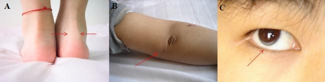
This composite clinical photograph illustrates hallmark physical manifestations of Familial Hypercholesterolemia (FH). Panel A presents a posterior view of the ankles, demonstrating bilateral Achilles tendon thickening (tendon xanthoma), indicated by red arrows. Panel B shows the extensor surface of the elbow featuring cutaneous xanthomas, which appear as raised, yellowish, irregularly shaped plaques on the skin surface. Panel C provides a close-up clinical view of the eye, revealing arcus corneae (arcus senilis), characterized by a distinct grayish-white opaque ring or arc at the periphery of the cornea. These findings are classic clinical markers for severe hyperlipidemia, often associated with genetic mutations in the LDL receptor. The image serves as an educational tool for identifying the external stigmata of lipid metabolism disorders in a pediatric or adult patient.
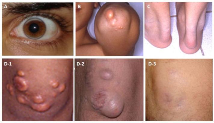
A multi-panel clinical photograph collection documenting physical manifestations of homozygous familial hypercholesterolemia (HoFH) and the effects of lipoprotein apheresis. Image A shows arcus lipoides (corneal arcus), characterized by a hazy, whitish-gray lipid ring at the corneal periphery. Image B demonstrates tuberous xanthomas on the extensor surface of the elbow, appearing as firm, yellowish-orange nodules. Image C illustrates tendon xanthomas, specifically bilateral thickening and prominence of the Achilles tendons. Panels D-1 through D-3 provide a longitudinal view of cutaneous xanthomas on the extensor surface of the knee. D-1 shows multiple eruptive and tuberous xanthomas before starting apheresis. D-2 shows the progression after a three-year discontinuation of apheresis, with significant enlargement and confluence of the nodules. D-3 demonstrates near-complete resolution of the xanthomas following three years of regular apheresis, with only residual skin hyperpigmentation. These images collectively serve as educational material for the dermatological and ophthalmological signs of severe hyperlipidemia and the visible efficacy of lipid-lowering interventions.
Diagnosis of Familial Hypercholesterolemia (FH)
Step 1 - Raise Clinical Suspicion
- An adult has LDL-C persistently >190 mg/dL with relatively normal triglycerides
- A child has LDL-C consistently elevated (>95th percentile for age)
- There is a family history of premature atherosclerotic cardiovascular disease (ASCVD) - defined as men <55 years or women <65 years in a first-degree relative
- There is a family history of a first-degree relative with known FH or very high LDL-C (>190 mg/dL)
- Physical stigmata are present (see below)
| Secondary Cause | Examples |
|---|---|
| Endocrine | Hypothyroidism, Cushing syndrome, estrogen deficiency |
| Renal | Nephrotic syndrome, chronic kidney disease |
| Hepatic | Biliary obstruction |
| Drugs | Glucocorticoids, cyclosporine, anticonvulsants, anabolic steroids, diuretics |
| Metabolic | Obesity, poorly controlled diabetes |
Step 2 - Physical Examination
| Sign | Description | Significance |
|---|---|---|
| Tendinous xanthomas | Firm cholesterol deposits in Achilles, patellar, and extensor tendons of hands | Most specific; scored highest in Dutch criteria (6 pts) |
| Tuberous/tuberoeruptive xanthomas | Yellow-orange nodules over knees, elbows, buttocks | More prominent in homozygous FH |
| Xanthelasma palpebrarum | Yellowish plaques on eyelids | Less specific; can occur in normal lipidemia |
| Corneal arcus | White-grey opaque ring at corneal periphery | Significant if present before age 45; scores 4 pts in Dutch criteria |
| Planar xanthomas | Flat yellow deposits, including interdigital webs | Suggest homozygous FH when present in childhood |
Step 3 - Laboratory Investigations
| Marker | Heterozygous FH | Homozygous FH |
|---|---|---|
| LDL-C | Usually >190-220 mg/dL | >400 mg/dL (often 600-1000 mg/dL) |
| Total cholesterol | Markedly elevated | Severely elevated |
| Triglycerides | Normal - a key distinguishing feature | Normal |
| HDL-C | Usually normal | Usually normal |
Step 4 - Formal Clinical Diagnostic Criteria
A. Dutch Lipid Clinic Network (DLCN) - Most widely used
| Category | Criterion | Points |
|---|---|---|
| LDL-C level | ≥8.5 mmol/L (≥328 mg/dL) | 8 |
| 6.5-8.4 mmol/L | 5 | |
| 5.0-6.4 mmol/L | 3 | |
| 4.0-4.9 mmol/L | 1 | |
| Personal clinical history | Premature coronary artery disease (<55M / <60F) | 2 |
| Premature cerebrovascular or peripheral artery disease | 1 | |
| Physical examination | Tendinous xanthoma (patient) | 6 |
| Corneal arcus before age 45 | 4 | |
| Family history | First-degree relative with tendinous xanthoma or premature ASCVD | 2 |
| First-degree relative with LDL-C ≥7.5 mmol/L or FH | 1 | |
| Genetic testing | Pathogenic LDLR, APOB, or PCSK9 mutation | 8 |
B. Simon Broome Register (UK)
-
Definite FH: Total cholesterol >7.5 mmol/L in an adult (or >6.7 mmol/L in a child <16 years) AND either:
- Tendinous xanthomas in the patient or a first- or second-degree relative, OR
- DNA evidence of pathogenic LDLR, APOB, or PCSK9 mutation
-
Possible FH: Same cholesterol threshold PLUS family history of:
- MI before age 50 in a second-degree relative, or before age 60 in a first-degree relative, OR
- Total cholesterol >7.5 mmol/L in a first- or second-degree relative
C. US MEDPED Program (Make Early Diagnosis - Prevent Early Deaths)
Step 5 - Genetic Testing
| Gene | Inheritance | Frequency | Mechanism |
|---|---|---|---|
| LDLR | Semidominant | 80-90% | >2000 loss-of-function mutations impair LDL uptake |
| APOB | Semidominant | 5-10% | R3500Q missense reduces LDL-receptor binding |
| PCSK9 | Semidominant | <2% | Gain-of-function mutations increase LDLR degradation |
| LDLRAP1 | Recessive | <1% | Impairs LDLR recycling (causes autosomal recessive FH) |
- LDL-C >8 mmol/L (pathogenic variant found in >95% of cases)
- To definitively confirm diagnosis and enable cascade screening
- When insurance/payer requires genetic confirmation before approving PCSK9 inhibitors
- Pediatric cases where early treatment decisions are needed
Step 6 - Distinguish Heterozygous vs. Homozygous FH
| Feature | Heterozygous FH | Homozygous FH |
|---|---|---|
| Prevalence | ~1 in 250 | ~1 in 500,000 |
| LDL-C | 190-400 mg/dL | >400 mg/dL (often >600) |
| ASCVD onset | Men: 30s-40s; Women: 40s-50s | Childhood |
| Xanthomas | Appear 2nd-3rd decade | Infancy/childhood, sometimes at birth |
| LDLR activity | ~50% residual | <2% (receptor-negative) or 2-25% (receptor-defective) |
| Aortic stenosis | Uncommon | Can occur from supravalvular cholesterol deposits |
| Statin response | Generally good | Poor; often requires apheresis |
Step 7 - Screening Strategy
| Population | Recommendation |
|---|---|
| General children | Universal fasting or non-fasting lipid panel at ages 8-11 years |
| High-risk children | Screen earlier if family history of LDL-C >190 mg/dL or premature ASCVD (men <55, women <65) in a first-degree relative |
| Adults | Lipid panel by age 20 at the latest |
| Cascade screening | Once an index case is identified, screen all first-degree relatives - as an autosomal dominant disorder, 50% of first-degree relatives will be affected |


Analyse and give comments