
what is the pathology behind heart block and it's types
I'll read the medical library skill first before answering this medical question.
Reading File
Now let me search the medical library for heart block pathology and types in parallel.
Finding Sources
Finding Sources
Excellent results. Now let me read the key sections from Braunwald's Heart Disease and Goldman-Cecil in parallel for comprehensive coverage.
Reading File
Reading File
Reading File
I have the classification section. Let me now read more of the detailed content on each type of heart block from Braunwald's, plus look for ECG images and etiology content.
Reading File
Finding Sources
Excellent. Now let me get the Fuster's Heart pathophysiology section and the Frameworks for Internal Medicine detailed causes section, plus fetch some ECG images.
Reading File
Reading File
Loading Image
Let me get more content from Frameworks on Mobitz types and third-degree block, plus the ECG image for the heart block classification diagram.
Reading File
Loading Image
Let me get the rest of the Frameworks content covering Mobitz II and third-degree block, and also the second-degree block section from Braunwald's.
Reading File
Reading File
I now have comprehensive material to deliver a detailed answer. Let me also fetch the Mobitz I and II ECG images to include them visually.
Loading Image
Loading Image
I now have comprehensive textbook material and ECG images to compose a thorough answer. Here it is:
Heart Block - Pathology and Types
What is Heart Block?
Heart block is a disturbance of impulse conduction - distinct from interference, which is a normal physiologic phenomenon caused by refractoriness after a preceding impulse. Block can occur at multiple levels: between the sinus node and atrium (SA block), between atria and ventricles (AV block), within the atria (intra-atrial), or within the ventricles (intraventricular). The term "heart block" most commonly refers to atrioventricular (AV) block.
AV block exists when an atrial impulse is conducted with delay or is not conducted at all to the ventricle, at a time when the AV junction is not physiologically refractory. The site of block can be in the AV node, the His bundle, or the bundle branches. - Braunwald's Heart Disease, p. 1315
Classification Overview
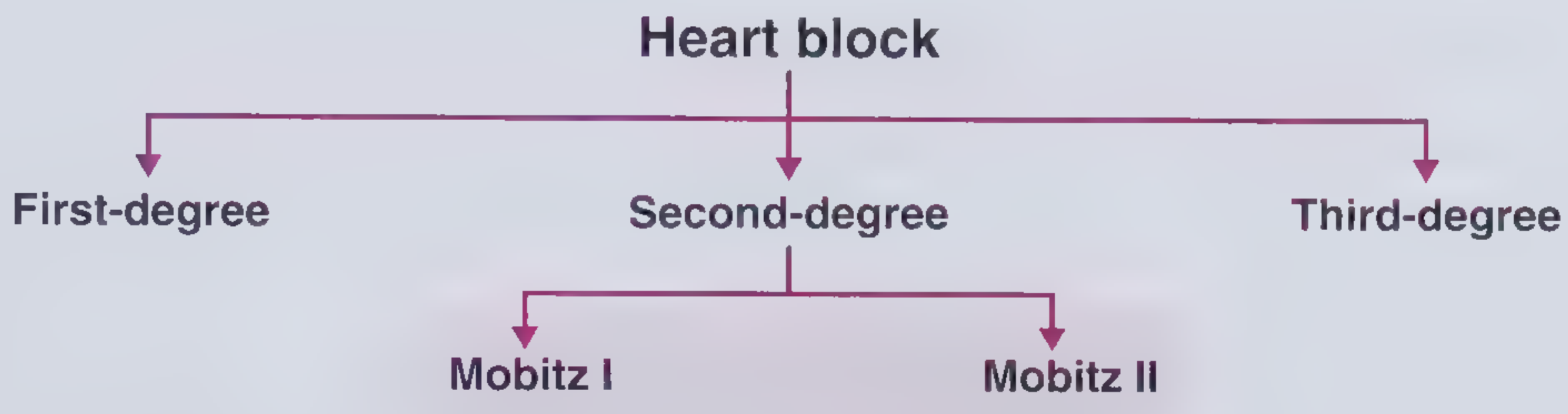
AV block is classified by severity into three degrees:
1. First-Degree AV Block
Pathophysiology
Every atrial impulse still reaches the ventricles, but conduction is delayed. The PR interval exceeds 0.20 seconds in adults (values as long as 1.0 second have been recorded). The delay can reside in:
- The AV node (prolonged A-H interval) - most common
- The His-Purkinje system (prolonged H-V interval)
- Both sites simultaneously
- Rarely, intra-atrial conduction delay
If the QRS is normal in contour, the AV delay almost always sits in the AV node. If the QRS shows a bundle branch block (BBB) pattern, delay may be at the AV node or His-Purkinje system, and a His bundle electrogram is needed to localize it. - Braunwald's Heart Disease, p. 1315
Causes
- Idiopathic degeneration of the conduction system (Lenegre's disease, Lev's disease)
- Drugs: beta-blockers, calcium channel blockers, digoxin
- Enhanced vagal tone (e.g., trained athletes)
- Inferior MI (right coronary artery territory, which supplies the AV node)
- Electrolyte disturbances (e.g., hypokalemia)
- Infections: Lyme disease, endocarditis, myocarditis
- Muscular dystrophies (myotonic muscular dystrophy)
- Infiltrative disease: amyloidosis, sarcoidosis
- Post-ablation procedures - Frameworks for Internal Medicine, p. 47
ECG
Constant PR interval >200 ms; every P wave followed by a QRS.
Prognosis
Generally benign. Some evidence of slightly increased risk of atrial fibrillation and more serious conduction abnormalities over time. Pacemaker is only considered if PR >300 ms with hemodynamic symptoms.
2. Second-Degree AV Block
Some (but not all) atrial impulses reach the ventricles. There are two subtypes:
2a. Mobitz Type I (Wenckebach Block)
ECG
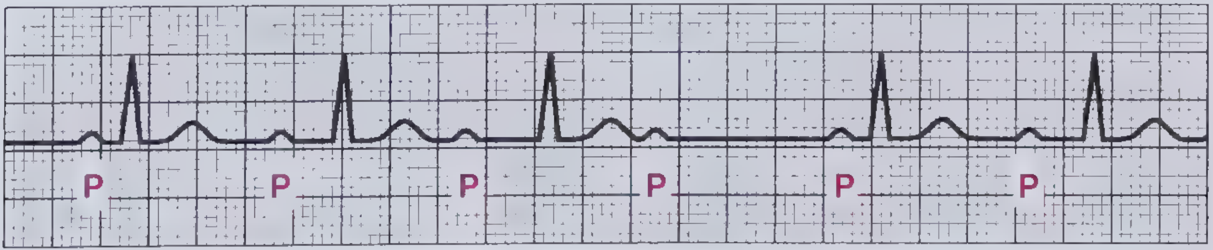
Progressive lengthening of the PR interval with each successive beat until a P wave is completely blocked (dropped QRS). After the dropped beat, the PR interval resets to its shortest value and the cycle repeats. The RR interval progressively shortens before the dropped beat. - Frameworks for Internal Medicine, p. 48
Originally described by Wenckebach using jugular venous tracings (A-C prolongation leading to dropped beats).
Pathophysiology
- Block location: within the AV node (suprahisian)
- The AV node progressively fatigues with each impulse until it completely fails to conduct, then recovers
- Associated with inferior MI (RCA territory, which supplies the AV node in ~90% of people) - look for ST elevation in leads II, III, aVF
Symptoms
Usually asymptomatic. Dyspnea and light-headedness are rare.
Prognosis
Generally benign; rarely progresses to complete block. Vagal-mediated forms are physiologic (athletes, during sleep).
2b. Mobitz Type II Block
ECG
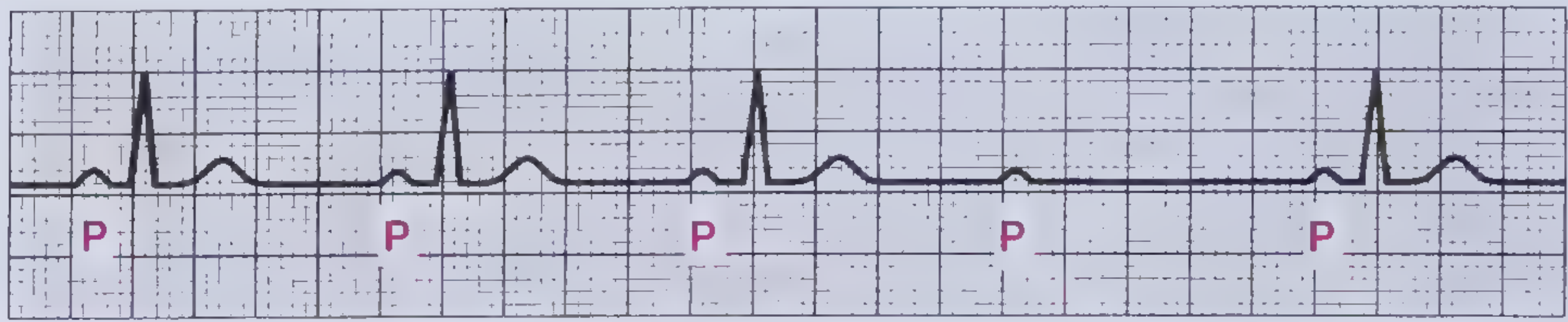
Conducted beats with a constant PR interval followed by a sudden dropped QRS - no preceding PR prolongation. The QRS is often wide because block is below the His bundle. - Frameworks for Internal Medicine, p. 48
Pathophysiology
- Block location: below the AV node, within the His-Purkinje system or bundle branches (infrahisian)
- Associated with anterior MI (LAD territory, which supplies the His-Purkinje system and bundle branches) - look for ST elevation in V1-V4
- Because block is infrahisian, it carries a much worse prognosis than Mobitz I
Symptoms
Frequently symptomatic: dyspnea, palpitations, light-headedness, syncope.
Prognosis
High rate of progression to complete (third-degree) AV block and increased mortality. Permanent pacemaker is typically indicated.
2c. High-Grade (Advanced) AV Block
When two or more consecutive P waves are blocked. The term distinguishes intermittent severe block from complete block, as some ventricular conduction still occurs. - Braunwald's Heart Disease, p. 1317
2d. 2:1 AV Block
Every other P wave is blocked - a special case of second-degree block where Mobitz I vs. II cannot be distinguished from the ECG pattern alone (consecutive PR intervals are not available for comparison). His bundle electrogram is needed to localize.
3. Third-Degree (Complete) AV Block
Definition
No atrial impulses reach the ventricles. The atria and ventricles beat completely independently - AV dissociation. P-P intervals are regular; R-R intervals are regular; but there is no relationship between P waves and QRS complexes. - Braunwald's Heart Disease, p. 1317
Pathophysiology
Block can be at three levels:
| Level | Block site | QRS | Escape rate | Notes |
|---|---|---|---|---|
| AV nodal | Above His bundle | Narrow (normal) | 40-60 bpm | Usually congenital; atropine increases rate |
| Intrahisian | Within His bundle | Can be narrow or wide | 40-60 bpm | Rarely identified without invasive studies |
| Infrahisian | Below His bundle (bundle branches) | Wide (abnormal) | <40 bpm | Usually acquired; worse prognosis |
In acquired complete block (most common in adults), block is distal to the His bundle due to trifascicular conduction disease. Each P wave is followed by a His deflection, but ventricular escape complexes are NOT preceded by a His deflection. - Braunwald's Heart Disease, p. 1317
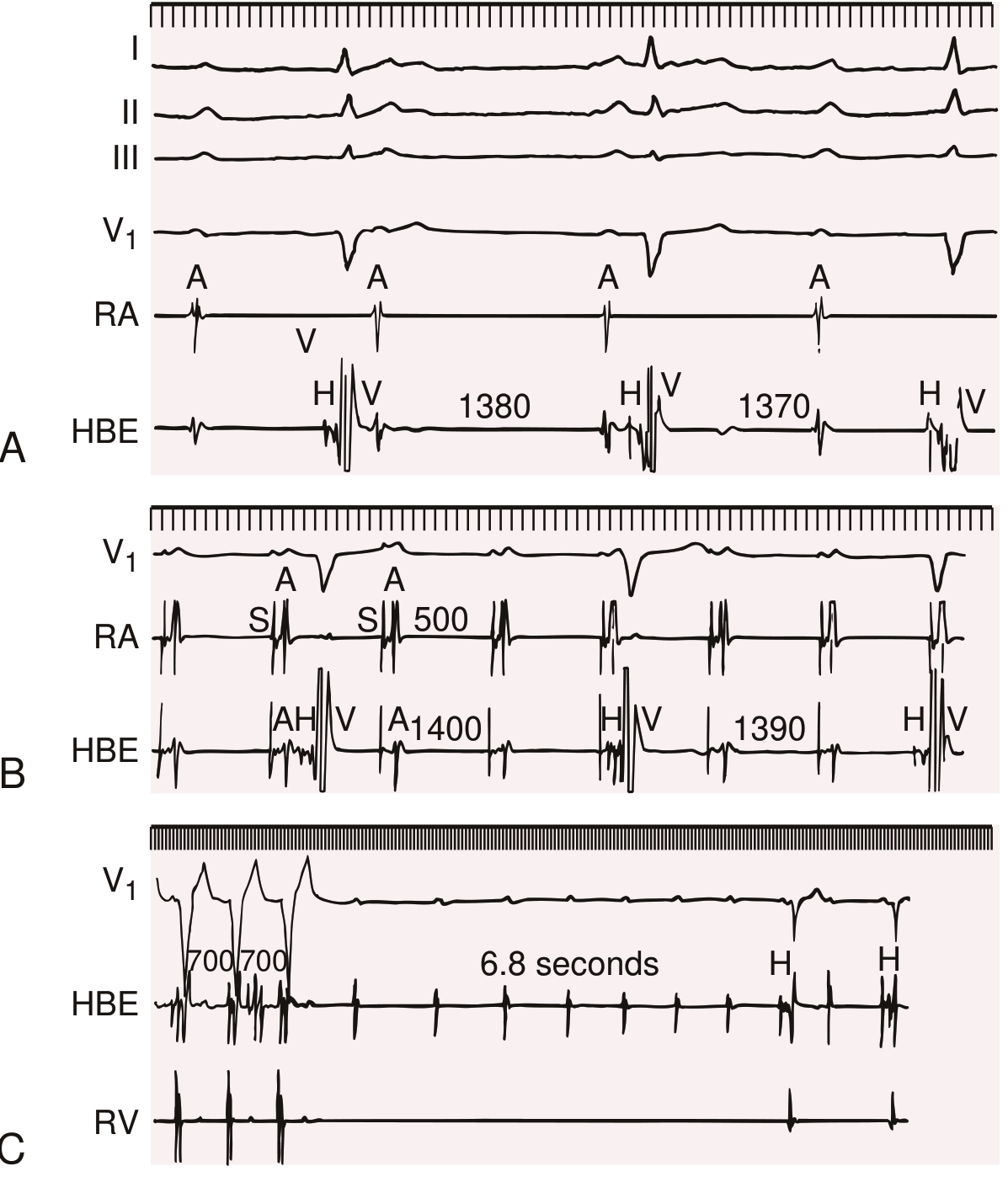
Above: His bundle electrograms in complete AV block, showing the A (atrial), H (His), and V (ventricular) deflections with long intervals (1380-1390 ms escape intervals visible) and eventually complete ventricular standstill (6.8 second pause in panel C). - Braunwald's Heart Disease
Causes of Complete AV Block
- Degenerative fibrosis of the conduction system (idiopathic - most common in elderly)
- Ischemic heart disease (inferior MI: AV nodal block; anterior MI: infrahisian block)
- Infiltrative disease: amyloidosis, sarcoidosis, hemochromatosis
- Infectious: Chagas disease, Lyme disease, rheumatic fever
- Surgical trauma: post-cardiac surgery (especially congenital heart disease repair)
- Congenital (associated with maternal anti-Ro/SSA antibodies in neonates; also with L-loop TGA, AV septal defects)
- Drugs: digoxin toxicity, beta-blockers, calcium channel blockers, amiodarone
- Tumors, polymyositis, myxedema, calcific aortic stenosis
- Hereditary: SCN5A gene mutations (same gene responsible for LQT3) - Braunwald's Heart Disease, p. 1317-1318
Symptoms
Syncope (Stokes-Adams attacks), near-syncope, fatigue, dyspnea, heart failure symptoms.
Treatment
Permanent pacemaker implantation is indicated.
Summary Table
| Type | PR Interval | Dropped QRS? | Location of Block | Associated MI Territory | Prognosis |
|---|---|---|---|---|---|
| 1st degree | >200 ms, constant | None | AV node (usually) | Inferior (mild) | Benign |
| 2nd degree - Mobitz I | Progressively lengthens | Yes (periodic) | AV node | Inferior (RCA) | Usually benign |
| 2nd degree - Mobitz II | Constant, then sudden drop | Yes (sudden) | His-Purkinje | Anterior (LAD) | High risk - pacemaker |
| High-grade | Variable | 2+ consecutive P waves blocked | Variable | Variable | Serious |
| 3rd degree (complete) | AV dissociation | All P waves blocked | AV node / His / infrahisian | Both | Emergency - pacemaker |
Key Pathological Mechanisms Underlying Heart Block
- Fibrosis / degeneration of the conduction system (Lenegre's disease = primary conduction fibrosis; Lev's disease = calcification spreading from aortic/mitral valves)
- Ischemia/infarction: AV node is supplied by the RCA (occlusion causes nodal block); His-Purkinje system is supplied by the septal perforators of the LAD (occlusion causes infrahisian block)
- Infiltration: amyloid, sarcoid granulomas directly disrupt conduction tissue
- Inflammation: Lyme carditis can cause all degrees of AV block; autoimmune (anti-Ro antibodies in congenital block)
- Drug effects: Drugs that slow AV nodal conduction (beta-blockers, CCBs, digoxin, adenosine)
- Genetic channelopathies: SCN5A mutations impair sodium channel function in conduction tissue
- Phase 3 / Phase 4 block: Rate-dependent block due to incomplete repolarization recovery or spontaneous diastolic depolarization in diseased tissue - Braunwald's Heart Disease, p. 1318
Sources: Braunwald's Heart Disease (10th ed.), Frameworks for Internal Medicine, Goldman-Cecil Medicine
This is a shared conversation. Sign in to Orris to start your own chat.