Lacrimal apparatus
Lateral wall of nose
Muscle of mastication
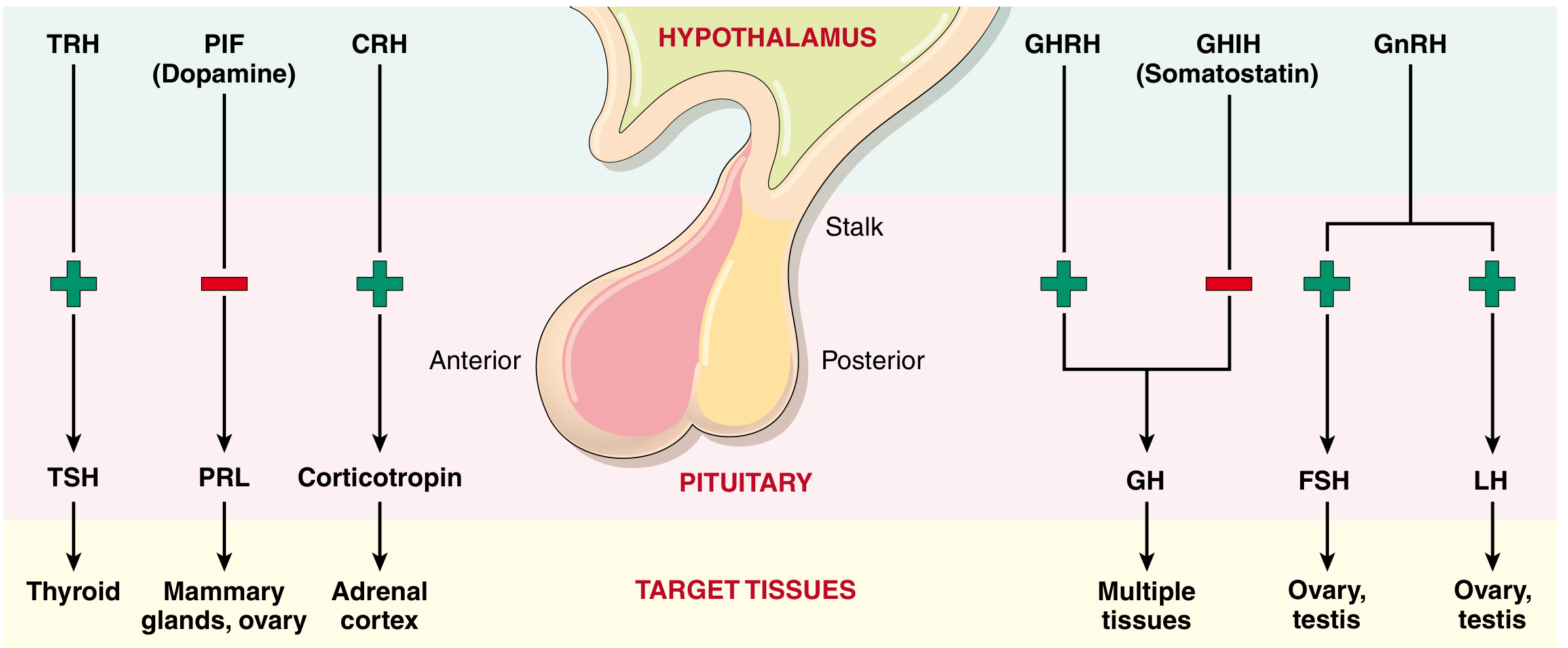
Pitutary
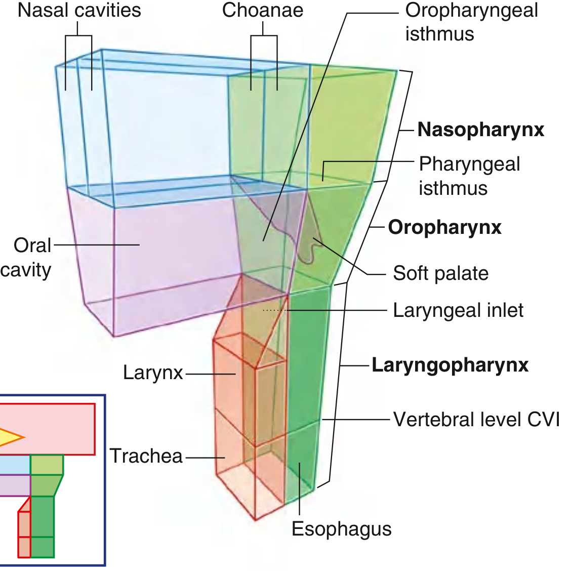
Pharynx
Reading File
These are all anatomy topics. Let me clarify the scope before diving in.
Asking for Preferences
What would you like for these anatomy topics? · Which level is this for?
Detailed MBBS-level notes for all five topics. Let me pull authoritative textbook content for each simultaneously.
Finding Sources
Finding Sources
Finding Sources
Finding Sources
Finding Sources
Good results. Now let me read all the relevant textbook sections in parallel, and also search for "lateral wall of nose" with a different query.
Reading File
Finding Sources
Reading File
Reading File
Reading File
Good content on lacrimal apparatus, mastication, pharynx, and pituitary. I need to read more sections and search for lateral wall of nose.
Reading File
Finding Sources
Reading File
Reading File
Good. Now I have Bailey and Love's source for lateral wall of nose. Let me read that section, the lacrimal innervation continuation, more pharynx content, and also read the muscles of mastication from a more anatomy-focused source.
Reading File
Reading File
Reading File
Excellent. I now have rich content for all five topics. Let me get the lateral wall of nose content more specifically and read the pharynx muscles table continuation.
Reading File
Reading File
I now have comprehensive content for all five topics. Let me also get the muscles of mastication from Gray's Anatomy and a bit more on the nasolacriml duct drainage site for completeness.
Finding Sources
Reading File
Reading File
I now have all the content needed. Let me update the todos and compile the comprehensive notes.Here are your detailed MBBS-level anatomy notes for all five topics:
1. LACRIMAL APPARATUS
(Source: Gray's Anatomy for Students)
Definition and Components
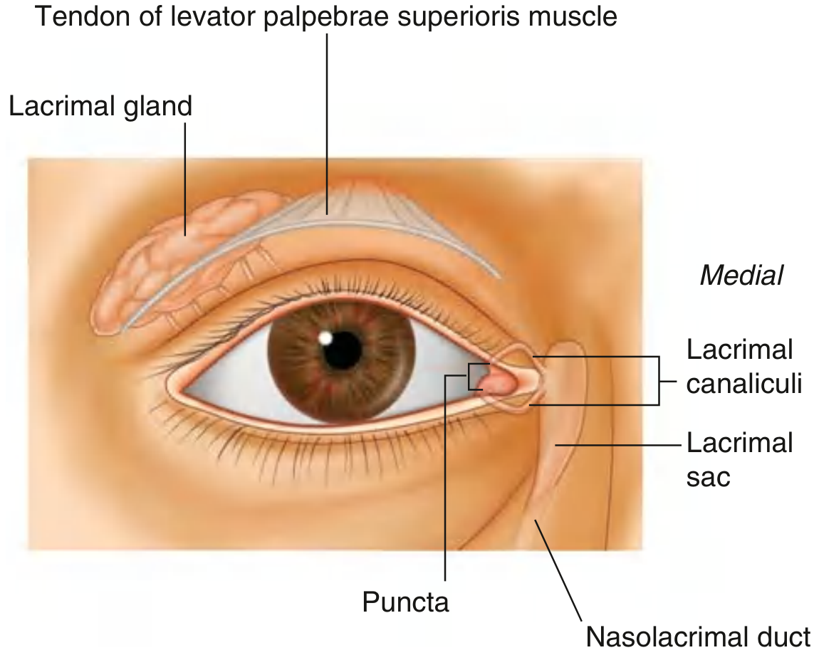
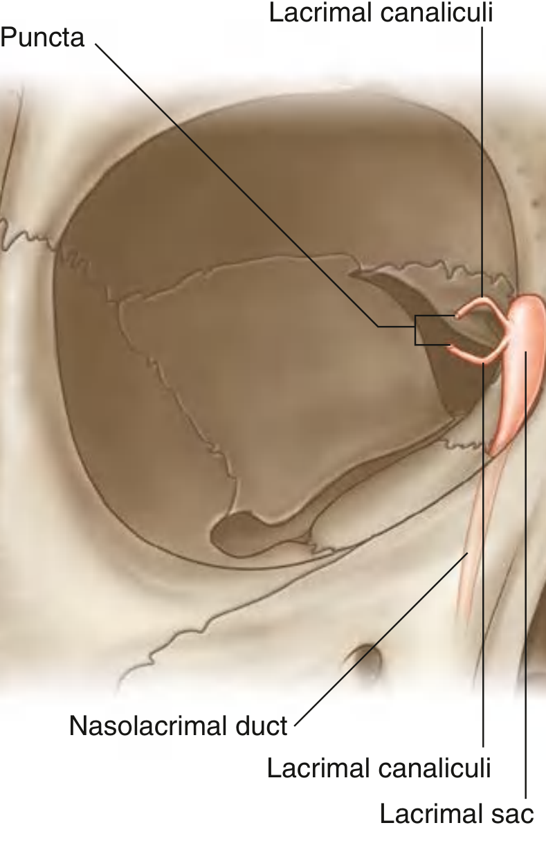
The lacrimal apparatus is responsible for the production, movement, and drainage of fluid from the surface of the eyeball. It consists of:
Lacrimal gland + its ducts
Lacrimal canaliculi
Lacrimal sac
Nasolacrimal duct
Lacrimal Gland
Located anteriorly in the superolateral region of the orbit
Divided into two parts by the levator palpebrae superioris:
Orbital part (larger) - lies in the lacrimal fossa in the frontal bone
Palpebral part (smaller) - lies inferior to the levator palpebrae, in the superolateral eyelid
Numerous ducts empty secretions into the lateral part of the superior fornix of the conjunctiva
Flow of Lacrimal Fluid
Gland secretes fluid continuously
Eyelid blinking moves fluid lateral to medial across the eyeball
Fluid accumulates in the lacrimal lake (medial angle)
Drains through lacrimal punctum (opening) into lacrimal canaliculus (one per eyelid)
Canaliculi join the lacrimal sac - located between the anterior and posterior lacrimal crests, posterior to the medial palpebral ligament
The lacrimal part of orbicularis oculi, when contracting during blinking, may dilate the lacrimal sac and draw tears in via the canaliculi
Lacrimal sac drains into the nasolacrimal duct → opens into the inferior meatus of the nasal cavity
Innervation of the Lacrimal Gland
Three components:
1. Sensory
Lacrimal branch of the ophthalmic nerve [V1]
2. Secretomotor (Parasympathetic)
Pathway (follow carefully):
Pre-ganglionic neurons leave in facial nerve [VII]
Enter greater petrosal nerve (branch of VII)
Form nerve of the pterygoid canal (greater petrosal + deep petrosal)
Post-ganglionic fibers from superior cervical ganglion
Travel along internal carotid artery plexus
Leave as deep petrosal nerve → joins nerve of pterygoid canal
Pass through pterygopalatine ganglion (without synapsing)
Follow same path as parasympathetic to lacrimal gland
Blood Supply
Arterial: Branches from the ophthalmic artery
Venous: Drained by ophthalmic veins
2. LATERAL WALL OF THE NOSE (Lateral Nasal Wall)
(Source: Bailey and Love's Short Practice of Surgery; Scott-Brown's Otorhinolaryngology)
Bony Framework
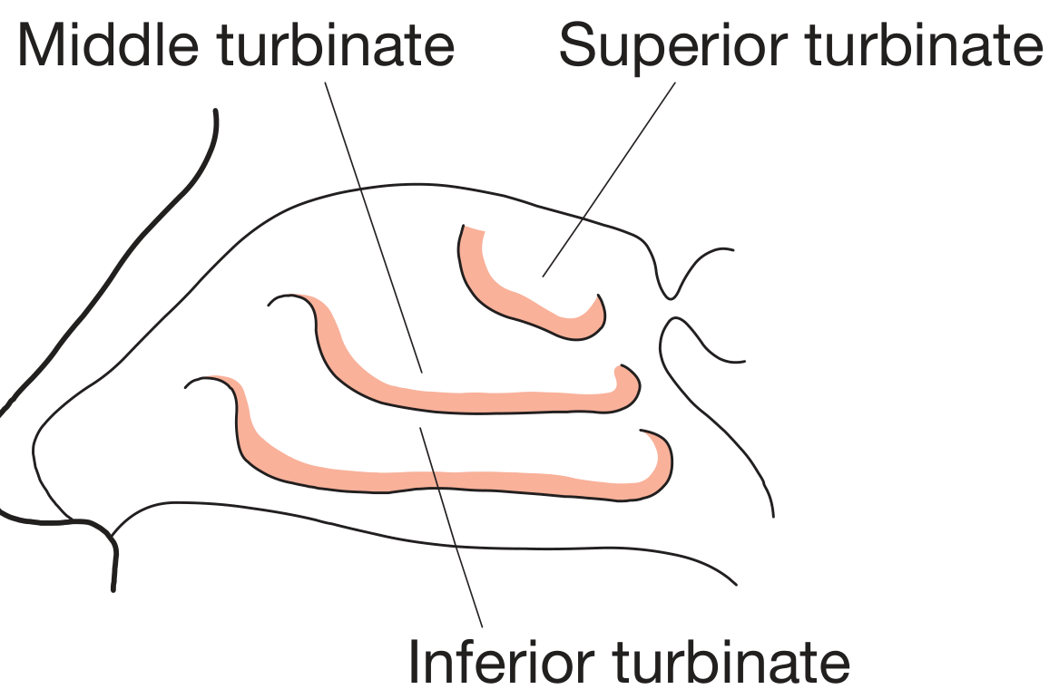
The lateral wall of the nasal cavity carries three scroll-like projections called turbinates (conchae):
Superior turbinate
Middle turbinate
Inferior turbinate
These turbinates warm and moisten the inspired air, increasing the surface area of the nasal cavity.
Meatuses (Spaces Under Turbinates)
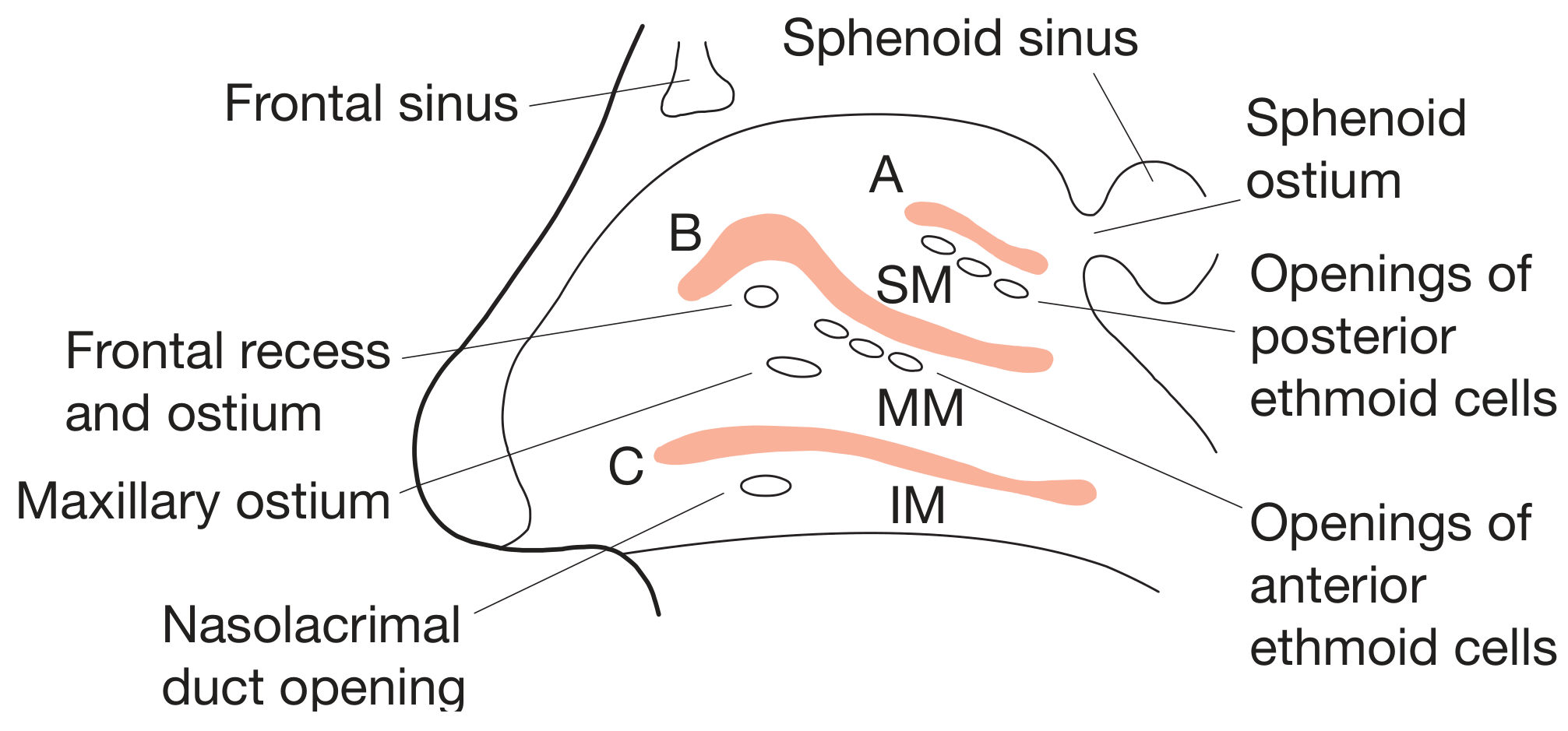
Each turbinate overhangs a meatus:
Meatus
Location
Structures Opening Into It
Inferior meatus
Below inferior turbinate
Nasolacrimal duct (tears drain here)
Middle meatus
Below middle turbinate
Frontal sinus, maxillary sinus, anterior ethmoid sinuses (via the hiatus semilunaris and infundibulum)
Superior meatus
Below superior turbinate
Posterior ethmoid sinuses
Sphenoethmoidal recess
Between superior turbinate and septum
Sphenoid sinus
The anterior nasal sinuses (frontal, maxillary, and anterior ethmoid) all drain into the middle meatus - clinically the most important.
Blood Supply of the Nasal Cavity / Lateral Wall
Dual supply from both external and internal carotid arteries:
Artery
Source
Area Supplied
Sphenopalatine artery
External carotid → maxillary
Interior of nose (main supply)
Greater palatine artery
External carotid → maxillary
Anteroinferior septum via incisive canal
Anterior ethmoidal artery
Internal carotid → ophthalmic
Anterior nasal cavity
Posterior ethmoidal artery
Internal carotid → ophthalmic
Posterior nasal cavity
All these anastomose at Kiesselbach's plexus (Little's area) on the anterior nasal septum - the most common site of epistaxis.
Venous drainage: Via ophthalmic and facial veins, pterygoid and pharyngeal plexuses. Intracranial drainage into the cavernous sinus via ophthalmic vein is clinically important (risk of intracranial spread of nasal sepsis).
3. MUSCLES OF MASTICATION
(Source: Gray's Anatomy for Students; Cummings Otolaryngology)
General Points
All four muscles of mastication are derived from the first pharyngeal (branchial) arch
All are innervated by the mandibular nerve [V3] (anterior trunk)
They act on the temporomandibular joint (TMJ)
The Four Muscles
Muscle
Origin
Insertion
Nerve
Function
Masseter
Zygomatic arch and maxillary process of zygomatic bone
Lateral surface of ramus of mandible
Masseteric nerve (anterior trunk V3)
Elevation and protrusion of mandible
Temporalis
Bone of temporal fossa + temporal fascia
Coronoid process of mandible and anterior margin of ramus
Deep temporal nerves (anterior trunk V3)
Elevation and retraction of mandible
Medial pterygoid
Deep head: medial surface of lateral plate of pterygoid process + pyramidal process of palatine bone; Superficial head: tuberosity of maxilla
Medial surface of mandible near angle
Nerve to medial pterygoid (V3)
Elevation and side-to-side movement
Lateral pterygoid
Upper head: roof of infratemporal fossa; Lower head: lateral surface of lateral plate of pterygoid process
Capsule of TMJ (articular disc) and pterygoid fovea on neck of mandible
Nerve to lateral pterygoid (anterior trunk V3 or buccal branch)
Protrusion and side-to-side movements; lower head also depresses mandible
Movements of the Mandible and Their Muscles
Movement
Muscles Responsible
Elevation (jaw closing)
Temporalis, masseter, medial pterygoid + superior head of lateral pterygoid
Alternate contraction of medial and lateral pterygoids
The masseter is the most powerful muscle of mastication and the most easily palpable.
Key Clinical Point
Trismus (lockjaw) = spasm of masticatory muscles; can occur in tetanus, peritonsillar abscess, TMJ arthritis
Mastication is a rhythmic reflex controlled by CPG circuits in the brainstem, but can be voluntarily overridden
4. PITUITARY GLAND (HYPOPHYSIS CEREBRI)
(Source: Robbins Pathologic Basis of Disease)
Location and Relations
Small, bean-shaped structure at the base of the brain within the sella turcica of the sphenoid bone
Connected to the hypothalamus by a stalk containing:
Axons from the hypothalamus
Hypophyseal portal venous plexus
Two Lobes
Anterior Lobe (Adenohypophysis) - 80% of the gland
Derived from Rathke's pouch (oral ectoderm/foregut epithelium)
Produces trophic hormones
Regulated by hypothalamic releasing and inhibiting factors carried via the hypophyseal portal venous plexus
Cell types and their hormones:
Cell Type
Staining
Hormone Produced
Somatotrophs
Acidophil
GH (Growth Hormone)
Mammosomatotrophs
Acidophil
GH + Prolactin
Lactotrophs
Acidophil
Prolactin (PRL)
Corticotrophs
Basophil
ACTH (Corticotropin)
Thyrotrophs
Basophil
TSH (Thyroid-Stimulating Hormone)
Gonadotrophs
Basophil
FSH + LH
Chromophobes
Poorly staining
Degranulated cells
Transcription factors: Somatotrophs, mammosomatotrophs, lactotrophs, and thyrotrophs are derived from a common precursor expressing POU1F1 (PIT-1). Corticotrophs are derived from a different precursor.
Posterior Lobe (Neurohypophysis)
Extension of the hypothalamus
Contains pituicytes (modified glial cells) and axonal terminals from hypothalamic nuclei (supraoptic and paraventricular nuclei)
Stores and releases two hormones (synthesized in hypothalamus, transported down axons):
The pharynx is a musculofascial half-cylinder that links the oral and nasal cavities in the head to the larynx and esophagus in the neck. It is a common pathway for air and food.
Extends from: Base of skull (superiorly) to the level of vertebra C6 (inferiorly) where it becomes the esophagus
Anteriorly attached to margins of the nasal cavities, oral cavity, and larynx
Separated from the vertebral column posteriorly by the retropharyngeal space (loose connective tissue)
Oropharyngeal isthmus (posterior opening of oral cavity)
Contains palatine tonsils; posterior 1/3 of tongue visible
Laryngopharynx (Hypopharynx)
Laryngeal inlet (aditus)
Continues into esophagus at C6; pyriform fossa laterally
Skeletal Framework / Attachments
Superior attachment: Irregular C-shaped line on base of skull - runs from posterior margin of medial pterygoid plate, medial to pharyngotympanic tube, across petrous temporal bone, to the pharyngeal tubercle on the occipital bone.
Posterior midline: Two sides joined by the pharyngeal raphe (descends from pharyngeal tubercle to C6).
Anterior vertical attachments (three parts):
First part: Medial pterygoid plate → pterygoid hamulus → pterygomandibular raphe → mandible (this raphe also gives attachment to buccinator anteriorly)
Second part: Stylohyoid ligament → lesser horn of hyoid → upper surface of greater horn of hyoid
Third part: Superior tubercle of thyroid cartilage → oblique line → inferior tubercle → cricoid cartilage
Muscles of the Pharynx
Constrictor Muscles (Circular orientation)
Three pairs - named by position; overlap like stacked flower pots (inferior overlaps middle overlaps superior):
Muscle
Anterior Attachment
Function
Superior constrictor
Pterygomandibular raphe and related structures
Constricts upper pharynx during swallowing
Middle constrictor
Greater and lesser horns of hyoid, stylohyoid ligament
Constricts mid-pharynx
Inferior constrictor
Oblique line of thyroid cartilage + cricoid cartilage
Constricts lower pharynx; blends with esophagus inferiorly
All unite posteriorly at the pharyngeal raphe.
Longitudinal Muscles (Vertical orientation) - Elevators of pharynx
Muscle
Origin
Innervation
Function
Stylopharyngeus
Medial base of styloid process
Glossopharyngeal nerve [IX] (only muscle supplied by CN IX)
Elevation of pharynx
Salpingopharyngeus
Inferior end of pharyngotympanic tube cartilage
Vagus nerve [X]
Elevation of pharynx
Palatopharyngeus
Upper surface of palatine aponeurosis
Vagus nerve [X]
Elevation of pharynx; closes oropharyngeal isthmus
Gaps in the Pharyngeal Wall (Clinically Important)
Gap
Structures Passing Through
Above superior constrictor
Levator veli palatini; tensor veli palatini (around pterygoid hamulus)
Between superior and middle constrictors (oropharyngeal triangle)
Stylopharyngeus muscle; lingual nerve; hypoglossal nerve; vessels to tongue
Elevates: Closes pharyngeal isthmus → seals nasopharynx from oropharynx (during swallowing, to prevent nasal regurgitation)
Depresses: Closes oropharyngeal isthmus → seals oral cavity from oropharynx
Nasopharynx - Key Details
Roof: Sloping base of skull (posterior body of sphenoid + basal occipital bone) - forms a domed vault, always open
Pharyngeal isthmus separates nasopharynx from oropharynx; closed during swallowing by soft palate elevation + palatopharyngeal sphincter (part of superior constrictor)
Pharyngotympanic tube opens into lateral wall of nasopharynx
Sources:
Gray's Anatomy for Students - Lacrimal apparatus, Pharynx, Muscles of mastication
Bailey and Love's Short Practice of Surgery - Lateral wall of nose
Robbins Pathologic Basis of Disease - Pituitary gland
Cummings Otolaryngology Head and Neck Surgery - Muscles of mastication (functional roles)
mkdir -p /tmp/workspace/anatomy-notes && python3 << 'PYEOF'
from docx import Document
from docx.shared import Pt, Inches, RGBColor
from docx.enum.text import WD_ALIGN_PARAGRAPH
from docx.oxml.ns import qn
from docx.oxml import OxmlElement
doc = Document()
# ── Page margins ──────────────────────────────────────────────────────────────
from docx.shared import Cm
section = doc.sections[0]
section.top_margin = Cm(2.0)
section.bottom_margin = Cm(2.0)
section.left_margin = Cm(2.5)
section.right_margin = Cm(2.5)
# ── Base font ─────────────────────────────────────────────────────────────────
normal_style = doc.styles["Normal"]
normal_style.font.name = "Arial"
normal_style.font.size = Pt(10.5)
# ── Helper: coloured heading ──────────────────────────────────────────────────
NAVY = RGBColor(0x1F, 0x39, 0x7A) # deep blue for H1
TEAL = RGBColor(0x00, 0x70, 0x6E) # teal for H2
ORANGE = RGBColor(0xC0, 0x50, 0x00) # burnt-orange for H3
for lvl, color, size in [(1, NAVY, 16), (2, TEAL, 13), (3, ORANGE, 11.5)]:
s = doc.styles[f"Heading {lvl}"]
s.font.name = "Arial"
s.font.bold = True
s.font.color.rgb = color
s.font.size = Pt(size)
s.paragraph_format.space_before = Pt(10 if lvl == 1 else 6)
s.paragraph_format.space_after = Pt(4)
# ── Helper: shade a table row ─────────────────────────────────────────────────
def shade_row(row, hex_color="D9E2F3"):
for cell in row.cells:
tc = cell._tc
tcPr = tc.get_or_add_tcPr()
shd = OxmlElement("w:shd")
shd.set(qn("w:val"), "clear")
shd.set(qn("w:color"), "auto")
shd.set(qn("w:fill"), hex_color)
tcPr.append(shd)
def make_table(doc, headers, rows, col_widths=None):
t = doc.add_table(rows=1, cols=len(headers))
t.style = "Table Grid"
hdr_row = t.rows[0]
shade_row(hdr_row, "1F397A")
for i, h in enumerate(headers):
cell = hdr_row.cells[i]
cell.text = h
run = cell.paragraphs[0].runs[0]
run.bold = True
run.font.color.rgb = RGBColor(0xFF, 0xFF, 0xFF)
run.font.size = Pt(9.5)
run.font.name = "Arial"
cell.paragraphs[0].paragraph_format.space_before = Pt(2)
cell.paragraphs[0].paragraph_format.space_after = Pt(2)
for ri, row_data in enumerate(rows):
row = t.add_row()
if ri % 2 == 1:
shade_row(row, "EEF3FC")
for ci, val in enumerate(row_data):
cell = row.cells[ci]
cell.text = val
for run in cell.paragraphs[0].runs:
run.font.size = Pt(9.5)
run.font.name = "Arial"
cell.paragraphs[0].paragraph_format.space_before = Pt(1)
cell.paragraphs[0].paragraph_format.space_after = Pt(1)
if col_widths:
for row in t.rows:
for ci, w in enumerate(col_widths):
row.cells[ci].width = Inches(w)
return t
def add_bullet(doc, text, bold_prefix=None):
p = doc.add_paragraph(style="List Bullet")
p.paragraph_format.space_after = Pt(1)
if bold_prefix:
run = p.add_run(bold_prefix + ": ")
run.bold = True
run.font.size = Pt(10.5)
run.font.name = "Arial"
run2 = p.add_run(text)
run2.font.size = Pt(10.5)
run2.font.name = "Arial"
def add_note(doc, text, color=RGBColor(0xC0, 0x50, 0x00)):
p = doc.add_paragraph()
p.paragraph_format.left_indent = Inches(0.3)
p.paragraph_format.space_after = Pt(4)
run = p.add_run("Note: " + text)
run.italic = True
run.font.color.rgb = color
run.font.size = Pt(10)
run.font.name = "Arial"
# ══════════════════════════════════════════════════════════════════════════════
# TITLE PAGE
# ══════════════════════════════════════════════════════════════════════════════
p_title = doc.add_paragraph()
p_title.alignment = WD_ALIGN_PARAGRAPH.CENTER
run = p_title.add_run("MBBS Anatomy")
run.bold = True; run.font.size = Pt(26); run.font.color.rgb = NAVY; run.font.name = "Arial"
p2 = doc.add_paragraph()
p2.alignment = WD_ALIGN_PARAGRAPH.CENTER
run2 = p2.add_run("Quick Revision Notes")
run2.font.size = Pt(18); run2.font.color.rgb = TEAL; run2.font.name = "Arial"
doc.add_paragraph()
p3 = doc.add_paragraph()
p3.alignment = WD_ALIGN_PARAGRAPH.CENTER
topics = ["1. Lacrimal Apparatus", "2. Lateral Wall of Nose",
"3. Muscles of Mastication", "4. Pituitary Gland", "5. Pharynx"]
for t in topics:
r = p3.add_run(t + "\n")
r.font.size = Pt(12); r.font.name = "Arial"; r.font.color.rgb = RGBColor(0x40,0x40,0x40)
doc.add_page_break()
# ══════════════════════════════════════════════════════════════════════════════
# 1. LACRIMAL APPARATUS
# ══════════════════════════════════════════════════════════════════════════════
doc.add_heading("1. Lacrimal Apparatus", level=1)
doc.add_heading("Components", level=2)
for item in ["Lacrimal gland + ducts",
"Lacrimal canaliculi (superior & inferior)",
"Lacrimal sac",
"Nasolacrimal duct (drains into inferior meatus of nose)"]:
add_bullet(doc, item)
doc.add_heading("Lacrimal Gland", level=2)
add_bullet(doc, "Superolateral region of orbit", "Position")
add_bullet(doc, "Divided by levator palpebrae superioris into:", "Division")
add_bullet(doc, "Orbital part (larger) - lies in lacrimal fossa of frontal bone")
add_bullet(doc, "Palpebral part (smaller) - lies in superolateral eyelid")
add_bullet(doc, "Open into lateral part of superior fornix of conjunctiva", "Ducts")
doc.add_heading("Flow of Lacrimal Fluid", level=2)
steps = [
"Fluid secreted laterally by lacrimal gland",
"Blinking sweeps fluid LATERAL → MEDIAL across eyeball",
"Accumulates in lacrimal lake (medial canthus)",
"Enters lacrimal punctum → lacrimal canaliculus (one per eyelid)",
"Canaliculi → lacrimal sac (posterior to medial palpebral ligament, between anterior & posterior lacrimal crests)",
"Lacrimal sac → nasolacrimal duct → opens in INFERIOR MEATUS of nose",
"Orbicularis oculi (lacrimal part) dilates lacrimal sac during blinking - pumping action"
]
for i, s in enumerate(steps):
p = doc.add_paragraph()
p.paragraph_format.left_indent = Inches(0.3)
p.paragraph_format.space_after = Pt(1)
r = p.add_run(f"Step {i+1}: {s}")
r.font.size = Pt(10.5); r.font.name = "Arial"
add_note(doc, "Blockage of nasolacrimal duct → epiphora (overflow of tears onto cheek); treated by DCR (dacryocystorhinostomy).")
doc.add_heading("Innervation of Lacrimal Gland", level=2)
make_table(doc,
["Component", "Pathway"],
[
["Sensory", "Lacrimal branch of ophthalmic nerve [V1]"],
["Parasympathetic (secretomotor)",
"CN VII → greater petrosal nerve → nerve of pterygoid canal → pterygopalatine ganglion (synapse) → V2 → zygomatic nerve → zygomaticotemporal → lacrimal nerve → gland"],
["Sympathetic",
"Superior cervical ganglion → internal carotid plexus → deep petrosal nerve → nerve of pterygoid canal → pterygopalatine ganglion (no synapse) → follows same path as para → gland"],
],
col_widths=[1.6, 4.4]
)
doc.add_heading("Blood Supply", level=2)
add_bullet(doc, "Ophthalmic artery (branch of internal carotid)", "Arterial")
add_bullet(doc, "Ophthalmic veins", "Venous")
doc.add_page_break()
# ══════════════════════════════════════════════════════════════════════════════
# 2. LATERAL WALL OF THE NOSE
# ══════════════════════════════════════════════════════════════════════════════
doc.add_heading("2. Lateral Wall of the Nose", level=1)
doc.add_heading("Turbinates (Conchae)", level=2)
doc.add_paragraph("Three bony projections on the lateral nasal wall that warm & moisten inspired air and increase mucosal surface area:")
for t in ["Superior turbinate", "Middle turbinate", "Inferior turbinate (largest; a separate bone)"]:
add_bullet(doc, t)
doc.add_heading("Meatuses and Their Drainage", level=2)
make_table(doc,
["Meatus", "Location", "Structures Draining Into It"],
[
["Inferior meatus", "Below inferior turbinate", "Nasolacrimal duct"],
["Middle meatus", "Below middle turbinate", "Frontal sinus, Maxillary sinus, Anterior ethmoid sinuses (via hiatus semilunaris & infundibulum)"],
["Superior meatus", "Below superior turbinate", "Posterior ethmoid sinuses"],
["Sphenoethmoidal recess", "Between superior turbinate & nasal septum", "Sphenoid sinus"],
],
col_widths=[1.4, 1.8, 3.2]
)
add_note(doc, "All ANTERIOR sinuses (frontal, maxillary, anterior ethmoid) → MIDDLE meatus. Clinically key for sinusitis.")
doc.add_heading("Blood Supply", level=2)
make_table(doc,
["Artery", "Origin", "Area"],
[
["Sphenopalatine artery", "ECA → maxillary artery", "Main supply to interior of nose / lateral wall"],
["Greater palatine artery", "ECA → maxillary artery", "Anteroinferior septum (via incisive canal)"],
["Anterior ethmoidal artery", "ICA → ophthalmic artery", "Anterior nasal cavity"],
["Posterior ethmoidal artery", "ICA → ophthalmic artery", "Posterior nasal cavity"],
],
col_widths=[1.8, 2.0, 2.6]
)
add_note(doc, "Kiesselbach's plexus (Little's area): anastomosis of all 4 arteries on anterior nasal septum. MOST COMMON site of epistaxis (nosebleed).")
doc.add_heading("Venous Drainage", level=2)
for v in ["Ophthalmic and facial veins",
"Pterygoid plexus and pharyngeal plexus",
"Cavernous sinus (via ophthalmic vein) - risk of intracranial spread of nasal sepsis"]:
add_bullet(doc, v)
doc.add_heading("Nerve Supply", level=2)
add_bullet(doc, "Olfactory nerve (CN I) - olfactory epithelium on superior turbinate and roof of nasal cavity", "Special sensory")
add_bullet(doc, "Ophthalmic [V1] - anterior nasal cavity; Maxillary [V2] (via sphenopalatine ganglion) - posterior nasal cavity", "General sensory")
add_bullet(doc, "Parasympathetic (via pterygopalatine ganglion) - regulates secretions; Sympathetic - regulates turbinate congestion", "Autonomic")
doc.add_page_break()
# ══════════════════════════════════════════════════════════════════════════════
# 3. MUSCLES OF MASTICATION
# ══════════════════════════════════════════════════════════════════════════════
doc.add_heading("3. Muscles of Mastication", level=1)
doc.add_heading("Key Facts", level=2)
add_bullet(doc, "All derived from the 1st pharyngeal (branchial) arch", "Embryology")
add_bullet(doc, "All innervated by mandibular nerve [V3] (anterior trunk)", "Nerve supply")
add_bullet(doc, "Act on the temporomandibular joint (TMJ)", "Action")
add_bullet(doc, "There are 4 true muscles of mastication: masseter, temporalis, medial pterygoid, lateral pterygoid", "Number")
doc.add_heading("Individual Muscles", level=2)
make_table(doc,
["Muscle", "Origin", "Insertion", "Nerve", "Function"],
[
["Masseter",
"Zygomatic arch + maxillary process of zygomatic bone",
"Lateral surface of ramus of mandible",
"Masseteric nerve (anterior trunk V3)",
"Elevation (jaw closure) + protrusion"],
["Temporalis",
"Temporal fossa (bone + temporal fascia)",
"Coronoid process of mandible + anterior ramus",
"Deep temporal nerves (anterior trunk V3)",
"Elevation + retraction of mandible"],
["Medial pterygoid",
"Deep head: medial surface of lateral pterygoid plate + pyramidal process of palatine bone\nSuperficial head: tuberosity of maxilla",
"Medial surface of mandible near angle",
"Nerve to medial pterygoid (V3)",
"Elevation + side-to-side movements"],
["Lateral pterygoid",
"Upper head: roof of infratemporal fossa\nLower head: lateral surface of lateral pterygoid plate",
"Capsule/articular disc of TMJ + pterygoid fovea on neck of mandible",
"Nerve to lateral pterygoid (V3)",
"Protrusion + side-to-side; lower head depresses mandible"],
],
col_widths=[1.1, 1.6, 1.4, 1.3, 1.4]
)
doc.add_heading("Movements of the Mandible", level=2)
make_table(doc,
["Movement", "Muscles"],
[
["Elevation (jaw closing)", "Temporalis + Masseter + Medial pterygoid + Superior head of lateral pterygoid"],
["Depression (jaw opening)", "Digastric + Geniohyoid + Mylohyoid + (gravity) + Lateral pterygoid"],
["Protraction (forward)", "Lateral pterygoid (main) + Medial pterygoid (assists)"],
["Retraction (backward)", "Posterior fibres of temporalis"],
["Side-to-side (grinding)", "Alternate medial + lateral pterygoids"],
],
col_widths=[1.8, 4.4]
)
add_note(doc, "Masseter is the most POWERFUL muscle of mastication and the most easily PALPABLE (at the angle of jaw). Trismus = masseter spasm - seen in tetanus, peritonsillar abscess, TMJ disorders.")
doc.add_heading("Extra: Muscles of Mastication vs Muscles of Facial Expression", level=2)
make_table(doc,
["Feature", "Muscles of Mastication", "Muscles of Facial Expression"],
[
["Arch origin", "1st pharyngeal arch", "2nd pharyngeal arch"],
["Nerve supply", "V3 (trigeminal)", "CN VII (facial)"],
["Deep fascia", "Enclosed in deep fascia", "No deep fascia"],
["Function", "Move mandible", "Move skin/face"],
],
col_widths=[1.6, 2.5, 2.3]
)
doc.add_page_break()
# ══════════════════════════════════════════════════════════════════════════════
# 4. PITUITARY GLAND
# ══════════════════════════════════════════════════════════════════════════════
doc.add_heading("4. Pituitary Gland (Hypophysis Cerebri)", level=1)
doc.add_heading("Location & Relations", level=2)
add_bullet(doc, "Lies in the sella turcica of the sphenoid bone at the base of the brain", "Position")
add_bullet(doc, "Connected to hypothalamus by pituitary stalk (axons + hypophyseal portal venous plexus)", "Connection")
add_bullet(doc, "Optic chiasm lies ANTEROSUPERIOR - expanding tumours → bitemporal hemianopia", "Key relation")
add_bullet(doc, "Cavernous sinus on each side - contains CN III, IV, V1, V2, VI and internal carotid artery", "Lateral relation")
doc.add_heading("Two Lobes", level=2)
doc.add_heading("Anterior Pituitary (Adenohypophysis) - 80% of gland", level=3)
add_bullet(doc, "Rathke's pouch (oral ectoderm / pharyngeal roof of stomodeum)", "Embryological origin")
add_bullet(doc, "Via hypophyseal portal venous plexus from hypothalamic nuclei", "Regulated by")
make_table(doc,
["Cell Type", "Staining", "Hormone", "Hypothalamic Control"],
[
["Somatotrophs", "Acidophil", "GH (Growth Hormone)", "GHRH (+) / Somatostatin (-)"],
["Lactotrophs", "Acidophil", "Prolactin (PRL)", "Dopamine = PIF (-)"],
["Mammosomatotrophs", "Acidophil", "GH + PRL", ""],
["Corticotrophs", "Basophil", "ACTH", "CRH (+)"],
["Thyrotrophs", "Basophil", "TSH", "TRH (+)"],
["Gonadotrophs", "Basophil", "FSH + LH", "GnRH (+)"],
["Chromophobes", "Pale/poor", "Degranulated - little/none", ""],
],
col_widths=[1.4, 0.9, 1.6, 2.3]
)
add_note(doc, "Mnemonic for acidophils: GPA - GH, Prolactin, Acidophil. Basophils: FLAT - FSH, LH, ACTH, TSH.")
doc.add_heading("Posterior Pituitary (Neurohypophysis)", level=3)
add_bullet(doc, "Extension of hypothalamus; contains pituicytes (modified glia) + axon terminals from supraoptic and paraventricular nuclei", "Structure")
add_bullet(doc, "Hormones SYNTHESIZED in hypothalamus, STORED & RELEASED from posterior pituitary", "Key point")
make_table(doc,
["Hormone", "Stimulus for Release", "Main Actions"],
[
["ADH (Vasopressin / AVP)",
"↑ plasma osmolality; ↓ BP (baroreceptors in atria/carotid)",
"Water retention (collecting duct); vasoconstriction; ↓ urine output"],
["Oxytocin",
"Cervical dilation; nipple stimulation",
"Uterine contraction (labor); milk ejection (let-down reflex)"],
],
col_widths=[1.5, 2.2, 2.7]
)
doc.add_heading("Clinical Manifestations", level=2)
make_table(doc,
["Condition", "Cause", "Key Features"],
[
["Hyperpituitarism", "Pituitary neuroendocrine tumour (adenoma)", "Excess hormone - e.g. acromegaly (GH), Cushing's (ACTH), galactorrhoea/amenorrhoea (prolactinoma)"],
["Hypopituitarism", "Ischaemia (Sheehan's), surgery, radiation, tumour mass effect", "Deficiency of trophic hormones - pan-hypopituitarism"],
["Bitemporal hemianopia", "Suprasellar extension compresses optic chiasm", "Loss of lateral visual fields in both eyes (crossing nasal fibres affected)"],
["Diabetes insipidus", "ADH deficiency (central) or resistance (nephrogenic)", "Large volumes of dilute urine; polydipsia"],
],
col_widths=[1.5, 2.1, 2.8]
)
doc.add_page_break()
# ══════════════════════════════════════════════════════════════════════════════
# 5. PHARYNX
# ══════════════════════════════════════════════════════════════════════════════
doc.add_heading("5. Pharynx", level=1)
doc.add_heading("Overview", level=2)
add_bullet(doc, "Musculofascial half-cylinder; common pathway for AIR and FOOD", "Definition")
add_bullet(doc, "Base of skull (superiorly) → level of C6 vertebra where it becomes the oesophagus", "Extent")
add_bullet(doc, "Anteriorly attached to margins of nasal cavities, oral cavity, and larynx", "Attachments")
add_bullet(doc, "Retropharyngeal space (loose connective tissue) separates pharynx from vertebral column posteriorly", "Relations")
doc.add_heading("Three Subdivisions", level=2)
make_table(doc,
["Region", "Anterior Opening", "Key Contents / Features"],
[
["Nasopharynx\n(above soft palate)",
"Choanae (posterior apertures of nasal cavities)",
"Always open; pharyngeal tonsil (adenoids) on roof; pharyngotympanic (Eustachian) tube opens on lateral wall; auditory tube"],
["Oropharynx\n(soft palate → epiglottis)",
"Oropharyngeal isthmus (posterior oral opening)",
"Palatine tonsils in tonsillar fossa; posterior 1/3 of tongue; palatoglossal & palatopharyngeal arches"],
["Laryngopharynx / Hypopharynx\n(epiglottis → C6)",
"Laryngeal inlet (aditus laryngis)",
"Pyriform fossae laterally; becomes oesophagus at C6; recurrent laryngeal nerve at risk here"],
],
col_widths=[1.6, 1.8, 3.0]
)
doc.add_heading("Muscles of the Pharynx", level=2)
doc.add_heading("Constrictor Muscles (Circular - 3 pairs)", level=3)
doc.add_paragraph("Overlap like stacked flower pots (inferior overlaps middle overlaps superior). All attach posteriorly to the pharyngeal raphe.")
make_table(doc,
["Muscle", "Anterior Attachments", "Nerve", "Function"],
[
["Superior constrictor",
"Pterygomandibular raphe + medial pterygoid plate + pterygoid hamulus + mandible",
"Vagus [X] via pharyngeal plexus",
"Constricts upper pharynx; forms Passavant's ridge during swallowing"],
["Middle constrictor",
"Greater & lesser horns of hyoid bone + stylohyoid ligament",
"Vagus [X] via pharyngeal plexus",
"Constricts mid-pharynx"],
["Inferior constrictor",
"Oblique line of thyroid cartilage + cricoid cartilage",
"Vagus [X] via pharyngeal plexus; recurrent laryngeal nerve",
"Constricts lower pharynx; lower fibres continuous with oesophagus"],
],
col_widths=[1.3, 2.2, 1.5, 1.8]
)
add_note(doc, "Killian's dehiscence = gap between thyropharyngeus and cricopharyngeus parts of inferior constrictor. Site of pharyngeal (Zenker's) diverticulum formation.")
doc.add_heading("Longitudinal Muscles (Vertical - Elevators)", level=3)
make_table(doc,
["Muscle", "Origin", "Nerve", "Function"],
[
["Stylopharyngeus", "Medial base of styloid process", "Glossopharyngeal [IX] - ONLY muscle supplied by CN IX", "Elevates pharynx"],
["Salpingopharyngeus", "Inferior end of pharyngotympanic tube cartilage", "Vagus [X]", "Elevates pharynx; opens Eustachian tube"],
["Palatopharyngeus", "Upper surface of palatine aponeurosis", "Vagus [X]", "Elevates pharynx; closes oropharyngeal isthmus"],
],
col_widths=[1.5, 1.9, 2.1, 1.9]
)
add_note(doc, "Stylopharyngeus is supplied by CN IX - only muscle with this nerve. Important exam fact.")
doc.add_heading("Important Gaps in the Pharyngeal Wall", level=2)
make_table(doc,
["Gap Location", "Structures Passing Through"],
[
["Above superior constrictor (pharyngeal fascia only)", "Levator veli palatini; tensor veli palatini; auditory tube"],
["Between superior & middle constrictors (oropharyngeal triangle)", "Stylopharyngeus; lingual nerve; hypoglossal nerve [XII]; vessels to tongue"],
["Between middle & inferior constrictors", "Internal laryngeal nerve + superior laryngeal vessels (→ thyrohyoid membrane)"],
["Below inferior constrictor", "Recurrent laryngeal nerve + inferior laryngeal vessels"],
],
col_widths=[2.4, 4.0]
)
doc.add_heading("Soft Palate as Flutter Valve", level=2)
add_bullet(doc, "Elevates → seals pharyngeal isthmus → separates nasopharynx from oropharynx (prevents nasal regurgitation during swallowing)")
add_bullet(doc, "Depresses → seals oropharyngeal isthmus → separates oral cavity from oropharynx")
doc.add_heading("Blood Supply of Pharynx", level=2)
add_bullet(doc, "Ascending pharyngeal artery (branch of ECA) - main supply")
add_bullet(doc, "Tonsillar branch of facial artery")
add_bullet(doc, "Ascending palatine artery (facial artery)")
add_bullet(doc, "Greater palatine artery and lesser palatine artery")
doc.add_heading("Nerve Supply of Pharynx", level=2)
add_bullet(doc, "Motor: Pharyngeal plexus (CN IX + CN X + sympathetics) - all pharyngeal muscles EXCEPT stylopharyngeus (CN IX alone)", "Motor")
add_bullet(doc, "Nasopharynx: maxillary nerve [V2]; Oropharynx: glossopharyngeal [IX]; Laryngopharynx: vagus [X] via internal laryngeal nerve", "Sensory")
# ══════════════════════════════════════════════════════════════════════════════
# QUICK MNEMONICS / MEMORY AIDS PAGE
# ══════════════════════════════════════════════════════════════════════════════
doc.add_page_break()
doc.add_heading("Quick Memory Aids", level=1)
mnemonics = [
("Lacrimal secretomotor path",
"VII → Greater petrosal → Nerve of pterygoid canal → Pterygopalatine ganglion → V2 → Zygomatic → Zygomaticotemporal → Lacrimal nerve → Gland\n"
"Mnemonic: '7 Great Navigators Pass Zigzag Lines'"),
("Paranasal sinus drainage into MIDDLE meatus",
"Remember: 'FMA drains Middle' → Frontal, Maxillary, Anterior ethmoid → Middle meatus"),
("Kiesselbach's plexus",
"4 arteries anastomose on anterior nasal septum: 2 from ICA (anterior + posterior ethmoidal) + 2 from ECA (sphenopalatine + greater palatine)"),
("Muscles of mastication - all V3",
"TMML: Temporalis, Masseter, Medial pterygoid, Lateral pterygoid - all V3, all 1st arch"),
("Jaw movements",
"CLOSE: Temporalis + Masseter + Medial pterygoid\nOPEN: Digastric + Geniohyoid + Mylohyoid\nPROTRACT: Lateral pterygoid\nRETRACT: Posterior temporalis"),
("Anterior pituitary cell mnemonics",
"Acidophils = GH + PRL (GPA: GH, Prolactin, Acidophil)\nBasophils = ACTH + TSH + FSH + LH (FLAT)\nChromophobes = depleted/inactive cells"),
("Posterior pituitary hormones",
"ADH = Antidiuretic (water retention); Oxytocin = Orgasmic/labour (uterine contraction + milk let-down)\nBoth SYNTHESIZED in hypothalamus, RELEASED from posterior pituitary"),
("Stylopharyngeus - only CN IX muscle",
"Styloid process → Stylopharyngeus → CN IX (Glossopharyngeal)\nAll other pharyngeal muscles = Vagus [X] via pharyngeal plexus"),
("Pharynx extends",
"Skull base → C6 (becomes oesophagus)\nDivisions: Naso (above soft palate) → Oro (soft palate to epiglottis) → Laryngo/Hypo (epiglottis to C6)"),
("Killian's dehiscence",
"Gap between thyropharyngeus and cricopharyngeus (inferior constrictor) → site of Zenker's (pharyngeal) diverticulum"),
]
for title, content in mnemonics:
p = doc.add_paragraph()
r1 = p.add_run(title + ": ")
r1.bold = True; r1.font.color.rgb = NAVY; r1.font.size = Pt(10.5); r1.font.name = "Arial"
r2 = p.add_run(content)
r2.font.size = Pt(10.5); r2.font.name = "Arial"
p.paragraph_format.space_after = Pt(6)
# ── Footer ────────────────────────────────────────────────────────────────────
from docx.oxml.ns import qn as oqn
for section in doc.sections:
footer = section.footer
fp = footer.paragraphs[0]
fp.alignment = WD_ALIGN_PARAGRAPH.CENTER
run = fp.add_run("MBBS Anatomy Quick Revision Notes | Sources: Gray's Anatomy for Students | Bailey & Love | Robbins Pathology | Cummings Otolaryngology")
run.font.size = Pt(8)
run.font.name = "Arial"
run.font.color.rgb = RGBColor(0x80, 0x80, 0x80)
# ── Save ──────────────────────────────────────────────────────────────────────
out_path = "/tmp/workspace/anatomy-notes/MBBS_Anatomy_Quick_Revision.docx"
doc.save(out_path)
print(f"Saved: {out_path}")
PYEOF
Running Command
python3 -c "
from docx import Document
doc = Document('/tmp/workspace/anatomy-notes/MBBS_Anatomy_Quick_Revision.docx')
headings = [p.text for p in doc.paragraphs if p.style.name.startswith('Heading')]
print('Headings found:')
for h in headings:
print(' -', h)
print(f'Total paragraphs: {len(doc.paragraphs)}')
print(f'Total tables: {len(doc.tables)}')
"
Your Word document is ready. Here's a summary of what's inside:
MBBS Anatomy Quick Revision
Word Document · DOCX
Document contents:
Title page with all 5 topics listed
5 topic chapters, each with:
Structured headings (H1 navy blue, H2 teal, H3 orange)
Colour-coded tables with shaded header rows and alternating row colours
Bullet-point summaries for quick scanning
Highlighted clinical notes in italic orange text
13 formatted tables covering:
Lacrimal innervation pathway
Meatal drainage (which sinus drains where)
Blood supply of the nose
Full muscles of mastication (origin, insertion, nerve, function)
Mandibular movements
Pituitary cell types, hormones, and hypothalamic factors
Quick Memory Aids page with 10 mnemonics and memory shortcuts (e.g., GPA/FLAT for pituitary cells, FMA for middle meatus, CN IX = stylopharyngeus only, Killian's dehiscence, etc.)
Footer on every page with source citations
This is a shared conversation. Sign in to Orris to start your own chat.