
Myoglobinuria 5 mark answer
Myoglobinuria
Definition
Pathophysiology
Causes
- Crush injury, compartment syndrome
- Electrical injury
- Vigorous exertion (e.g., marathon running, military training)
- Status epilepticus
- Hyperthermia / heat stroke
- Myonecrosis from prolonged coma or immobilization
- Glycogenoses (e.g., McArdle disease - phosphorylase deficiency)
- Lipid disorders (e.g., Carnitine palmitoyl transferase-2 / CPT-2 deficiency - precipitated by exercise, cold, or fever)
- Mitochondrial myopathies (cytochrome B deficiency)
- Hypokalemia, hypophosphatemia
- Malignant hyperthermia (RYR1 mutations)
- Dermatomyositis / polymyositis
- Viral / bacterial myositis
- Drugs and toxins (statins, alcohol)
Clinical Features
- Dark red to brown ("coca-cola" or "tea-colored") urine appearing within 1-2 days after the precipitating event
- Muscle tenderness, weakness, and cramps
- Oliguria or anuria if AKI develops
- Systemic features: fever, nausea, malaise
Laboratory Diagnosis
| Parameter | Finding in Myoglobinuria |
|---|---|
| Urine color | Red to brown |
| Urine dipstick (heme) | Strongly positive |
| Urine microscopy | Few/no RBCs (distinguishes from hematuria) |
| Serum CK | Markedly elevated (most sensitive marker) |
| Serum aldolase | Elevated |
| Serum color | Normal (myoglobin cleared rapidly; unlike hemoglobinuria where serum is pink) |
| Serum haptoglobin | Normal (unlike hemoglobinuria where it is low) |
| Serum creatinine | May be elevated (AKI) |
- Urine dipstick tests positive for heme in only up to 50% of rhabdomyolysis cases (myoglobin is rapidly cleared), so a negative dipstick does not rule out the diagnosis. Serum CK is the more reliable test.
- Accurate quantification can be done by urine myoglobin immunoassay.
- Hematuria: many RBCs on microscopy, normal serum
- Hemoglobinuria: pink serum, low haptoglobin, occasional RBCs
- Myoglobinuria: normal serum color, normal haptoglobin, markedly elevated CK, few RBCs on microscopy
Complications
- Acute Kidney Injury (AKI) - the most serious complication; myoglobin precipitates in tubules (especially in acidic urine), causes direct oxidative tubular injury and vasoconstriction
- Electrolyte abnormalities: hyperkalemia, hyperphosphatemia, hypocalcemia
- Disseminated intravascular coagulation (DIC) in severe cases
- Compartment syndrome (may be both a cause and a consequence)
Management
- Aggressive IV fluid resuscitation - maintain urine output of 1-2 mL/kg/h to flush myoglobin from tubules; continue until serum CK is less than 5 times normal or urine myoglobin returns to normal
- Urinary alkalinization - sodium bicarbonate may be added to increase urine pH > 6.5, reducing myoglobin precipitation (used in some centers)
- Correct electrolyte abnormalities - especially hyperkalemia
- Monitor for and treat AKI - may require dialysis in severe cases
- Treat the underlying cause (e.g., immobilization, stop offending drugs)
- Patients with myoglobinuria from electrical injury should be considered for fasciotomy if compartment syndrome is suspected
TRALI 10 mark answer
TRALI transfusion related acute lung injury chest X-ray bilateral infiltrates
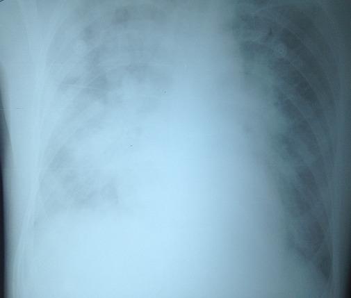
This diagnostic image is a front-facing chest X-ray (radiography) demonstrating extensive bilateral pulmonary opacities. The lungs exhibit a mixed alveolar-interstitial pattern characterized by diffuse, fluffy infiltrates and reticular markings. The opacities are distributed throughout both lung fields, with a more pronounced 'white-out' appearance in the lower zones and a noted asymmetric severity where the left lung appears more densely involved than the right. Notably, the cardiac silhouette remains within normal size limits, showing no evidence of cardiomegaly or significant distortion of the heart borders. These radiographic findings, when associated with acute respiratory distress post-transfusion, are highly suggestive of non-cardiogenic pulmonary edema, such as Transfusion-Related Acute Lung Injury (TRALI). The image serves as a clinical example of pulmonary parenchymal involvement without cardiac failure, essential for differentiating TRALI from Transfusion-Associated Circulatory Overload (TACO) in critical care and hematology education.
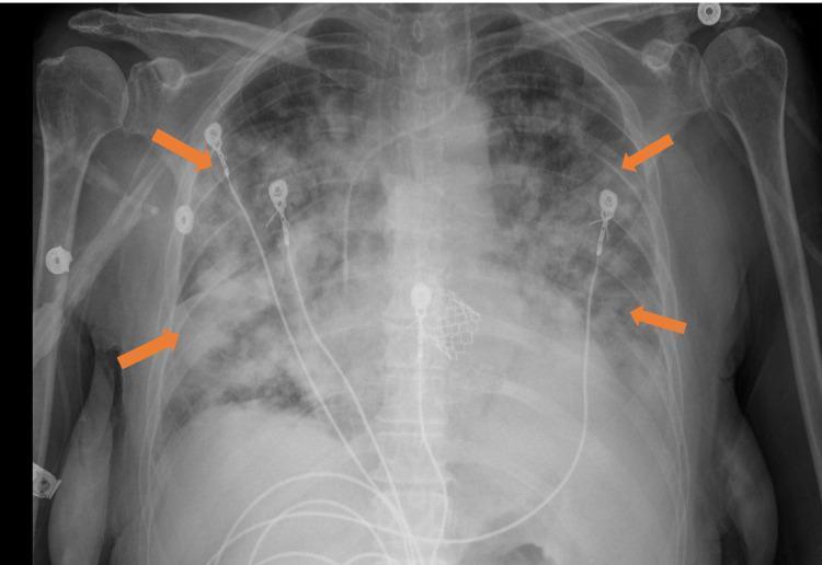
This anteroposterior (AP) chest X-ray illustrates bilateral, diffuse alveolar airspace opacities throughout both lung fields (indicated by orange arrows). The opacities present as patchy, confluent infiltrates characteristic of acute pulmonary edema or transfusion-related acute lung injury (TRALI). The distribution is relatively uniform across the lung parenchyma, obscuring underlying vascular markings. Several medical devices are visible: multiple ECG leads, a central venous catheter entering from the right, and a radiopaque cardiac device in the midline mediastinum, likely representing a transcatheter aortic valve replacement (TAVR) prosthesis. The cardiac silhouette is obscured by the overlying pulmonary infiltrates. This diagnostic image is used to teach radiographic recognition of acute respiratory distress syndrome (ARDS) or acute pulmonary congestion following medical intervention such as fluid resuscitation or transfusion.
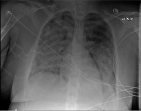
This diagnostic image is an anteroposterior (AP) chest X-ray demonstrating severe, diffuse bilateral pulmonary infiltrates. The infiltrates are characterized by extensive, fluffy, alveolar opacities distributed throughout both lung fields, consistent with Acute Respiratory Distress Syndrome (ARDS) or Transfusion-Related Acute Lung Injury (TRALI). Notable medical devices are visible, including an endotracheal tube (ETT) for mechanical ventilation and a central venous catheter, indicating a critically ill patient receiving advanced life support. Electrocardiogram (ECG) leads are also superimposed over the thorax. The cardiac silhouette appears within normal limits for size, which, combined with the diffuse pulmonary edema-like appearance, suggests a non-cardiogenic origin of the pulmonary infiltrates. This image serves as a clinical example of rapid-onset pulmonary complications following medical procedures, emphasizing the radiological presentation of acute lung injury in a critical care setting.
TRALI - Transfusion-Related Acute Lung Injury
Definition
Blood Products Implicated
- Fresh Frozen Plasma (FFP) - highest plasma volume
- Apheresis platelets - high plasma content
- Whole blood
- Packed Red Blood Cells (pRBCs)
- Cryoprecipitate (less common)
Epidemiology
- Historically, TRALI occurred in approximately 1 in 5,000-10,000 plasma-containing product transfusions
- True incidence is likely underestimated due to under-reporting and difficulty distinguishing TRALI from other causes of respiratory distress in critically ill patients
- After implementation of male-donor plasma policies, the incidence decreased from ~2.57 per 10,000 units to ~0.81 per 10,000 units
- Occurs equally in both sexes and all age groups
- Multiparous female donors are the most commonly implicated donors, due to HLA antibody formation from exposure to fetal leukocyte antigens during pregnancy
Pathogenesis - The Two-Hit Model
Hit 1: Neutrophil Priming
Hit 2: Neutrophil Activation by Transfused Products
- Donor plasma contains anti-HLA class I or class II antibodies or anti-Human Neutrophil Antigen (HNA) antibodies (especially anti-HNA3a)
- These antibodies react with recipient leukocytes (neutrophils, monocytes) and pulmonary endothelium
- Neutrophil degranulation and reactive oxygen species release damage the alveolar-capillary membrane
- Most commonly from multiparous female donors who develop HLA antibodies during pregnancy
- Stored blood products accumulate bioactive lipids (e.g., lysophosphatidylcholine) and CD40 ligand - both capable of activating primed neutrophils
- Older stored units contain more of these substances
- This mechanism explains TRALI in the absence of donor antibodies
Clinical Features
| Feature | Frequency |
|---|---|
| Acute respiratory distress / dyspnea | ~100% |
| Hypoxemia (SpO2 < 90% on room air) | 100% (required for diagnosis) |
| Bilateral pulmonary infiltrates on CXR | 100% (required) |
| Fever | ~33% of cases |
| Hypotension | ~32% of cases |
| Cyanosis | ~25% of cases |
| Pink frothy sputum (high albumin - permeability edema) | Present in severe cases |
- Chest X-ray shows bilateral alveolar opacities with a normal cardiac silhouette and no pleural effusions
- Approximately 70% of patients require mechanical ventilation at presentation
- The illness tends to resolve within 48 hours in most cases
- Chest radiograph typically returns to normal within 4 days
Diagnostic Criteria (2019 International Consensus - TRALI Type I & II)
- No evidence of acute lung injury before transfusion
- Acute onset hypoxemia: PaO2/FiO2 ≤ 300 mmHg or SpO2 < 90% on room air
- Bilateral infiltrates on chest imaging
- No evidence of left atrial hypertension (or if present, not judged to be the primary cause)
- Onset within 6 hours of blood product transfusion
- Respiratory status was stable in the 12 hours before transfusion
- Transfusion judged to have caused the acute deterioration
The 2019 consensus panel removed the older term "possible TRALI" and replaced it with this Type I/Type II classification.
Differentiation: TRALI vs TACO vs ARDS
| Feature | TRALI | TACO | ARDS |
|---|---|---|---|
| Onset | Within 6 h of transfusion | Within 6 h of transfusion | Variable |
| Mechanism | Immune/inflammatory - non-cardiogenic | Cardiogenic (fluid overload) | Variable |
| Pulmonary edema fluid | High protein (exudate) | Low protein (transudate) | High protein |
| JVP/CVP | Normal or low | Elevated | Variable |
| Response to diuretics | Poor | Good | Poor |
| BNP/NT-proBNP | Normal or mildly elevated | Markedly elevated | Variable |
| CXR - cardiac silhouette | Normal | Enlarged | Normal |
| Hypotension | Common | Uncommon (hypertension more likely) | Variable |
| Treatment | Supportive (O2, ventilation) | Diuresis | Lung-protective ventilation |
Management
Immediate Steps:
- Stop the transfusion immediately if TRALI is suspected
- Keep the IV line open; do NOT discard the blood bag (send for investigation)
- Notify the blood bank - quarantine all products from the implicated donor
Respiratory Support:
- Supplemental oxygen - high-flow O2 via facemask as first step
- Mechanical ventilation in ~70% of patients - use lung-protective strategy (same as ARDS: low tidal volumes 6 mL/kg, PEEP titration, plateau pressure < 30 cmH2O)
- ECMO may be used in severe refractory cases (limited evidence)
Hemodynamic Support:
- Avoid aggressive diuresis (unlike TACO) - TRALI involves capillary leak, and diuresis may worsen hypotension
- IV fluid resuscitation may be needed to maintain end-organ perfusion
Pharmacological:
- Corticosteroids: No convincing evidence of benefit; most providers avoid if lung injury is >14 days old (risk of worsening as in late-phase ARDS)
- Vasopressors if hypotension is refractory
Laboratory Investigation:
- Test donor for anti-HLA (class I and II) and anti-HNA antibodies (especially anti-HNA3a)
- If donor has antibodies against recipient's leukocyte antigens: defer donor from future high-plasma donations
- Note: TRALI is a clinical diagnosis - absence of antibodies does not exclude it
Prognosis
- Estimated mortality: ~5% (Murray & Nadel's)
- Most patients recover within 48-96 hours with supportive care
- Patients who recover from TRALI are not at increased risk for recurrence if future transfusions avoid products from the implicated donor
Prevention
| Strategy | Details |
|---|---|
| Male-only plasma policy | Most plasma products sourced from male donors in US, UK, Germany - has significantly reduced TRALI incidence |
| HLA antibody screening of female donors | Females with pregnancy history screened; if anti-HLA antibodies found, deferred from high-plasma products |
| Donor deferral | Donors implicated in prior TRALI cases deferred from donating plasma-rich products |
| Leukoreduction | Reduces leukocyte load in stored products; may reduce priming mediators |
| Minimize unnecessary transfusion | Restrictive transfusion strategies reduce TRALI exposure |
| Avoid older stored products | Older units accumulate more bioactive lipids (controversial) |