
Suicidal case forensic reports and images
What is the context of your question about suicidal case forensic reports and images?
hanging suicide ligature mark neck forensic autopsy
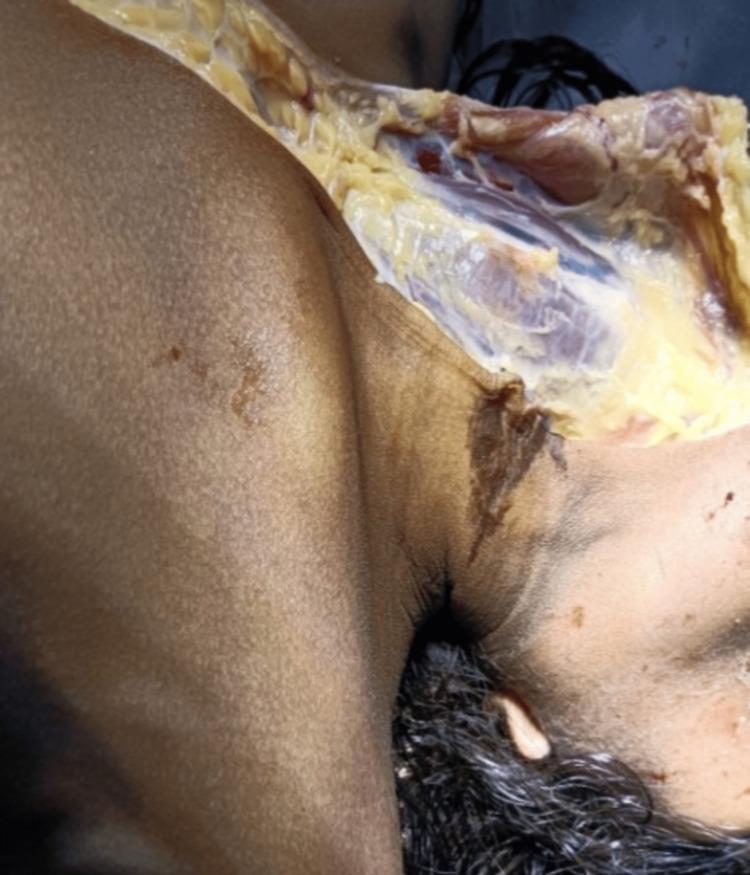
This forensic clinical photograph depicts an autopsy examination of a human neck, illustrating findings consistent with suspension hanging. A prominent, dark brown, parchment-like ligature mark is visible on the right lateral aspect of the neck. The mark exhibits an oblique, upward orientation toward the mastoid region, characteristic of antemortem compression. A surgical dissection flap has been reflected to expose the underlying subcutaneous tissue and superficial fascia. This underlying tissue appears white, glistening, and yellowish, without evidence of significant subcutaneous or intramuscular hemorrhage, which is a key diagnostic feature in certain forensic assessments of ligature-related injuries. The image serves as an educational resource for forensic pathology, specifically demonstrating the external morphology of a ligature furrow and the corresponding internal examination of neck structures during a medicolegal postmortem.
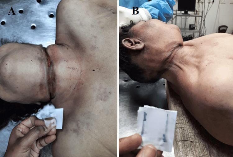
Clinical photograph from a forensic autopsy showing two views (A and B) of a ligature mark on the neck of a deceased adult. View A displays the anterior aspect of the neck, revealing a well-defined, horizontal, and incomplete ligature furrow located above the level of the thyroid cartilage. The mark is reddish-brown with a parchment-like, depressed appearance, indicating focal pressure. View B shows the lateral aspect of the neck, illustrating the oblique and ascending trajectory of the mark as it travels toward the point of suspension. The surrounding skin exhibits postmortem changes, but the mark itself is clearly demarcated and does not encircle the neck completely, a finding consistent with typical hanging. These images are used in forensic medicine to demonstrate classic external signs of mechanical asphyxia, highlighting the morphology, location, and directionality of ligature marks essential for determining the mechanism and manner of death.
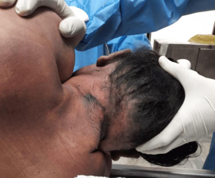
This forensic clinical photograph depicts the posterior aspect of the neck and head of a deceased individual during a medicolegal autopsy. The primary finding is a prominent, broad ligature mark located high on the neck, above the level of the thyroid cartilage. The mark is characterized by dark, parchment-like skin discoloration and bruising, appearing oblique and discontinuous at the back, which is a classic finding in cases of hanging where the knot creates a gap in the impression. The skin surrounding the furrow remains largely intact without significant laceration. The image illustrates the external signs of mechanical asphyxia, serving as an educational resource for forensic pathology and trauma assessment. Key features include the ligature’s width, its anatomical position relative to the laryngeal structures, and the characteristic lack of complete encirclement.
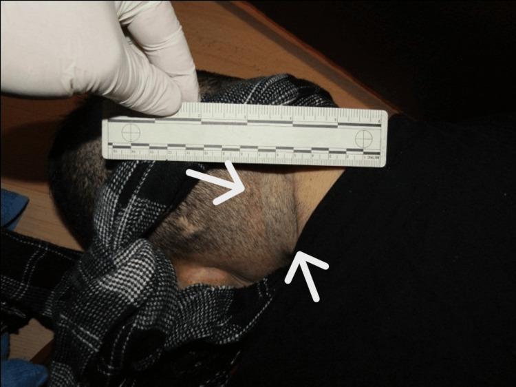
This clinical photograph from a forensic pathology context demonstrates a ligature mark on the neck of a deceased individual. The image shows a well-defined, slightly depressed negative imprint on the upper neck, positioned above the level of the thyroid cartilage. The mark follows an oblique, rising course towards the posterior aspect of the neck, which is characteristic of suspension in hanging cases. White arrows highlight the location and trajectory of the furrow. A forensic ruler is held against the neck by a gloved hand to provide a scale for the width and positioning of the mark. The surrounding skin appears pale, and part of the head is covered by a patterned textile (scarf). This visual serves as an educational example of external findings in mechanical asphyxiation, specifically demonstrating the morphology and distribution of a ligature furrow used to distinguish between different types of neck constriction.
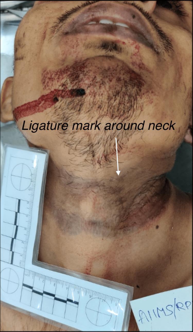
This forensic clinical photograph depicts the head and neck of a male decedent. A prominent ligature mark is visible encircling the neck, characterized as a wide, horizontal band of pressure abrasion and parchment-like skin discoloration. The mark appears more pronounced on the left anterolateral aspect of the neck, showing a reddish-brown hue consistent with vital or perimortem compression. In the facial region, a linear streak of dried blood extends obliquely across the right cheek and chin, with additional punctate blood staining noted within the beard and mustache areas. The subject's skin appears pale. A forensic L-shaped measurement scale is positioned adjacent to the left side of the neck to provide a size reference for the injuries. This image serves as a diagnostic tool in forensic pathology for documenting evidence of hanging or strangulation and associated secondary trauma in a medico-legal context.
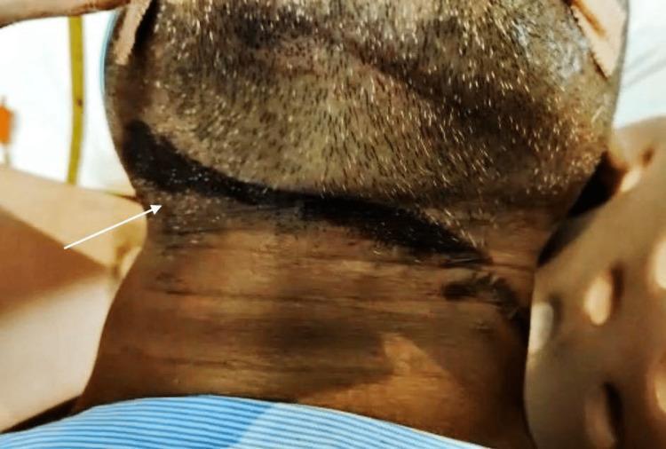
This clinical photograph shows a close-up, inferior-to-superior view of the neck and submental region of an intubated patient. A prominent, dark-pigmented ligature mark is visible, indicated by a white arrow. The mark is a circumferential, superficial abraded furrow that follows an oblique path across the neck. It is situated at the level of the cricoid cartilage, extending from the right mandibular angle and slanting across the anterior cervical region. The skin within the furrow appears dark, dried, and parchment-like, which is characteristic of ligature marks in cases of hanging. The patient is shown with medical adjuncts, including adhesive tape on the chin and a striped garment at the bottom of the frame, indicating a hospital or forensic clinical setting. This image serves as an educational example in forensic medicine and emergency trauma for identifying and documenting ligature injuries associated with near-hanging or strangulation events.
suicidal incised wound hesitation cuts wrist forensic
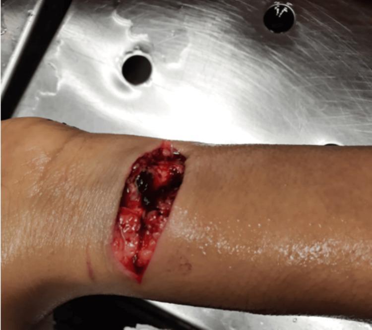
Clinical photograph of the ventral aspect of the left wrist during a medicolegal autopsy. The image displays two distinct incised wounds. A primary deep, gaping incision (approximately 7 cm x 2 cm) is oriented transversely across the wrist. The wound edges are clean-cut, revealing exposure of the subcutaneous adipose tissue, underlying tendons, and superficial blood vessels, with clotted blood visible within the wound cavity. Distal to this larger wound is a secondary, superficial incision (approximately 2 cm x 0.1 cm) oriented parallel to the primary injury, characteristic of a 'hesitation mark.' These findings are significant in forensic pathology for evaluating self-inflicted injuries and suicidal behavior. The photograph is taken against a stainless steel background typical of a morgue or clinical setting, focusing on the traumatic skin lesions and underlying structural damage.
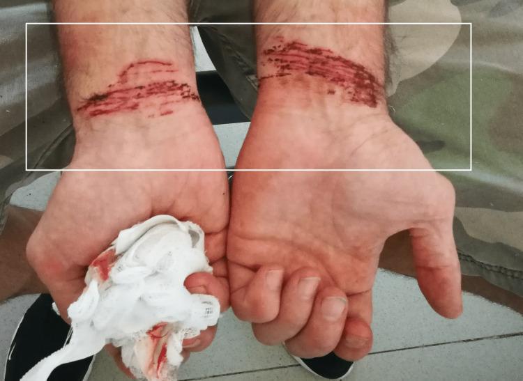
This clinical photograph displays the bilateral palmar wrists of a patient, showing multiple self-inflicted injuries. The lesions are characterized as parallel, linear abrasions and superficial incised wounds. These injuries exhibit varying depths, with evidence of fresh blood and minor oozing. The wounds are concentrated in a horizontal orientation across the wrist crease, with a higher density and severity noted on the left wrist compared to the right. In the foreground, the patient's left hand is holding a blood-stained white gauze pad. The image serves as a forensic and psychiatric educational tool for identifying patterns of non-suicidal self-injury (NSSI) or manipulative self-harm. The visual presentation of parallel, uniform, and accessible-site injuries is highly characteristic of intentional self-infliction rather than accidental trauma. The clinical context involves forensic examination to differentiate between genuine suicidal intent and secondary gain motivation.
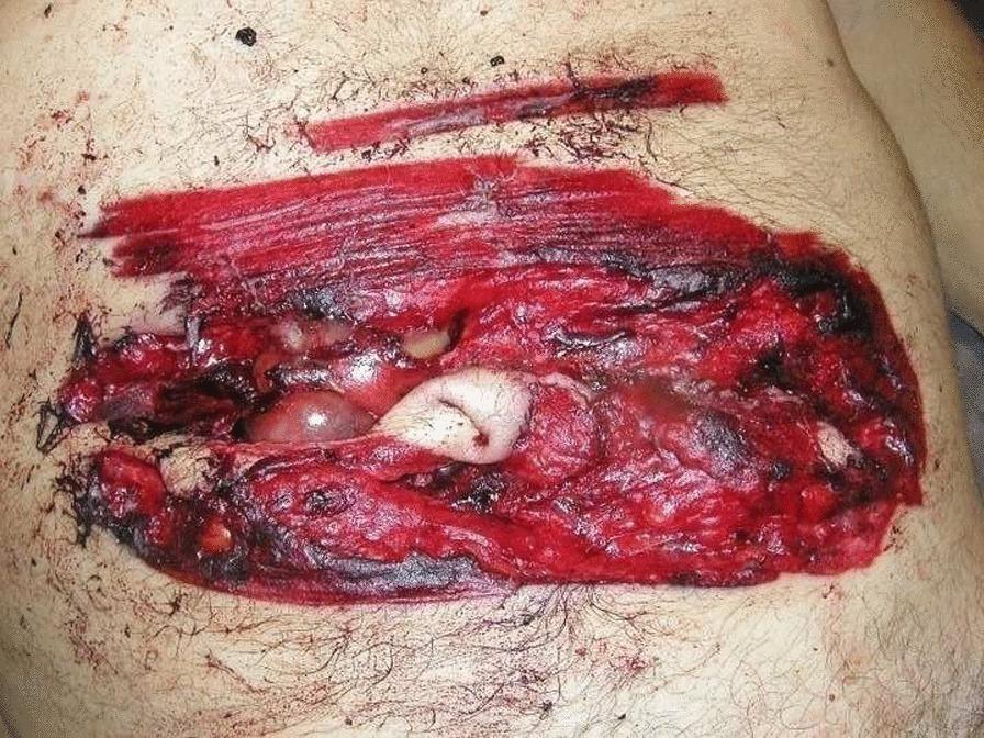
This clinical photograph from a forensic pathology context displays a large, complex abdominal wound. The primary lesion is a horizontal, deep incised wound located in the mesogastrium. The wound morphology is characterized by irregular, torn edges with focal areas of grazing, differing from a typical clean surgical incision. Centrally, the wound penetrates through the skin and subcutaneous fat layers, exposing internal structures including glistening, pinkish-white intestinal loops. Surrounding the deep defect is an extensive, well-demarcated area of reddish-brown abrasion and cutaneous bruising, measuring approximately 23 cm by 12 cm. There is evidence of peripheral blackish discoloration and hemorrhagic infiltration within the exposed tissue. Superior to the main lesion, a smaller, superficial linear 'trial' or hesitation wound is visible. The image illustrates severe abdominal trauma and is used in medical education to differentiate between standard incised wounds and those caused by high-velocity mechanical instruments like chainsaws.
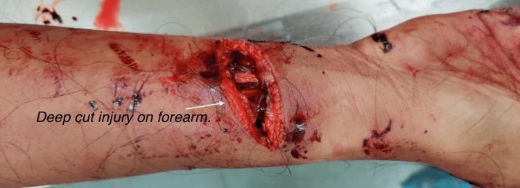
Clinical photograph of a deep, transverse incised wound located on the lower one-third of the volar aspect of the right forearm, near the wrist. The wound is gaping and exhibits sharply defined but blood-soaked margins, characteristic of a sharp force injury. Within the depth of the wound, anatomical structures are visible, including the severed ends of the flexor carpi radialis tendon and the radial artery. Significant hemorrhage is evident, with extensive blood staining, spatter, and streaks across the surrounding skin. Several small, punctate abrasions and dried blood clots are scattered distal to the primary injury towards the palm and wrist. This image serves as a forensic and clinical example of a life-threatening vascular injury resulting from sharp force trauma, demonstrating complete severance of a major peripheral artery and associated tendinous damage.
contact gunshot wound entrance exit forensic autopsy firearm suicide
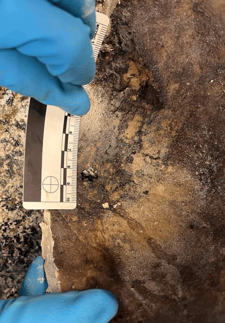
This forensic clinical photograph depicts the internal view of a human skull specimen during autopsy, specifically showcasing a gunshot exit wound. The image displays the internal bony plate (tabula interna) which exhibits a small, irregular, dark perforating fracture approximately 0.9–1 cm in diameter. Notable features include external beveling characteristic of a firearm exit wound, where the exit side of the bone is wider than the entry side. The surrounding bone shows significant postmortem changes, including mottled discoloration with tan, beige, and dark brown-to-black shades, suggesting advanced decomposition and staining from organic material. A dark reddish-brown to black granular substance is adhered to the internal bony plate, consistent with a decomposed hematoma or blood collection. The bone texture appears porous and uneven with scattered fine debris. A forensic metric ruler is placed alongside the lesion for scale, held by a gloved examiner to document the dimensions of the fracture for medicolegal investigation. The specimen demonstrates key principles of forensic osteology and ballistics in the context of a skeletonized or partially decomposed cadaver.
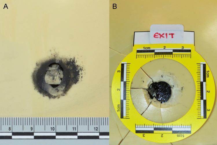
Educational forensic pathology specimens demonstrating terminal ballistics on a bone model. Image A displays an entrance wound characterized by a circular perforation with inward-protruding margins and a prominent ring of dark soot deposition, indicating a close-range or contact discharge. A metric scale shows the entrance diameter is approximately 10mm. Image B illustrates an exit wound with characteristic external beveling and radiating linear fractures extending from the circular defect. A yellow circular forensic scale is placed around the exit site for measurement. These specimens serve to educate on the differentiating morphological features between entrance and exit gunshot wounds, specifically focusing on marginal soot, beveling orientation, and fracture patterns used in forensic investigations to determine projectile trajectory and range of fire.
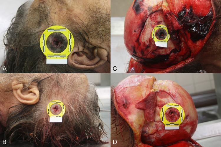
A series of four forensic clinical photographs (A-D) documenting a penetrating gunshot wound to the human head. Panel A displays the entrance wound in the right temporal region, characterized by burned-intruding margins, soot deposition, and a distinct muzzle imprint, indicating a loose contact discharge. Panel B shows the corresponding exit wound in the left temporal region, presenting with irregular, protruding margins and an absence of soot or thermal injury. Panel C illustrates the entrance site following soft tissue dissection; a circular defect in the right temporal bone is visible with significant soot deposition concentrated around the osseous opening. Panel D reveals the exit site on the left temporal bone, showing characteristic external beveling of the outer table, where the defect is wider on the exterior than the interior. Each panel includes a yellow forensic scale for size reference. This series demonstrates the classic diagnostic features used in forensic pathology to differentiate between entry and exit ballistic trauma, as well as to determine range of fire.
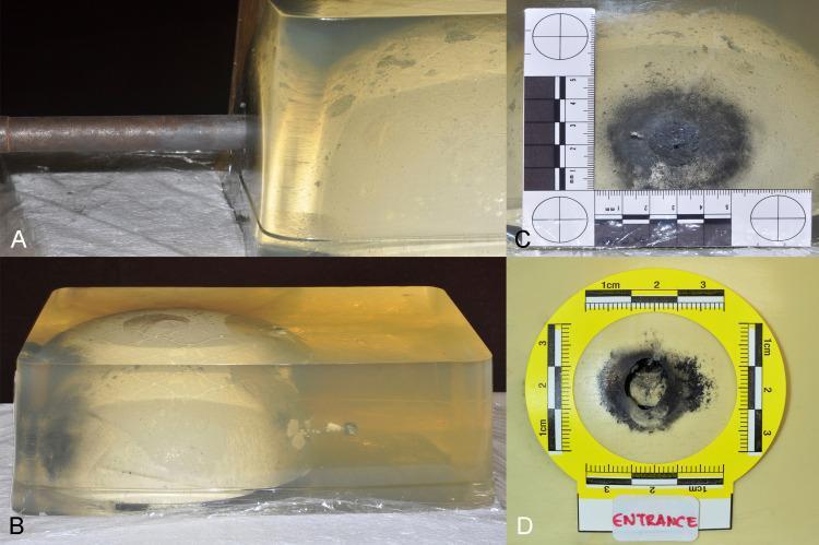
This forensic pathology experimental image set demonstrates a ballistics reconstruction of a penetrating gunshot wound using a gelatin-based head model with an embedded synthetic bone structure. Frame A shows a contact-range test shot being fired into the model using a handmade firearm. Frame B provides a lateral profile of the ballistic gelatin block, revealing a linear projectile trajectory from left to right; a permanent wound cavity is visible, which is widest near the entry point and tapers toward the exit, representing energy dissipation and tissue displacement. Frames C and D are close-up views of the entrance wound on the gelatin surface and the underlying bone model, respectively. Both detail a central circular defect surrounded by extensive, dark, irregular soot deposition and charring, characteristic of a contact-range discharge. Each frame includes forensic scaling and labeling to quantify the diameter of the soot pattern and the projectile entrance. This model serves as a simulation for cranial ballistics, demonstrating the relationship between muzzle distance and forensic markers like soot deposition and internal beveling.
Forensic Pathology: Suicidal Deaths - Autopsy Findings, Manner of Death Determination, and Forensic Reports
1. Manner of Death: Definitions and Certification
- Natural
- Suicide
- Homicide
- Accident
- Undetermined
"The manner of death can be changed if subsequent information that alters the circumstances surrounding the death is discovered." - DiMaio's Forensic Pathology
2. Forensic Autopsy Report Structure (Suicidal Cases)
| Component | Details |
|---|---|
| Authority | Who ordered the autopsy |
| Deceased identity | Name, age, sex, date/time/place of examination, mode of identification |
| External examination | Detailed description of all injuries (location, size, character, age), postmortem changes, clothing |
| Internal examination | Positive AND important negative findings; contents of stomach, GI, bladder; state of coronary arteries, lungs, brain |
| Photographs/radiographs | Objective record that enhances report value |
| Laboratory results | Toxicology, histology, chemical analysis |
| Opinion | Cause of death, manner of death, approximate time since death |
| Signature | Qualifications and designation of medical officer |
"This 45-year-old man died as a result of perforating gunshot wound of the head. Information available at the time of examination combined with the finding of entry wound with visible powder residues on the roof of the mouth support the conclusion that the manner of death was suicide."
3. Suicidal Hanging: Autopsy Findings
External Findings
- Ligature mark: Oblique, directed upward toward the point of suspension, forming a classic inverted-V configuration
- Mark is incomplete - does not encircle the neck fully; there is a gap at the knot site with no skin damage in the gap
- Position: usually above the thyroid cartilage
- Character: Dry, parchment-like, glistening white subcutaneous tissue on dissection (in contrast to strangulation, where subcutaneous tissue shows ecchymosis)
- Face is usually pale (not congested)
- Saliva may run vertically from the angle of the mouth down the chest
- Signs of struggle are absent in suicidal hanging
Internal Findings
- Fracture/fracture-dislocation of cervical vertebrae (especially in judicial-type hanging with a long drop)
- Fracture of hyoid bone - rare in hanging, more common in throttling (manual strangulation)
- Injury to carotid arteries: possible in cases with a long drop
- Absence of other fatal injuries and poisoning (confirmed only after full autopsy)
Classic Ligature Mark Images
4. Hanging vs. Strangulation (Differential Table)
| Feature | Hanging (Suicidal) | Strangulation (Homicidal) |
|---|---|---|
| Manner | Suicidal usually | Homicidal usually |
| Signs of struggle | Absent | Present |
| Ligature mark position | Above thyroid cartilage, oblique, incomplete | Below thyroid cartilage, horizontal, complete encirclement |
| Skin under mark | Dry, glistening, white | Ecchymosed subcutaneous tissue |
| Abrasions/bruises around mark | Rare | Common |
| Face | Usually pale | Congested, pronounced petechiae |
| Fracture of hyoid | Rare | Not rare (aged) |
| Fracture of laryngeal cartilages | Rare | Common |
| Bleeding from nose/mouth | Very rare | Common |
| Saliva | Runs vertically down chest | May be blood-tinged, not vertical |
5. Suicidal Incised Wounds: Hesitation Cuts
Features of Suicidal/Self-Inflicted Wounds (Hesitation Marks):
- Multiple and parallel (or nearly so) in the same area
- Uniform in depth and direction
- Relatively minor/superficial - often involving only skin
- Located on accessible areas: front of neck, wrists, groin, front of chest, occasionally back of legs
- Avoid vital and sensitive areas: eyes, lips, nose, ears
- Fatal wound is usually present alongside multiple trial wounds - the fatal incision is made with greater force and produces a large gaping wound
- When a safety razor blade is used: unintentional cuts on the fingers where the blade was gripped
Common Locations of Suicidal Incised Wounds:
- Throat (front of neck)
- Wrists (most common)
- Front of chest
- Ankles
Forensic Images: Hesitation Cuts and Suicidal Wrist Wounds

6. Suicidal Firearm Deaths: Autopsy Features
- Contact or close-range wound - soot (blackening), tattooing (powder stippling), muzzle imprint on skin
- Accessible site: temple, roof of mouth (intraoral), submental, anterior chest
- Entry wound characteristics: circular, punched-in, with blackening from soot and burning at contact range
- Entry wound into the skull: inner table beveling (entry side); exit shows external beveling (outer table wider)
- Consistency with self-infliction: weapon must be reachable - check arm length vs. wound-to-muzzle distance
- Absence of defensive wounds
- Scene and circumstantial evidence: weapon found at scene, suicide note
Important Caveat (DiMaio's):
Firearm Forensic Images
7. Key Indicators of Suicide vs. Homicide at Scene/Autopsy
| Feature | Suicide | Homicide |
|---|---|---|
| Suicide note | Present (~25% of cases) | Usually absent |
| Scene | Secluded, no disturbance | Evidence of struggle |
| Prior suicidal ideation/attempts | Often present | N/A |
| Wound location | Accessible, self-inflicted sites | Often on back, multiple sites |
| Hesitation marks | Present in sharp force cases | Absent (defense wounds instead) |
| Weapon found at scene | Yes (accessible) | May be absent |
| Contact/close-range GSW | Yes (suicidal firearm) | Variable |
| Ligature mark (hanging) | Oblique, incomplete, no struggle | Horizontal, complete, struggle present |
| Dress/clothing | Usually tidy | May be disheveled |
8. Medicolegal Masquerades
- Hanging arranged to appear homicidal: the mode of suspension can simulate throttling if the ligature is placed differently
- Suicide by drowning after poisoning: the primary cause may be masked
- Suicide note may be forged (in rare cases to conceal homicide)
- Presence of additional injuries (e.g., bruises from fall during suicide attempt by hanging) can cause confusion
- DiMaio's Forensic Pathology, 3rd Ed. - Cause, Manner and Mechanism of Death
- Parikh's Textbook of Medical Jurisprudence, Forensic Medicine and Toxicology - Autopsy Report Structure
- The Essentials of Forensic Medicine and Toxicology, 36th Ed. (2026) - Incised Wounds, Asphyxia
- P.C. Dikshit's Textbook of Forensic Medicine and Toxicology - Hanging and Strangulation, Ligature Marks