
MALE AGED 60 YEARS WHO IS A CHRONIC SMOKER PRESENTED WITH HISTORY OF SLOWLY INCREASING SEVERE EXERTIONAL DYSPNOEA AND WEIGHT LOSS, HE IS BARREL-CHESTED AND DYSPNEIC WITH PROLONGED EXPIRATION, SITS FORWARD IN A HUNCHED-OVER POSITION AND BREATHS THROUGH PURSED LIPS (2+4+4=10) (A) WHAT IS THE PROBABLE DIAGNOSIS (B) WRITE THE MAJOR TYPES (CLASSIFICATION) WITH APPROPRIATE DIAGRAMS (C) DISCUSS THE PATHOGENESIS OF THE LESION
emphysema types centriacinar panacinar diagram comparison

This diagnostic image consists of four high-resolution computed tomography (HRCT) axial sections of the lung parenchyma, illustrating the primary radiological patterns of emphysema in Chronic Obstructive Pulmonary Disease (COPD). Images A and B demonstrate centriacinar emphysema, characterized by small, focal areas of low attenuation (lucent airspaces) located in the center of the secondary pulmonary lobules, typically surrounding the central bronchovascular bundle. Image C displays panacinar emphysema, showing a more uniform and diffuse destruction of the entire acinus, resulting in generalized lung hyperlucency and attenuation of the pulmonary vasculature. Image D illustrates paraseptal emphysema (distal acinar emphysema), where focal lucent airspaces are distributed preferentially along the subpleural surfaces and interlobular septa, often demarcated by thin walls. Key educational concepts include the distinction between lobular distributions and the resulting architectural distortion and vascular attenuation associated with progressive alveolar wall destruction.
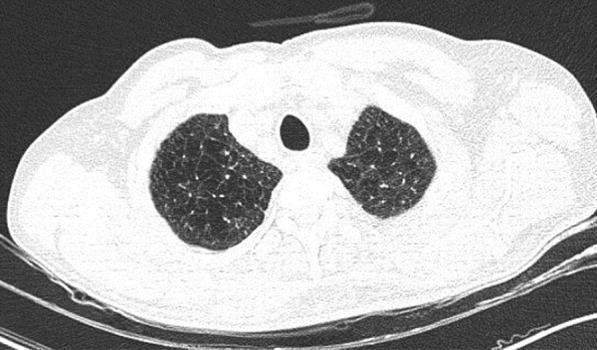
This diagnostic image is an axial non-contrast high-resolution computed tomography (HRCT) scan of the thorax at the level of the upper lobes. The image demonstrates bilateral, diffuse, and homogeneous areas of abnormally decreased lung attenuation (hyperlucency). These findings are characteristic of panacinar (panlobular) emphysema, where there is a uniform destruction of the pulmonary acinus. Key features include a simplified lung architecture with a notable paucity of pulmonary vascular markings throughout the affected parenchyma. Unlike centriacinar emphysema, which often shows focal areas of low attenuation, this presentation shows a generalized darkening of the lung fields without well-defined walls or cystic structures. No acute infiltrates, ground-glass opacities, or consolidations are present, which in a clinical context (such as COVID-19 screening) helps differentiate chronic obstructive pulmonary disease (COPD) from acute infectious processes. The image serves as a classic educational example of end-stage alveolar destruction and hyperinflation seen in severe emphysema.
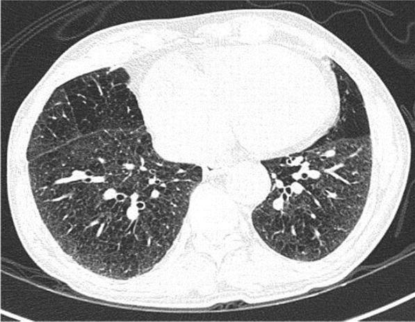
This diagnostic image is an axial non-contrast chest CT scan at the level of the lower lobes, demonstrating characteristic features of panacinar emphysema. The lung parenchyma exhibits a diffuse, generalized decrease in attenuation (hypodensity) bilaterally, resulting in a significantly darker appearance than normal lung tissue. Structurally, there is extensive destruction of the alveolar walls, leading to the formation of numerous small, round, and irregularly shaped airspaces that give the parenchyma a coarse, 'swiss cheese' or porous texture. Unlike centriacinar emphysema which typically affects the upper lobes, these changes are distributed uniformly across the visible lung segments without regional predilection. Key landmarks include the central mediastinum with a visible heart and descending aorta, and the bilateral major fissures. There is a notable absence of peripheral ground-glass opacities or consolidations, distinguishing this chronic obstructive pulmonary disease (COPD) manifestation from acute infectious processes like COVID-19 bronchopneumonia.
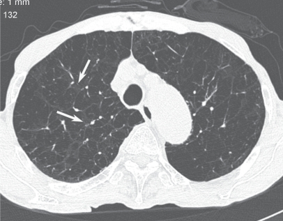
**Imaging Modality:** High-resolution computed tomography (HRCT), axial section. **Anatomical Region:** Thorax, specifically the upper lobes of the lungs. **Observed Pathology:** Advanced centriacinar (centrilobular) emphysema. **Characteristic Visual Features:** The image demonstrates diffuse, bilateral areas of low attenuation representing parenchymal destruction and airspace enlargement. These lucencies lack discernible walls, distinguishing them from cystic lung disease. While the disease is extensive, there are focal areas of preserved, higher-attenuation lung parenchyma immediately adjacent to the central bronchovascular bundles (indicated by arrows). This "sparing" of the immediate perilobular area suggests a centrilobular origin of the destructive process. The pulmonary vasculature appears attenuated and distorted within the areas of emphysematous change. **Key Diagnostic Features:** The presence of small, discrete lucencies centered on the core of the secondary pulmonary lobule is characteristic of centriacinar emphysema. In this advanced stage, the coalescence of these lucencies mimics panacinar emphysema; however, the identification of residual normal lung tissue surrounding the central bronchovascular structures serves as a critical differentiating feature for centriacinar classification.
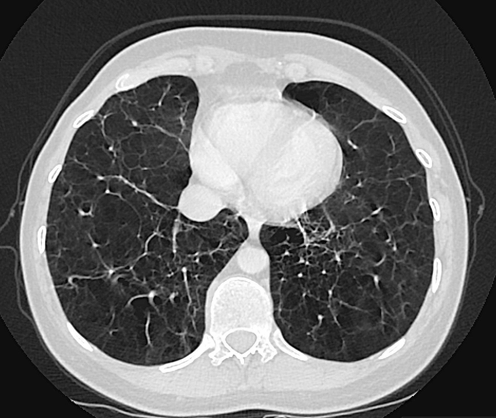
This diagnostic image is a transverse (axial) CT scan of the thorax, specifically focusing on the pulmonary parenchyma in a lung window. The imaging reveals widespread, severe emphysematous changes characterized by diffuse areas of abnormally low attenuation (lucency) and rarefaction of the lung tissue. The findings include both centriacinar and panacinar emphysema patterns, leading to significant destruction of the alveolar walls and a visible reduction in normal vascular markings. These changes are particularly pronounced in the lower lobes, manifesting as large, confluent areas of air entrapment and parenchymal volume loss. Additionally, localized regions of increased density suggest secondary features such as atelectasis or underlying fibrosis, and tractional bronchiectasis is evident within the distorted lung architecture. Clinically, this pattern of basilar-predominant emphysema is a hallmark radiological finding of alpha-1 antitrypsin deficiency (AATD), demonstrating the progressive lung destruction associated with this genetic protease-antiprotease imbalance.
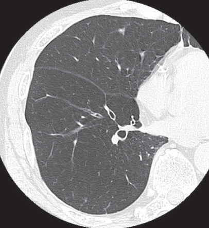
**Imaging Modality:** High-resolution computed tomography (HRCT) of the chest. **Anatomical Region:** Axial section of the right lung at the level of the pulmonary hilum. **Observed Pathology:** Panacinar (panlobular) emphysema. **Characteristic Visual Features:** The image demonstrates diffuse, uniform low attenuation of the lung parenchyma. There is a notable absence of discrete, focal low-attenuation areas (LAA) or visible cyst walls, which differentiates this from centriacinar or paraseptal subtypes. The pulmonary vasculature appears attenuated and sparse, appearing as thin, simplified branching structures within the hyperlucent lung field. The interlobular septa and bronchovascular bundles remain visible but are set against a background of generalized parenchymal destruction. **Key Diagnostic Features:** Global involvement of the secondary pulmonary lobule rather than focal destruction. This pattern of widespread lung simplification and uniform decrease in attenuation is characteristic of panacinar emphysema, often associated with alpha-1 antitrypsin deficiency. No significant pleural effusion or consolidations are present in the visualized field.
(A) Probable Diagnosis: Pulmonary Emphysema (COPD - Emphysematous Type / "Pink Puffer")
| Feature | Significance |
|---|---|
| Chronic smoker, 60-year-old male | Primary etiological factor; centriacinar emphysema |
| Slowly progressive severe exertional dyspnoea | Progressive loss of functional alveolar surface area and air trapping |
| Weight loss | Increased work of breathing, hypermetabolic state |
| Barrel chest | Hyperinflation - increased AP diameter due to air trapping |
| Prolonged expiration | Loss of elastic recoil - small airways collapse on expiration |
| Hunched-forward position (tripod) | Accessory muscle use; fixes shoulder girdle |
| Pursed-lip breathing | Generates positive end-expiratory pressure (auto-PEEP) to keep airways open and slow expiration - prevents airway collapse |
(B) Major Types (Classification) of Emphysema
Diagram: Major Patterns of Emphysema
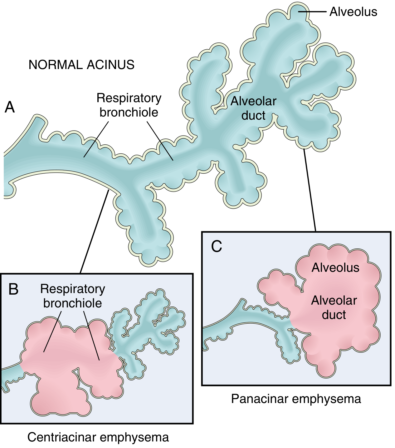
Type 1: Centriacinar (Centrilobular) Emphysema
- Most common type - >95% of clinically significant cases
- The central/proximal parts of the acinus (respiratory bronchioles) are destroyed; distal alveoli are spared
- Both emphysematous and normal airspaces exist within the same acinus
- Upper lobe predominance, especially the apical segments
- Cause: Strongly associated with cigarette smoking - this is the type in the patient above
- In severe disease, distal acinus is also involved, making distinction from panacinar difficult
Type 2: Panacinar (Panlobular) Emphysema
- The entire acinus is uniformly enlarged - from the respiratory bronchiole all the way to the terminal blind alveoli
- Lower lobe and anterior lung margin predominance (most severe at bases)
- Cause: Associated with α1-antitrypsin (AAT) deficiency; exacerbated by smoking
- About 1% of emphysema patients have AAT deficiency (encoded by the Pi locus on chromosome 14)
Type 3: Distal Acinar (Paraseptal) Emphysema
- The proximal acinus is normal; the distal part (adjacent to pleura, lobular septa) is predominantly affected
- Forms bullae (enlarged cystic spaces), especially in the upper lungs
- Cause: Unknown
- Clinical significance: Most common cause of spontaneous pneumothorax in young adults; not typically a cause of COPD
Type 4: Irregular (Paracicatricial) Emphysema
- Acinus is irregularly involved, almost always associated with scarring
- Usually occurs in small foci and is clinically insignificant
CT Imaging: Radiological Patterns of Emphysema
Summary Table: Types of Emphysema
| Feature | Centriacinar | Panacinar | Distal Acinar | Irregular |
|---|---|---|---|---|
| Part of acinus affected | Central (resp. bronchiole) | Entire acinus | Distal (alveolus) | Irregular |
| Location in lung | Upper lobes | Lower lobes/bases | Subpleural/septa | Around scars |
| Main cause | Smoking | AAT deficiency | Unknown | Scarring |
| Clinical COPD | Yes (most common) | Yes | No | No |
| Special complication | - | - | Spontaneous pneumothorax | - |
(C) Pathogenesis of Emphysema
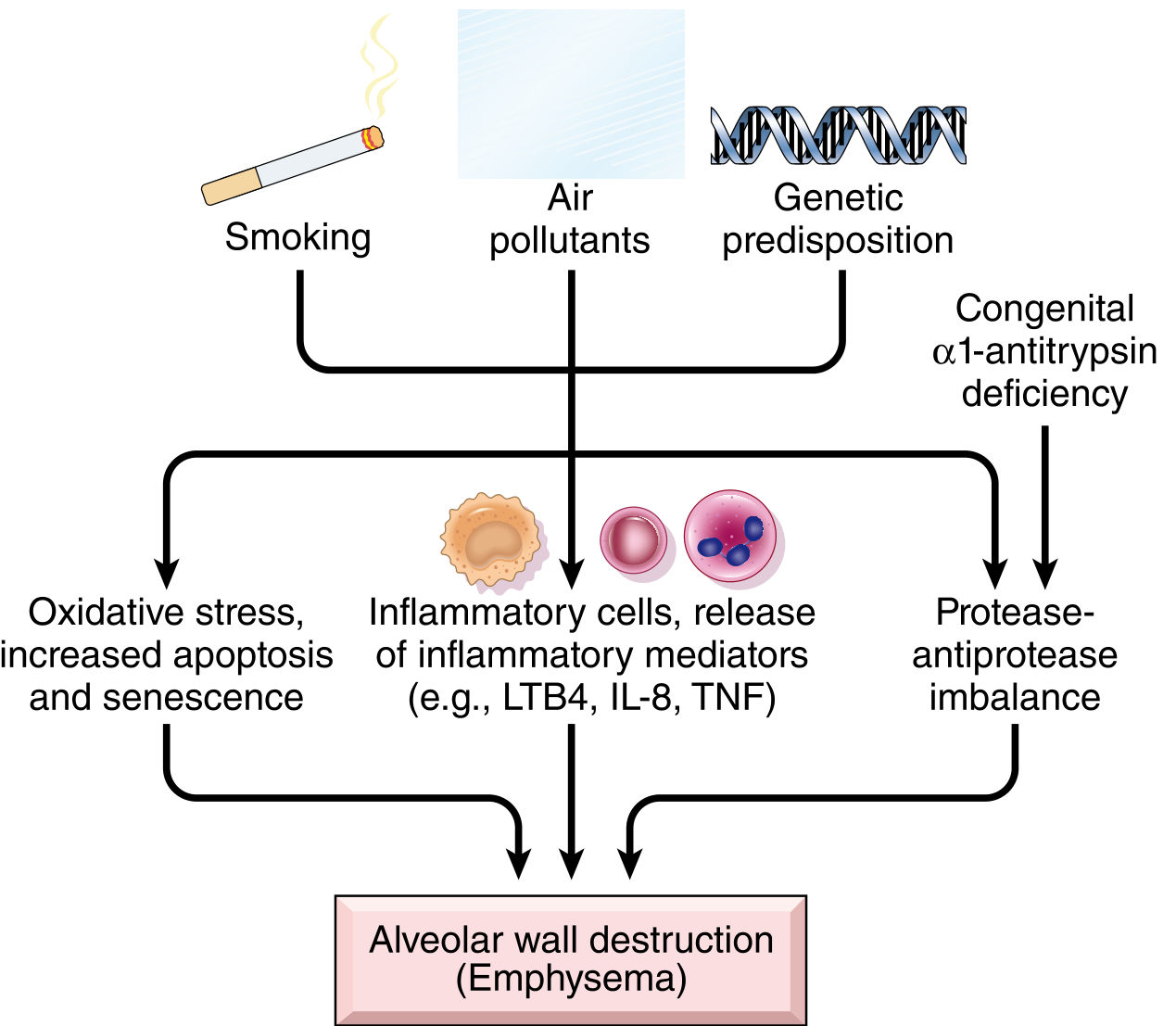
Step-by-Step Pathogenesis
Step 1: Inhalation of Noxious Stimuli
Step 2: Toxic Injury and Inflammation
- Inhaled particles damage airway epithelial cells and resident macrophages, which release chemokines and inflammatory mediators including:
- Leukotriene B4 (LTB4)
- IL-8 (interleukin-8)
- TNF (Tumor Necrosis Factor)
- These mediators recruit neutrophils and macrophages from the circulation into the lung parenchyma
- Chronic inflammation leads to accumulation of T and B lymphocytes in affected areas (role of adaptive immunity still being clarified)
Step 3: Protease-Antiprotease Imbalance (Central Mechanism)
- Recruited neutrophils and macrophages release proteases (especially neutrophil elastase, matrix metalloproteinases)
- These enzymes break down elastin and other connective tissue components of alveolar walls
- Normally, α1-antitrypsin (AAT) - present in serum, tissue fluids, and macrophages - inhibits neutrophil elastase, protecting the lung
- In smokers, two things happen simultaneously:
- Increased protease activity (from inflammation)
- Decreased antiprotease activity - cigarette smoke oxidizes and inactivates AAT
- The net result is unchecked elastase activity → destruction of alveolar walls
- In patients with congenital AAT deficiency (Pi locus, chromosome 14 - ZZ homozygotes), this imbalance is even more severe, causing panacinar emphysema
Step 4: Oxidative Stress
- Tobacco smoke contains massive amounts of reactive oxygen species (ROS)
- Inflammatory cells also produce oxidants upon activation
- Oxidants cause:
- Direct tissue damage to cell membranes and proteins
- Endothelial dysfunction
- Inactivation of antiproteases (especially AAT) - compounding the protease-antiprotease imbalance
- Increased cell apoptosis and senescence
- The transcription factor NRF2 (encoded by NFE2L2) normally senses intracellular oxidants and upregulates protective genes; genetic variants in this pathway increase susceptibility to smoking-related lung disease
Step 5: Alveolar Wall Destruction and Functional Consequences
- Progressive destruction of alveolar walls and the elastic tissue network of the lung leads to:
- Permanent enlargement of airspaces distal to terminal bronchioles
- Loss of elastic recoil - the lung can no longer recoil after inspiration; air is trapped
- Small airway collapse on expiration (no radial traction from surrounding parenchyma) → prolonged expiration, wheeze
- Bullae formation - coalescence of destroyed airspaces
- Reduced surface area for gas exchange → hypoxia with exertion
- Pulmonary hyperinflation → barrel chest, diaphragm flattened
Step 6: Compensatory Mechanisms (explaining clinical signs)
- Pursed-lip breathing: Creates back-pressure (auto-PEEP) that splints open collapsible small airways during expiration
- Tripod/forward-leaning position: Optimizes the length-tension relationship of accessory respiratory muscles (sternocleidomastoid, scalene) by fixing the shoulder girdle
- Pink puffer physiology: The patient increases respiratory drive and rate to maintain near-normal PaO2, at the cost of high work of breathing and weight loss