
In detail about vagotomy and complications
vagotomy types truncal selective highly selective diagram surgery
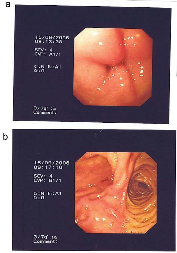
Two-panel clinical photograph displaying upper gastrointestinal endoscopic views of a gastrojejunostomy site in a patient with a history of truncal vagotomy. Image (a) focuses on the anastomotic site, showing clear signs of mucosal inflammation and significant edema. The surrounding tissue is swollen and erythematous with scattered white spots suggesting mucus or superficial exudate. Image (b) shows the view beyond the anastomosis into the efferent loop. The intestinal lumen appears patent with visible circular folds (plicae circulares) of the jejunum and no evidence of immediate mechanical obstruction or stricture at the surgical site. These images are clinically significant for evaluating postoperative complications such as anastomotic inflammation or ruling out early-onset gastric outlet obstruction following peptic ulcer surgery.
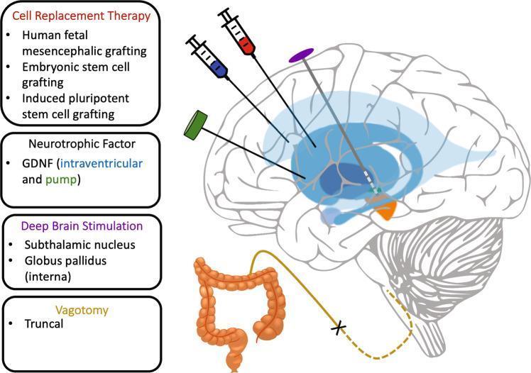
This medical illustration outlines four surgical disease-modification strategies for neurodegenerative disorders, primarily Parkinson's disease. The central diagram is a sagittal cross-section of the human brain overlaid with neuroanatomical interventions. 'Cell Replacement Therapy' is shown via a syringe targeting the striatum for human fetal mesencephalic or stem cell grafting. 'Neurotrophic Factor' administration (e.g., GDNF) is illustrated using a syringe and an external pump delivering therapy into the ventricular system, shaded in blue. 'Deep Brain Stimulation (DBS)' is depicted with an electrode lead targeting specific deep nuclei, including the subthalamic nucleus and globus pallidus interna. Below the brain, 'Abdominal Vagotomy' is represented by a schematic of the large intestine and the vagus nerve, indicating a truncal vagotomy aimed at preventing the gut-to-brain spread of misfolded alpha-synuclein. The diagram effectively correlates invasive neuromodulation and regenerative medicine techniques with their respective anatomical targets (ventricles, basal ganglia, and enteric nervous system) to describe current surgical approaches in neurology and neurosurgery.
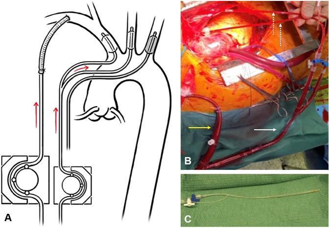
This composite educational resource illustrates techniques for bilateral selective antegrade cerebral perfusion (ACP) during cardiac surgery. Panel A is a schematic diagram showing ACP using a dedicated secondary pump circuit (e.g., cardioplegia pump) to deliver flow through balloon-tipped catheters placed in the supra-aortic branches. Panel B is an intraoperative clinical photograph demonstrating an alternative ACP setup where perfusion is diverted from the main arterial return line via a Y-connector (white arrow). It highlights the clamping of the right subclavian line (yellow arrow) to isolate cerebral flow, with dotted arrows indicating the small-bore perfusion catheters inserted into the carotid arteries. Panel C displays a specialized, small-caliber selective ACP catheter featuring a distal inflatable balloon for vessel occlusion and a proximal Luer-lock hub with a valve for balloon inflation. The content focuses on neuroprotection strategies during aortic arch surgery, emphasizing the use of balloon-occlusion catheters to avoid surgical trauma to fragile, potentially dissected vessel walls.
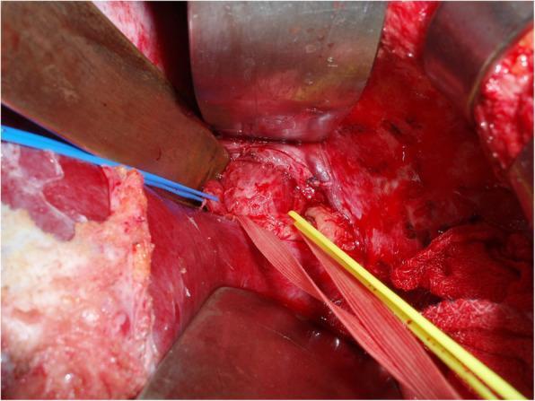
An intraoperative clinical photograph showing the surgical field of an upper-middle laparotomy for truncal vagotomy. The image highlights the subphrenic abdominal esophagus and the esophagogastric junction (EGJ) following cranial retraction of the liver's lateral segment using a wide, shiny metal retractor. A yellow vessel loop or surgical tape is seen encircling the abdominal esophagus, providing caudal traction to facilitate exposure and identification of the vagal trunks. The liver is visible in the lower-left quadrant, with a blue vessel loop placed nearby. The surgical field exhibits expected vascularity and significant exposure of the hiatus. The image illustrates the anatomical relationship between the distal esophagus, the proximal stomach remnant, and adjacent hepatic structures during a corrective surgical procedure for peptic ulcer disease following prior gastrectomy. This visualization is intended for surgical education regarding the identification of nerve trunks at the EGJ.
Vagotomy: A Detailed Overview
Historical Background
Physiology of Vagal Innervation
- Hepatic branches (from anterior trunk) - to liver and gallbladder
- Coeliac branches (from posterior trunk) - to small bowel and pancreas
- Nerves of Latarjet (anterior and posterior) - provide the terminal innervation to the stomach, running along the lesser curvature down to the "crow's foot" at the antrum
- "Criminal nerve of Grassi" - a posterior branch from the right vagus to the fundus that, if left intact, causes incomplete vagotomy and ulcer recurrence
Types of Vagotomy
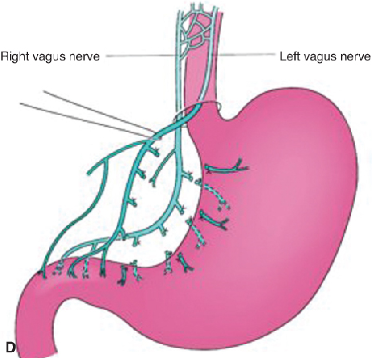
1. Truncal Vagotomy (TV)
- Heineke-Mikulicz pyloroplasty (most common): longitudinal incision across the pylorus closed transversely
- Jaboulay gastroduodenostomy: side-to-side anastomosis of stomach and duodenum
- Gastroenterostomy (GEJ): when duodenum is too scarred for pyloroplasty
2. Selective Vagotomy (SV)
3. Highly Selective Vagotomy (HSV) - also called Parietal Cell Vagotomy or Proximal Gastric Vagotomy
- Exposure and gastric mobilisation (gastrocolic omentum mobilisation)
- Dissection of the anterior leaf of the lesser omentum (dividing all branches from 6 cm proximal to the pylorus - the "crow's foot" - up to the cardia)
- Dissection of the posterior leaf of the lesser omentum
- Dissection of vagal fibres along the distal 3-5 cm of the oesophagus (including the "criminal nerve of Grassi")
4. Vagotomy and Antrectomy (TV/SV + Antrectomy)
- Billroth I reconstruction: gastroduodenostomy (preferred for benign disease)
- Billroth II reconstruction: gastrojejunostomy (used when duodenum is scarred)
- Roux-en-Y: avoids bile reflux
Outcomes Comparison Table
| Operation | Operative Mortality | Significant Side Effects | Recurrent Ulceration |
|---|---|---|---|
| Gastrectomy | 1-2% | 20-40% | 1-4% |
| Truncal vagotomy + drainage | <1% | 10-20% | 2-7% |
| Selective vagotomy + drainage | <1% | 10-20% | 5-10% |
| Highly selective vagotomy | <0.2% | <5% | 2-10% |
| Truncal vagotomy + antrectomy | 1% | 10-20% | 1% |
Complications of Vagotomy
A. Intraoperative / Early Complications
| Complication | Notes |
|---|---|
| Oesophageal perforation | Particularly during dissection of periesophageal fibres in HSV |
| Gastric perforation | Entry into mucosa during pyloromyotomy |
| Splenomegaly / splenic injury | Traction on gastrophrenic ligament |
| Bleeding | Injury to short gastric or left gastric vessels |
| Liver injury | Excessive retractor pressure on lateral segment |
B. Recurrent Ulceration
- Incomplete vagotomy - most common cause; the criminal nerve of Grassi is often missed
- Retained gastric antrum - residual G cells no longer exposed to acid, leading to hypergastrinaemia and marginal ulcer
- Persistent H. pylori infection
- NSAID use (surreptitious)
- Zollinger-Ellison syndrome (should have been excluded preoperatively)
C. Gastric Stasis / Gastroparesis
- Occurs when the drainage procedure fails to compensate for vagotomy-induced gastric motility disorder
- Patients retain food in the stomach for several hours
- Presents as nausea, bloating, early satiety, vomiting
- Diagnosis: Gastric emptying study (nuclear scintigraphy)
- Treatment: Prokinetics (metoclopramide, domperidone); dietary modification (small, frequent meals); rarely requires revision surgery
- More common after truncal vagotomy + drainage; rare after HSV
D. Dumping Syndrome
- Crampy abdominal pain, nausea, diarrhoea, belching
- Vasomotor symptoms: tachycardia, palpitations, diaphoresis, light-headedness, rarely syncope
- Mechanism: rapid emptying of hyperosmolar gastric contents into small bowel → fluid shift into gut lumen → plasma volume contraction → acute intestinal distension + release of vasoactive GI hormones (VIP, neurotensin, motilin)
- Predominantly vasomotor: light-headedness, diaphoresis, palpitations, syncope
- Mechanism: reactive hypoglycaemia from excessive insulin release triggered by rapid glucose absorption
- Dietary: small, multiple (6) meals; avoid simple carbohydrates and large fluid volumes with meals; avoid sucrose
- Antidiarrhoeals (loperamide, diphenoxylate), anticholinergics
- Guar/pectin (increase luminal viscosity)
- Acarbose (α-glucosidase inhibitor) for late dumping
- Octreotide 50 mcg SC TID (or long-acting depot monthly) for diet-refractory cases
- Surgical: Roux-en-Y conversion, reversal of jejunal segment (rarely needed)
E. Postvagotomy Diarrhoea
- Affects ~10% of patients seeking medical attention; most common after truncal vagotomy
- Intermittent, episodic diarrhoea 1-2 h after meals; rarely continuous and severe
- Mechanism: motility disorder from interruption of vagal fibres to the gut + increased bile acid excretion + decreased nutrient absorption + luminal secretagogue release
- Treatment:
- Loperamide or diphenoxylate
- Cholestyramine (bile salt binder) for severe cases
- Surgical: reversal of a 10-cm segment of jejunum can substantially improve bowel frequency in a subset of patients
F. Alkaline Reflux Gastritis (Bile Reflux Gastropathy)
- Reflux of bile into the gastric remnant
- Presents with epigastric pain, early satiety, nausea, and vomiting
- Endoscopy: mucosal erythema of gastric remnant; histology shows epithelial injury with minimal inflammation
- Treatment: Prokinetics, cholestyramine, sucralfate; severe cases may need Roux-en-Y conversion
G. Afferent Loop Syndrome (after Billroth II)
- Chronic (bacterial overgrowth): Stasis in afferent limb → bacterial overgrowth → postprandial abdominal pain, bloating, diarrhoea, malabsorption of fat and vitamin B12. Treat with antibiotics; refractory cases need surgical revision.
- Acute (complete obstruction): Bilious distension of afferent loop → severe epigastric pain, bilious vomiting → can progress to duodenal stump rupture (surgical emergency).
H. Retained Antrum Syndrome (after Billroth II)
- Incomplete antrectomy leaves residual G cells no longer exposed to gastric acid
- Leads to hypergastrinaemia → stimulation of acid production → marginal ulceration at the gastrojejunostomy
- Diagnosed by elevated fasting gastrin + scintigraphy
- Treatment: reoperation to excise retained antrum
I. Roux Stasis Syndrome
- After Roux-en-Y reconstruction, the Roux limb can develop abnormal motility
- Symptoms: postprandial pain, nausea, vomiting, weight loss
- Mechanism: disruption of normal intestinal pacemaker after jejunal transection + vagal denervation
J. Metabolic Complications
- Iron deficiency anaemia: Reduced gastric acid impairs iron absorption; also from bypassing duodenum (main iron absorption site)
- Vitamin B12 deficiency: Reduced intrinsic factor after gastric resection (more relevant with gastrectomy than vagotomy alone)
- Calcium malabsorption: Due to bypass of duodenum + reduced acid-mediated calcium solubilisation
- Osteoporosis / osteomalacia: Long-term risk in postgastrectomy patients
K. Gallstone Formation
- Truncal vagotomy denervates the gallbladder, causing reduced contractility and bile stasis
- Increased risk of cholecystitis and cholelithiasis after truncal vagotomy (not seen with HSV)
- Selective and highly selective vagotomies preserve hepatic branches and spare the gallbladder
L. Other Complications
- Small stomach syndrome: Sensation of early fullness and inability to eat large meals, particularly after gastrectomy combined with vagotomy
- Gastric remnant cancer: Long-term risk (>15 years) after partial gastrectomy with Billroth II; thought to be related to chronic bile reflux and gastritis
Laparoscopic Vagotomy
Key Clinical Points
- HSV has the lowest mortality (<0.2%) and fewest side effects (<5%) but highest recurrence (10-15%). It is the preferred elective procedure.
- Truncal vagotomy + antrectomy has the lowest recurrence (1%) but the highest side effect burden (20%).
- The "criminal nerve of Grassi" is the most common cause of incomplete vagotomy and must be sought during every HSV.
- Sham feeding test is the standard for diagnosing incomplete vagotomy in recurrent ulcer patients.
- With modern PPIs and H. pylori eradication, vagotomy is now almost exclusively reserved for emergency situations (bleeding, perforation, obstruction).
- Maingot's Abdominal Operations (Chapter 26, Vagotomy)
- Harrison's Principles of Internal Medicine, 22nd ed. (Chapter 335, PUD)
- Sabiston Textbook of Surgery, The Biological Basis of Modern Surgical Practice (Chapter 86)
- Bailey and Love's Short Practice of Surgery, 28th ed. (Chapter 67)
- Fischer's Mastery of Surgery, 8th ed. (Chapter 115)