
Classify goitres and management of primary thyrotoxicosis in detail according to standard ENT textbook and in simplified manner
classification of goitre thyroid enlargement diagram
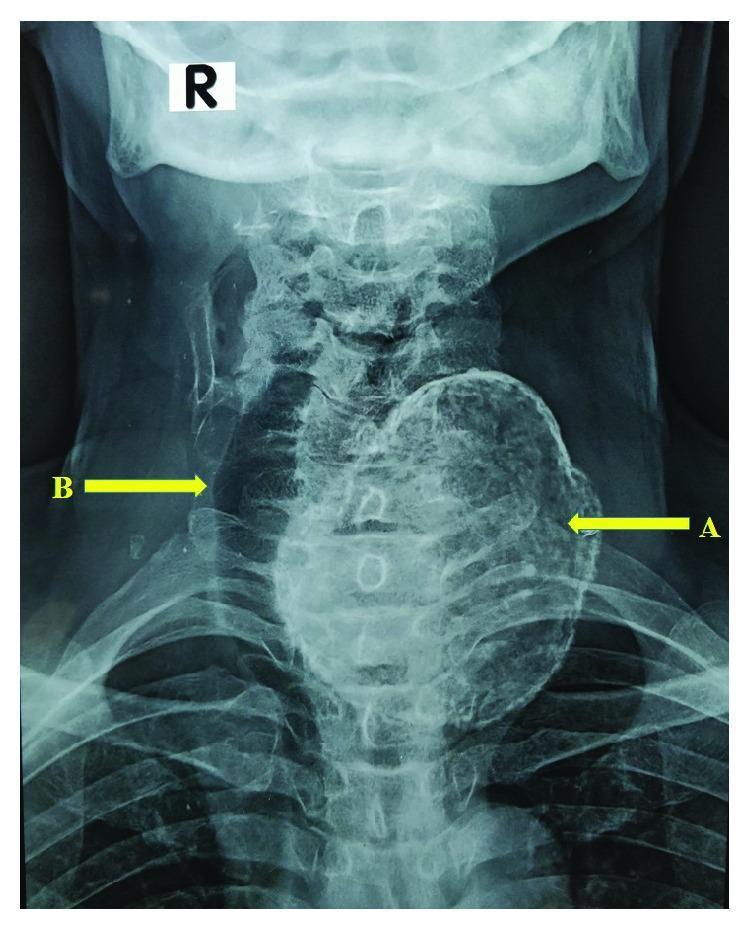
This anteroposterior (AP) neck X-ray demonstrates a diagnostic image of severe goitre with extensive dystrophic calcification. The primary finding is a large, dense, radio-opaque mass in the left cervical region, indicated by arrow A, which corresponds to a heavily calcified left thyroid lobe. The mass exhibits a characteristic 'eggshell' or coarse calcification pattern along its periphery. Significant mass effect is evident as the trachea, marked by arrow B, is markedly displaced and deviated toward the right side of the midline. The cervical vertebrae and upper thoracic ribs are visible in the background, providing anatomical context for the degree of tracheal shifting. This image illustrates the clinical consequences of long-standing multinodular goitre, specifically airway compression and deviation caused by calcified thyroid enlargement. It is an educational example for radiology and endocrinology students regarding the evaluation of cervical masses and their impact on adjacent midline structures.
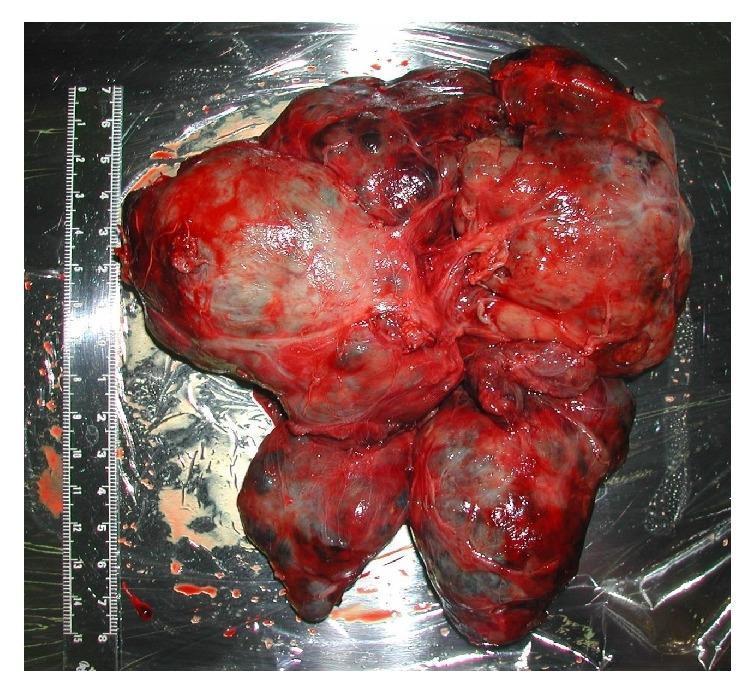
A clinical photograph of an extirpated gross surgical specimen, identified as a giant goitre (thyroid tumor) following total thyroidectomy. The specimen is a large, multi-nodular, and lobulated mass with an irregular morphology. It exhibits a predominantly dark red and fleshy appearance with variations in tissue color, suggesting heterogeneous vascularization and potentially areas of cystic change or hemorrhage. The surface is moist and glistening with evidence of adherent blood. A medical ruler placed alongside the specimen indicates its significant proportions, measuring approximately 15 cm in length and 13 cm in width, with a total weight of 515 g. This image demonstrates the massive enlargement of thyroid tissue capable of causing severe anatomical displacement, such as tracheal compression and glottic shift, which are critical considerations for anesthetic airway management. The educational focus is on gross pathology of benign thyroid disease and its surgical implications.
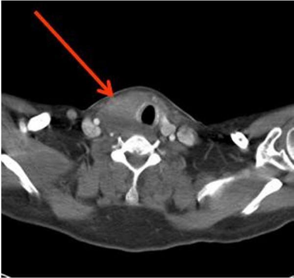
This diagnostic axial contrast-enhanced CT scan of the neck and thoracic outlet demonstrates a significant, asymmetric enlargement of the thyroid gland, indicated by a red arrow. The large thyroid mass predominantly involves the right lobe and exhibits heterogeneous enhancement. It exerts a clear mass effect, resulting in marked lateral deviation of the air-filled trachea toward the left side. Surrounding cervical anatomy is visible, including the cervical vertebrae and major vascular structures such as the carotid arteries and internal jugular veins, which appear in close proximity to the infiltrative mass. The image illustrates the clinical presentation of an invasive cervical process, consistent with pathology such as Riedel's thyroiditis or anaplastic carcinoma. This imaging modality is used to evaluate goitre progression, airway patency, and the involvement of adjacent neurovascular structures in the neck.
Graves disease exophthalmos thyrotoxicosis clinical photo
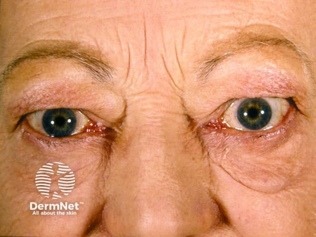
Imaging modality and technique: Clinical frontal photograph of the orbits and periorbital region in a living adult patient. Frontal view capturing both eyes demonstrates bilateral exophthalmos with mild periorbital edema and eyelid swelling. The ocular surfaces are visible with subtle scleral exposure and conjunctival vascularity not markedly prominent in this image. Anatomical localization centers on the orbital contents including extraocular muscles and orbital fat, with involvement of eyelids and periorbital soft tissues. The appearance is most consistent with thyroid ophthalmopathy (Graves’ orbitopathy), a form of autoimmune orbital inflammatory disease associated with hyperthyroidism or autoimmune thyroiditis. Pathophysiology involves infiltration and edema of orbital connective tissues and extraocular muscles, leading to proptosis, lid retraction, diplopia, and possible optic neuropathy in advanced cases. Pathognomonic imaging features in other modalities include enlargement of the rectus muscles, particularly the inferior and medial recti, with sparing of tendons; however this clinical image illustrates recognizable exophthalmos and periorbital edema rather than radiologic details. This image is clinically relevant for education, differential diagnosis, and patient counseling, as well as for correlating with thyroid function testing and orbital imaging when planning management such as immunotherapy, selenium supplementation, or orbital decompression in refractory cases. This photo supports patient diagnosis and monitoring in endocrinology, ophthalmology, and rheumatology contexts.
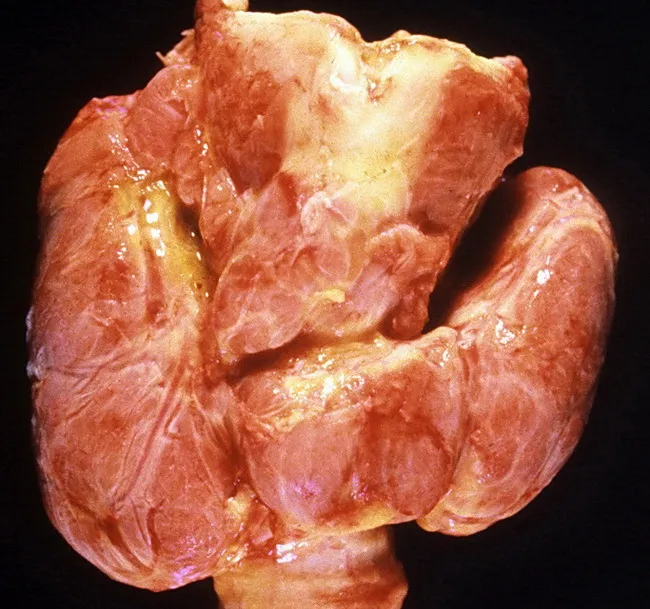
Imaging modality and technique: Gross autopsy photograph of the thyroid gland. The view is a frontal, macroscopic examination of the neck showing diffuse enlargement of both lobes with smooth surfaces and no discrete nodules. The tissue is pink to tan with subtle lobular architecture preserved. This gross appearance corresponds to diffuse hyperplasia and hypertrophy of thyroid follicles seen in Graves disease, producing a diffusely enlarged, symmetric goiter. There are no focal masses or inflammatory lesions evident in this specimen. Clinically, Graves disease is an autoimmune thyrotoxicosis characterized by endogenous hyperthyroidism, ophthalmopathy (exophthalmos), and dermopathy; the autopsy image reflects the thyroid component of that syndrome. Pathophysiology involves stimulation by thyroid-stimulating immunoglobulins that drive diffuse follicular hyperplasia and increased thyroid hormone synthesis. The gross findings support laboratory and clinical data such as suppressed TSH and elevated free T4/T3. Epidemiology notes peak incidence in females aged 20–40 years, with female-to-male ratio ≈10:1. This photograph is valuable for teaching gross pathology, correlating macroscopic thyroid changes with endocrinologic manifestations, and differentiating Graves disease from nodular goiters or thyroiditis in educational and research settings. It also highlights the importance of autopsy correlations in endocrine pathology. Educational utility.
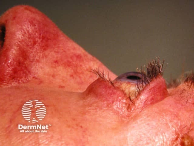
This clinical photograph is a high-resolution, lateral oblique view of the periocular region focusing on the right eye (periorbital skin). The image demonstrates marked periorbital edema with pronounced erythema of the eyelids and surrounding cutaneous tissue. The upper and lower eyelids appear swollen and thickened, with subtle skin redundancy. In thyroid-associated ophthalmopathy (Graves' orbitopathy), this presentation reflects inflammatory edema of orbital tissues, increased adipose and extraocular muscle volume, and skin involvement. The visible periorbital edema may precede or accompany exophthalmos, lid retraction, and diplopia; however, soft tissue contour is partially obscured by lighting. The photo lacks palpation data but serves as a qualitative depiction of orbital involvement. Clinically, these features require correlation with systemic thyroid status: hyperthyroidism or euthyroid Graves' disease; presence of TSH receptor autoantibodies; risk of progressive-eye involvement. This image supports differential diagnosis including periorbital cellulitis and dermatitis, but the chronic inflammatory pattern aligns with thyroid eye disease. For diagnostic significance, the image is valuable for medical education, patient counseling, and multidisciplinary planning—endocrinology, ophthalmology, and dermatology collaboration. Potential clinical use cases include screening for orbital involvement in Graves' disease, documenting disease progression, and teaching residents about periorbital edema etiologies and differential diagnoses.
Classification of Goitres and Management of Primary Thyrotoxicosis
PART 1 - WHAT IS A GOITRE?
- Isolated (Solitary) swelling: Discrete nodule in an otherwise impalpable gland
- Dominant swelling: Discrete nodule in a gland with clinical evidence of generalised abnormality elsewhere
PART 2 - CLASSIFICATION OF GOITRES
Standard Classification (Bailey & Love, Table 55.3)
| Category | Type | Subtypes |
|---|---|---|
| Simple Goitre (Euthyroid) | Diffuse Hyperplastic | Physiological, Pubertal, Pregnancy |
| Multinodular Goitre | - | |
| Toxic Goitre | Diffuse Toxic | Graves' disease |
| Toxic Multinodular | - | |
| Toxic Adenoma | Plummer's disease | |
| Neoplastic | Benign | Follicular adenoma |
| Malignant | Papillary, Follicular, Medullary, Anaplastic | |
| Inflammatory | Autoimmune | Chronic lymphocytic thyroiditis, Hashimoto's disease |
| Granulomatous | de Quervain's thyroiditis | |
| Fibrosing | Riedel's thyroiditis | |
| Infective | Acute (bacterial/viral), Chronic (TB, syphilitic) | |
| Other | Amyloid |
Simplified Memory Aid (by Function)
GOITRE
  │
  ├── EUTHYROID (normal thyroid function)
  │       ├── Diffuse Hyperplastic (simple) → physiological/pubertal/pregnancy
  │       └── Multinodular (colloid degeneration, cysts, adenomas)
  │
  ├── TOXIC (overactive thyroid)
  │       ├── Diffuse Toxic = GRAVES' DISEASE (PRIMARY thyrotoxicosis)
  │       ├── Toxic Multinodular Goitre (SECONDARY thyrotoxicosis)
  │       └── Toxic Adenoma / Plummer's Disease (SECONDARY thyrotoxicosis)
  │
  ├── NEOPLASTIC
  │       ├── Benign (follicular adenoma)
  │       └── Malignant (papillary, follicular, medullary, anaplastic)
  │
  └── INFLAMMATORY
          ├── Hashimoto's (autoimmune)
          ├── de Quervain's (granulomatous/viral)
          └── Riedel's (fibrosing)
A. Simple Goitre (Euthyroid)
- TSH stimulation of the thyroid (due to low circulating T3/T4)
- Most common cause: iodine deficiency (endemic in mountainous regions - Alps, Himalayas, Andes, Rockies; also Derbyshire/Yorkshire in UK)
- Daily iodine requirement: 0.1-0.15 mg/day
- Dyshormonogenesis: enzyme defects in thyroid hormone synthesis (sporadic goitre; often familial)
- Other growth factors (immunoglobulins) also stimulate follicular cell proliferation
- Seen physiologically in puberty and pregnancy
- Due to increased demand for thyroid hormone
- Results from heterogeneous structural/functional response - clones of cells with varying sensitivity to growth stimulation create nodularity
- Long natural history; may remain euthyroid for years
B. Toxic Goitre (Overactive)
Diffuse Toxic Goitre = Graves' Disease (PRIMARY thyrotoxicosis)
- Young women (F:M > 5:1), peak incidence in 20s-30s
- Autoimmune: circulating IgG autoantibodies (TSHR-Ab) against TSH receptor - activate cAMP, mimic TSH effect, stimulate thyroid hormone synthesis
- Associated with ophthalmopathy, dermopathy (pretibial myxoedema), acropachy
- 55% have family history of autoimmune endocrine disease
- Goitre is diffuse, symmetrical; may have a palpable thrill and audible bruit (increased blood flow)
- Smoking almost doubles the risk; worsens ophthalmopathy
Toxic Multinodular Goitre (SECONDARY thyrotoxicosis)
- Older patients (elderly); pre-existing nodular goitre present for years before toxicity develops
- Two or more autonomously functioning nodules secrete excess thyroid hormone
- May be precipitated by pharmacological iodine (contrast media) via Jod-Basedow effect
- Hyperfunction less marked than Graves' disease
- Cardiovascular symptoms predominate: palpitations, AF, tachyarrhythmias
- May have compressive symptoms: dysphagia, dyspnoea (worsens at night)
Toxic Adenoma / Plummer's Disease (SECONDARY thyrotoxicosis)
- Single autonomous nodule secreting excess thyroid hormone
- Usually grows to at least 3 cm before causing overt hyperthyroidism
- TSH receptor gene mutation in 20-83% of cases (somatic mutations in transmembrane domain)
- Accounts for ~5% of thyrotoxicosis cases
- Surrounding normal thyroid is suppressed
- Radionuclide scan: single "hot spot" with no other visible uptake
- No eye signs (unlike Graves' disease)
Retrosternal Goitre
- Arises from slow growth of MNG down into the mediastinum
- Causes: tracheal compression, dysphagia, airway symptoms
-
95% can be removed transcervically
- Requires cross-sectional imaging (CT) preoperatively
- Sternotomy needed if: malignant disease, posterior mediastinal extension, diameter > thoracic inlet
PART 3 - PRIMARY THYROTOXICOSIS (GRAVES' DISEASE) - MANAGEMENT IN DETAIL
Step 1 - Symptom Control (Immediate)
- Beta-blockers (propranolol 40 mg TDS or nadolol 320 mg OD): target-organ blockade
- Propranolol also inhibits peripheral conversion of T4 → T3
- Rapid clinical response; patient can be "clinically euthyroid" within days
- Does NOT reduce thyroid hormone synthesis
- Used as bridge until definitive treatment takes effect
Step 2 - Three Modalities of Definitive Treatment
| Feature | Antithyroid Drugs | Radioiodine (¹³¹I) | Surgery |
|---|---|---|---|
| Graves' disease | ✓ | ✓ | ✓ |
| Toxic nodular disease | - | ✓ | ✓ |
| Pregnancy | ✓ | ✗ | ✓ |
| Children | ✓ | ✗ | ✓ |
| Significant ophthalmopathy | ✓ | ✗ | ✓ |
| Definitive treatment | - | ✓ | ✓ |
| Non-invasive | ✓ | ✓ | - |
| Low cost | ✓ | ✓ | - |
| Risk of hypothyroidism | Low | High | High |
| Worsens ophthalmopathy | - | ✓ (risk) | - |
| Histological diagnosis | - | - | ✓ |
A. Antithyroid Drugs (Thionamides)
- Block synthesis of T4 and T3 by inhibiting thyroperoxidase enzyme
- Disrupt organification and oxidation of iodide
- PTU additionally inhibits peripheral T4 → T3 conversion (useful in thyroid storm)
- Carbimazole 30-40 mg/day initially
- Once euthyroid (after 8-12 weeks), reduce to 5 mg 8-hourly
- OR use "Block and Replace" regime: continue high-dose carbimazole (to block T3/T4) + add thyroxine 0.1-0.15 mg/day to prevent hypothyroidism
- Mild cases: 6 months
- Severe cases: up to 2 years
- ~30% achieve lasting remission after antithyroid drug course
- Failure rate is at least 55% (recurrence on stopping drugs)
- Antithyroid drugs cannot cure a toxic adenoma - the tissue is autonomous, and recurrence is certain on stopping.
B. Radioiodine (¹³¹I)
- Europe: Antithyroid drugs first (under 40 years); radioiodine for relapse
- USA: Radioiodine is first-line in Graves' disease and toxic MNG
- No surgery, no prolonged drug therapy
- Outpatient procedure
- Cost-effective
- Radiation thyroiditis risk (transient worsening)
- Patient must be quarantined while radiation levels are high
- Must defer pregnancy (typically for 6 months after treatment)
- Avoid close physical contact, especially with children
- Permanent hypothyroidism is the expected outcome (not a complication - acceptable trade-off)
- May worsen ophthalmopathy - therefore NOT used in significant Graves' ophthalmopathy
C. Surgery
Indications for Surgery in Primary Thyrotoxicosis:
- Large goitre (especially with compressive symptoms)
- Progressive eye signs (ophthalmopathy worsening)
- Patient refuses radioiodine
- Pregnancy (second trimester only)
- Relapse after antithyroid drugs / radioiodine failure
- Suspected malignancy
- Patients who prefer definitive cure without radiation
Surgical Options (Table 55.5 - Bailey & Love):
| Parameter | Total Thyroidectomy | Subtotal Thyroidectomy |
|---|---|---|
| Control of toxicity | Immediate | Immediate |
| Return to euthyroid | Immediate | Variable (up to 12 months) |
| Risk of recurrence | None | Up to 5% (lifelong) |
| Risk of thyroid failure | 100% | Up to 100% at 30 years |
| Permanent hypoparathyroidism | 5% | 1% |
| Follow-up required | Minimal | Lifelong |
Preoperative Preparation (MANDATORY before surgery):
- Carbimazole 30-40 mg/day; take 8-12 weeks to achieve euthyroid state
- Last dose given on evening before surgery
- Propranolol 40 mg TDS (up to 80 mg TDS or nadolol 320 mg OD if needed)
- Acts on target organs (not on the gland itself)
- Can achieve clinical euthyroid state within days - allows surgery within days rather than weeks
- Continue until 7-10 days post-operatively (to prevent rebound sympathetic surge)
- Given preoperatively in some centres to reduce vascularity of the gland
- Given for 10 days pre-surgery
- Cannot be used alone (not a treatment, only an adjunct)
Special Situation: Thyroid Eye Disease in Graves'
- Mild-to-moderate ophthalmopathy: often needs no treatment; may improve spontaneously
- Local discomfort: artificial tears, elevate head of bed (to reduce oedema)
- Severe ophthalmopathy: multidisciplinary team management with ophthalmologists
- High-dose glucocorticoids ± other immunosuppressants
- Orbital radiotherapy
- Rituximab (anti-CD20, depletes B-cells) - evidence emerging
- Surgical orbital decompression for compressive optic neuropathy
- Rapid deterioration in visual acuity
- Ocular nerve palsies
- Unacceptable cosmetic appearance
- Visual acuity < 6/18
- Decreased colour vision
- Corneal ulceration
PART 4 - QUICK SUMMARY TABLE (Primary vs Secondary Thyrotoxicosis)
| Feature | Primary (Graves') | Secondary (Toxic MNG / Adenoma) |
|---|---|---|
| Cause | Autoimmune (TSHR-Ab) | Autonomous nodule(s) |
| Age | Young women (20s-30s) | Middle-aged / elderly |
| Goitre type | Diffuse, smooth, vascular | Nodular |
| Eye signs | Present (50%) | Absent |
| Onset of goitre | Simultaneous with toxicity | Goitre long before toxicity |
| Bruit/thrill | Present | Absent |
| Severity | More severe | Less severe (especially in MNG) |
| Predominant symptoms | Full thyrotoxic picture | Cardiovascular (AF, palpitations) |
| Radioiodine | Used (caution with eye disease) | Preferred definitive treatment |
| Surgery | After antithyroid drugs / relapse | Often first-line for large MNG |
- Scott-Brown's Otorhinolaryngology Head & Neck Surgery, Vol 1 - Chapter 61 (Thyroid disorders), Table 61.5, Box 61.4
- Bailey & Love's Short Practice of Surgery, 28th Ed. - Chapter 55, Table 55.3, Table 55.5
Solitary thyroid nodule, Robotic assisted thyroid surgery, minimally invasive thyroid surgery in detail according to standard ENT textbook
robotic thyroid surgery transaxillary da Vinci
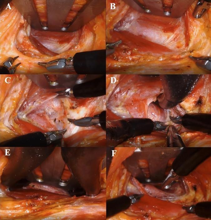
This composite of six intraoperative clinical photographs (A–F) demonstrates a 'two-step retraction' operative procedure for robotic transaxillary thyroidectomy using the da Vinci SP system. A-B: The initial stages show the surgical console view following the first docking, where a narrow skin flap is created and widened using a specialized Chung’s retractor and robotic graspers. C: Robotic instruments are seen dissecting and creating a working space between the sternal and clavicular heads of the sternocleidomastoid (SCM) muscle. D: Deeper dissection reveals the thyroid gland situated beneath the strap muscles. E-F: These frames illustrate the 'second docking' or redocking phase, where the retractor is repositioned deeper (beneath the strap muscles) to optimize the spherical working space for the robotic arms. The series highlights the progression from superficial flap dissection to deep anatomical exposure of the thyroid, emphasizing the minimally invasive approach through a single axillary incision and the specialized manipulation of neck musculature to achieve surgical access.
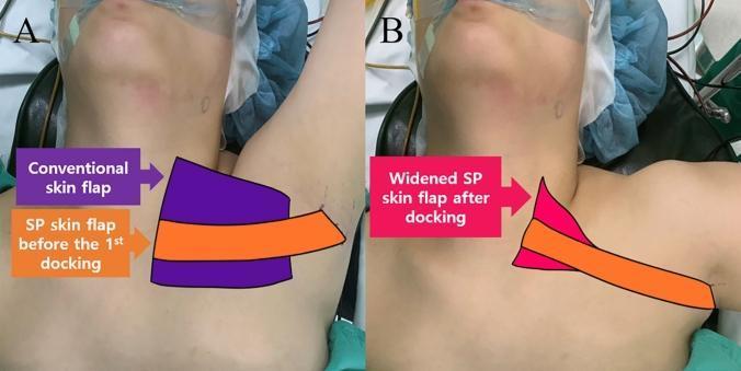
This clinical photograph set (Figures 4A and 4B) illustrates a comparison of skin flap dissection areas for robotic transaxillary thyroidectomy. The images show a patient in a supine position with surgical markings. In Figure 4A, a 'conventional skin flap' (purple) is depicted as a large, trapezoidal area extending from the axilla across the upper chest toward the sternal notch and lower neck. Overlaid on this is the 'SP (Single Port) skin flap before the 1st docking' (orange), which is significantly narrower and confined to a direct path from the axilla to the midline. Figure 4B demonstrates the 'widened SP skin flap after docking' (pink), showing a triangular expansion toward the superior neck and thyroid region compared to the initial orange tract. The series highlights the technical advancement of the da Vinci SP system, which allows for a more targeted, minimally invasive dissection compared to traditional robotic thyroidectomy approaches. The anatomical focus is the pectoralis major surface, sternocleidomastoid muscle, and the surgical access pathway from the axilla to the thyroid gland.
thyroid nodule ultrasound fine needle aspiration cytology
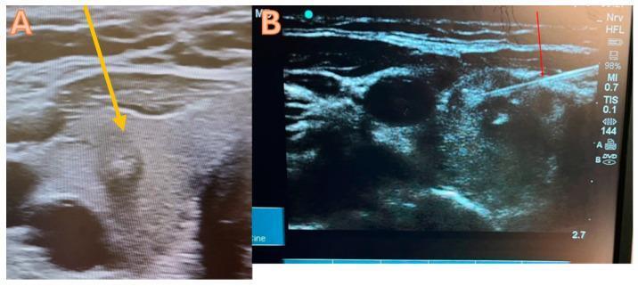
This dual-panel diagnostic image displays high-resolution ultrasound views of the thyroid gland, illustrating the procedure for ultrasound-guided Fine-Needle Aspiration Cytology (FNAC). Panel A shows a transverse ultrasound view of the right thyroid lobe containing a suspicious nodule marked with a yellow arrow. The nodule is roughly circular with heterogeneous echogenicity and irregular internal margins, categorized clinically as TI-RADS 4, indicating a moderate suspicion of malignancy. Panel B demonstrates the procedural phase, where a 21-gauge biopsy needle (red arrow) is visualized as a linear hyperechoic structure. The needle is shown utilizing a trans-isthmic approach at a shallow angle to accurately target the center of the hypoechoic nodule. These images serve as an educational resource for interventional endocrinology and radiology, highlighting the importance of real-time needle visualization during biopsy to ensure diagnostic accuracy and safety in managing potentially malignant thyroid lesions.
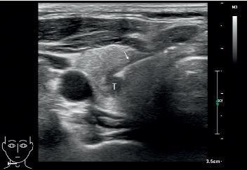
This diagnostic ultrasound image in the transverse plane demonstrates a Fine Needle Aspiration Biopsy (FNAB) of a thyroid nodule. The nodule, labeled 'T', presents as a well-defined structure with heterogeneous echogenicity, appearing predominantly hypoechoic compared to the surrounding isoechoic thyroid parenchyma. A biopsy needle, indicated by a white arrow, is visualized as a highly echogenic (bright) linear structure. The needle is shown using an in-plane approach, where it is positioned parallel to the long axis of the ultrasound probe, allowing for continuous visualization of the needle shaft and tip as it enters the nodule. In the lateral aspect of the thyroid lobe, a prominent anechoic (black) circular structure represents a major neck vessel, likely the carotid artery or internal jugular vein. Superficial to the thyroid, the strap muscles are visible as stratified, hypoechoic bands. This image serves as a clinical example of real-time ultrasound guidance to ensure precise needle placement and adequate tissue sampling in the evaluation of thyroid pathology.
Solitary Thyroid Nodule, Robotic-Assisted Thyroid Surgery, and Minimally Invasive Thyroid Surgery
PART 1 - SOLITARY THYROID NODULE (STN)
Definition and Epidemiology
- Palpable in 3-4% of the adult population in the UK and USA
- Three to four times more frequent in women than men
- ~70% of discrete swellings are clinically isolated; ~30% are dominant
- Ultrasonography and post-mortem studies detect far more nodules than clinical palpation (nodule prevalence rises with age)
Importance - Risk of Malignancy
| Type of Swelling | Risk of Malignancy | Risk of Follicular Adenoma |
|---|---|---|
| Truly isolated nodule | 15% | 30-40% |
| Dominant nodule | ~7-8% (half of isolated) | Still substantial |
| Remainder | Non-neoplastic (colloid, cysts, thyroiditis) | - |
Clinical Assessment - History and Examination
- Young age (especially children) or elderly males
- History of radiation exposure to head/neck (especially in childhood)
- Rapid nodule growth
- Dysphagia, dysphonia, dyspnoea (upper aerodigestive tract symptoms)
- Fixation of the nodule (tethered to surrounding structures)
- Hard, irregular, non-tender consistency
- Cervical lymphadenopathy
- Family history of medullary thyroid cancer or MEN-2
- Smooth, soft, mobile nodule
- Tenderness (suggests thyroiditis or haemorrhage into cyst)
- Long-standing, slowly growing lesion
Investigation of Solitary Thyroid Nodule
Step 1 - Thyroid Function Tests (TSH)
- TSH is the initial screening test
- Hypothyroidism/hyperthyroidism shifts workup away from malignancy toward functional disorders
- Suppressed TSH - radionuclide scan to rule out toxic hot nodule, Marine-Lenhart syndrome, or Graves' disease with concurrent cold nodule
- Elevated TSH - treat hypothyroidism, then perform FNAC
- Normal TSH (most patients) - proceed to FNAC
Step 2 - Autoantibody Titres
- Raised anti-thyroid peroxidase (anti-TPO) and anti-thyroglobulin antibodies suggest Hashimoto's thyroiditis
Step 3 - Fine-Needle Aspiration Cytology (FNAC)
- Reduced the number of patients requiring surgery by 35-75%
- Almost tripled the yield of malignancies in patients going to surgery
- Accuracy for papillary carcinoma: 99% with a false-positive rate of <1%
| Grade | Meaning | Malignancy Risk | Action |
|---|---|---|---|
| Thy 1 | Non-diagnostic | - | Repeat FNAC |
| Thy 1c | Non-diagnostic (cystic) | - | Repeat/US-guided FNAC |
| Thy 2 | Non-neoplastic / Benign | 1-6% (false negative) | Observe; surgery only for symptoms/growth |
| Thy 3 | Follicular / Indeterminate | ~20% | Surgery (diagnostic hemithyroidectomy) |
| Thy 4 | Suspicious of malignancy | - | Surgery |
| Thy 5 | Malignant | - | Surgery |
- ~15% of aspirates are non-diagnostic (from cystic, haemorrhagic, hypervascular or hypocellular colloid nodules) - repeat FNAC is mandatory; a non-diagnostic result must NEVER be interpreted as negative for cancer
- Surgical diagnoses after repeated non-diagnostic aspirations revealed malignancy in 4% of women and 29% of men
Step 4 - Ultrasonography
- Most valuable initial imaging; non-invasive and inexpensive
- Differentiates cystic from solid nodules
- Detects non-palpable nodules
- Identifies malignant US features: solid hypoechoic appearance, ill-defined margins, taller-than-wide shape, microcalcifications - all suggest papillary thyroid cancer (NPV 97%)
- Well-defined hyperechoic nodules with peripheral halo = likely follicular lesion
- Abnormal neck nodes (cystic or with microcalcification) - raise suspicion for thyroid cancer
- Guides FNA of non-palpable nodules and improves yield
Step 5 - Isotope Scanning (Radionuclide Scan)
- Indicated when TSH is suppressed (to localise hyperfunction)
- Hot nodule = functioning autonomously - low malignancy risk
- Cold nodule = does not take up isotope - higher malignancy risk; if FNAC shows follicular neoplasm + cold nodule on scan, surgery is indicated
- Replaced to a large extent by USS in routine evaluation
Step 6 - Molecular Marker Testing (for Indeterminate FNAC)
- Used for Thy3/indeterminate cytology
- Afirma Gene Expression Classifier: sensitivity 92%, NPV 93% (excellent rule-out test)
- ThyroSeq: good rule-in test (mutations in BRAF, RAS, RET/PTC, etc.)
- ThyGenX/ThyraMIR and Rosetta GX Reveal: less published data
Management Algorithm (Cummings Chapter 122)
Thyroid Nodule Discovered
         │
         ▼
History + Physical Exam
(High-risk features → Consider direct surgery)
         │
         ▼
TSH Level
 ┌───────┴───────────────────────┐
 │                               │
Suppressed TSH               Normal/High TSH
     │                           │
Radionuclide scan            FNAC ± Ultrasound
     │                           │
 Hot nodule = treat        ┌─────┴──────────────────────┐
 toxicity                  │                            │
 Cold nodule = FNAC    Benign (Thy2)           Follicular/Susp/Malignant
                           │                    (Thy3/4/5)
                        Observe                    Surgery
                 (surgery if symptoms/growth)
PART 2 - MINIMALLY INVASIVE THYROID SURGERY (MIVAT/MIT)
Introduction and Rationale
- Kocher's principles of thyroid surgery (early 20th century) remained unchanged for decades
- 71% of patients surveyed in a thyroid clinic would prefer not to have a neck scar (independent of sex and skin colour)
- Professionals consistently rate the aesthetics of a thyroid scar more favourably than patients themselves do
- Technological advances in endoscopes and energy-sealing devices enabled small-incision techniques
Types of Minimally Invasive Thyroid Surgery
- MIVAT (Minimally Invasive Video-Assisted Thyroidectomy) = Miccoli technique
- MIT (Minimally Invasive Thyroidectomy)
- EAT (Endoscopic-Assisted Thyroidectomy)
- Henry technique (insufflation method)
A. Miccoli Technique (MIVAT)
- 2-3 cm midline incision facilitates endoscopic dissection of the superior pole
- Superior pole delivered into the neck via the incision
- Lower pole dissection and completion thyroidectomy done conventionally once gland is delivered
- Uses a 30-degree 4 mm endoscope
- Requires specialised instruments: suction dissectors, spatulas, specially designed retractors
- Needs two competent assistants and video stacks
- Excellent visualization of recurrent laryngeal nerve, external laryngeal nerve, and parathyroids
- No insufflation required (avoids CO2 complications)
- Can perform total thyroidectomy through a single incision
- Superior cosmetic outcome (meta-analysis evidence)
- Solitary nodules up to 3 cm
- Total thyroid lobe volume ≤ 20 mL (equating to 14 mL nodule volume)
- Extended by experienced surgeons to: selected Graves' disease, selected thyroid cancer patients
- History of thyroiditis
- Cancer (except by highly skilled surgeons)
- Previous surgery (adhesions)
- Nodules > 3 cm
- Requires >50 cases before expert status is reached
- Only applicable to ~8% of a UK surgeon's thyroid practice
- 86% of BAETS (British Association of Endocrine and Thyroid Surgeons) members perform fewer than 50 thyroidectomies per year - explaining why few centres in the UK have adopted this
- Meta-analysis (Radford, 2011): 318 patients, complications no higher, superior cosmetic outcomes, at the expense of operative time. Scar cosmesis studied only in early post-operative period.
B. Henry Technique (Insufflation Method)
- Two 3 mm ports for instrumentation
- One 1 cm port for the insufflator endoscope
- Dissection occurs completely within the neck
- Delivery of the thyroid lobe through the endoscope port on completion
- Requires an assistant to hold and manipulate the endoscope
- Total thyroidectomy is NOT possible by this method
- Technically much more challenging than Miccoli method
- Better suited to parathyroidectomy than thyroidectomy
- CO2 insufflation risk: hypercarbia (potentially serious complication)
- Not widely adopted for thyroid surgery
- Long-term scar satisfaction comparison with conventional thyroidectomy shows little or no difference at 18 months after full healing (personal communication, JFH)
C. Scarless-in-the-Neck Techniques
- Transaxillary approach (incision in axilla)
- ABBA - Axillo-Bilateral-Breast Approach
- BABA - Bilateral Axillo-Breast Approach
- Facelift approach (incision behind the ear)
PART 3 - ROBOTIC-ASSISTED THYROIDECTOMY (RAT)
Historical Background and Rationale
- Da Vinci telerobot introduced in the late 1990s
- Originally conceived by NASA (remote surgery for astronauts) and the Ministry of Defence (bringing surgical expertise to the front line without endangering the surgeon)
- Patents acquired by Intuitive Surgical, Sunnyvale, California
- Feasibility of transatlantic surgery demonstrated by Marescaux et al. (2001)
- First lobectomy using da Vinci with insufflation: Lobe et al. (2005)
- Current transaxillary technique pioneered by Chung and colleagues, Yonsei University, Seoul - by 2013 they had experience with >3,000 patients
The Da Vinci Robotic System
- Surgeon Console - surgeon sits here remotely, controls all movements (master-slave system)
- Surgical Cart - docked with the patient; has four arms:
- One holding the stereoscopic 3D endoscope (8 mm or 12 mm)
- Three arms holding surgical instruments (right hand, left hand, assistant/retractor arm)
- Video Stack - containing camera control units (CCDs) and hardware
- 3D stereoscopic vision (two endoscopes within the casing replicate true 3D - gives depth of field)
- Magnified view
- Endoscope can be moved to alter magnification and avoid line-of-sight conflicts
- 7 degrees of freedom for instrument movement (wristed instruments that replicate hand and wrist movement)
- Tremor filtration (smooths out natural hand tremor)
- Surgeon is seated in an ergonomic console
Indications for RAT (Transaxillary Approach)
- Thin, narrow-chested female (body habitus important)
- Thyroid nodule up to 4 cm (hemithyroidectomy); nodules up to 6 cm can be handled by experienced surgeons
- Total thyroidectomy provided the contralateral lobe is "near normal"
- Patients with history of hypertrophic scarring or keloid tendency (valid reason to avoid neck scar)
- Personal preference to avoid neck scar
- Microcarcinoma in young patients (used cautiously)
- BMI < 30
Contraindications to RAT
- Obesity (BMI > 30)
- Degenerative shoulder pathology
- ASA category > 2 (significant comorbidity)
- Cancer (other than microcarcinoma in the young)
- Graves' disease
- Large cystic nodules > 6 cm (though aspiration at time of surgery can make some suitable)
The RAT Operation - Step-by-Step
Pre-operative Preparation
- No lines, BP cuffs, or ECG leads on the arm of the operative side
- Laryngeal nerve monitoring used routinely
- Mark the axillary incision (5-6 cm) while the patient is awake with the back of the hand touching the central forehead ("extended salute" position) - the only technique that identifies the best incision site
- Infiltrate incision site with 2% Xylocaine + 1:200,000 adrenaline
Patient Positioning
- Supine, head on pillow (NOT on shoulder roll - avoids overextending neck and moving superior pole away from robotic instruments)
- "Sniffing the morning air" position (subtle sniff position)
- Head of table dropped by ~20 degrees to widen angle between arm and chest
- Arm abducted and flexed with forearm pronated, back of hand resting on central portion of forehead - elevates and externally rotates the clavicle, shortening the distance from the incision to the thyroid gland
Cart Positioning (Docking)
- Cart placed opposite side of the operating table from the incision
- Docked at right angles to the operating table
- All four arms can be placed through a single axillary incision, OR the fourth arm through a periareolar incision (scar invisible once healed)
Preparation of Robotic Field (Flap Dissection)
- Medially in subfascial and subcutaneous planes above pectoralis major and over the clavicle
- Natural dehiscence between sternal and clavicular heads of sternomastoid is entered
- Access the plane of strap muscles
- Under the strap muscles to access the thyroid gland
- Sternothyroid muscle lower insertion into manubrium/first rib is divided, as is its superior attachment to thyroid cartilage - gives full access to superior pole
- Omohyoid muscle routinely divided as it extends over the internal jugular vein
- Strap muscles separated off the thyroid gland to ~1/3 of contralateral lobe
- Modified retractor (Chung, Kuppersmith, Modena, or Imperial) inserted - then robot docked
Robotic Dissection
- 30-degree down 12 mm stereoscopic endoscope placed at 220 degrees, inserted low laterally, extending high and upward medially toward the thyroid
- Fourth arm (Prograsp 8 mm) - assistant/retractor role
- First and third arms carry: Maryland forceps, Debakey forceps, Harmonic shears
- Identify RLN and parathyroids first using Maryland and Debakey forceps
- Once clearly defined, swap Debakey for Harmonic shears for haemostasis and dissection
Safety and Complications of RAT
- Cord palsy and hypocalcaemia rates no different from conventional thyroidectomy
- However: low degree of freedom, high heterogeneity, heavy weighting from Korean pioneering units, publication bias (complications of RAT tend not to get published)
- Brachial plexus dysfunction (from arm positioning) - unique to RAT, absent in conventional thyroidectomy
- Note: Chung's original method had arm fully extended over the head - this puts the brachial plexus at risk; Tolley modified this with the "hand on forehead" position
- Counterintuitively, less of a concern in RAT than conventional thyroidectomy - because the potential space for a haematoma to disseminate is far larger (the subfascial chest wall dissection space), so it cannot compress the airway as in conventional neck surgery
Why RAT is Less Common in the West vs. Korea
- Thyroid screening programmes - detecting small volume pathology early
- Reimbursement tariff for robotic vs. conventional surgery = 5:1 differential (economic incentive)
- National psyche for avoiding neck scars (feng shui cultural belief)
Evidence Base Summary - RAT
- Further prospective cohort studies are required before general conclusions can be made
- Currently considered an evaluation phase technology for thyroid surgery
- Mean tumour size excised in Korean series: 8 mm (microcarcinoma category) - caution extrapolating safety data to larger pathology
- RAT is safe for hemithyroidectomy, lobectomy, total thyroidectomy, and level VI central compartment dissection; increasingly extended to lateral compartment neck dissection through the axillary approach
QUICK COMPARISON TABLE
| Feature | MIVAT (Miccoli) | Henry Technique | RAT (Transaxillary) |
|---|---|---|---|
| Incision site | Neck (2-3 cm midline) | Neck (lateral ports) | Axilla (5-6 cm) |
| Neck scar | Yes (smaller) | Yes (port scars) | No neck scar |
| Gas insufflation | No (gasless) | Yes (CO2, 8 mmHg) | No (gasless) |
| Max nodule size | 3 cm | 3 cm | 4-6 cm |
| Total thyroidectomy | Possible | Not possible | Possible |
| 3D vision | No (2D endoscope) | No (2D) | Yes (3D stereo) |
| Degrees of freedom | Limited (2D) | Limited | 7 (wristed instruments) |
| Learning curve | >50 cases | Very steep | Very steep |
| Additional risks | Scar hypertrophy | Hypercarbia | Brachial plexus injury |
| Adoption (UK) | Very limited | Very rare | Niche/specialist centres |
| Pioneer | Miccoli, Pisa | Henry, Marseille | Chung, Yonsei/Tolley, UK |