
Hii Can you please explIn symptomatology in resp medi ine 1.cough 2.expectoration 3. Hemoptysis 4.Dyspnea 5. Chest pain wrt respi system Other that are important In this please try to tell causes but also explain how do these causes lead to particular symptom elaboratively What are important thing to keep in mind while taking history How ro rule out the causes Ho to arrive at diagnosis Please explain in more understandable language giving examples And simplified wordings easy to understand
respiratory symptoms cough hemoptysis dyspnea chest pain diagram
Respiratory Medicine - Symptomatology: A Complete Student Guide
How to Use This Guide
1. COUGH
What Is a Cough, Really?
The Mechanism (Step by Step)
-
Trigger (Sensor): An irritant (mucus, dust, acid from stomach, a tumor) touches sensitive nerve endings (called cough receptors) lining the throat, larynx, trachea, and bronchi. These nerve endings also exist in the ear canal (Arnold's nerve) and esophagus - which explains why ear wax or acid reflux can make you cough without any lung problem!
-
Signal (Alarm): The signal travels via the vagus nerve to the brainstem, which processes it as "urge to cough."
-
Response (Action): The vocal cords snap shut, the breathing muscles build up enormous pressure (up to 300 mmHg - like squeezing a bottle), and then the vocal cords suddenly open - the air shoots out at explosive speed, dragging mucus or particles with it. This is what clears the airway.
Types of Cough by Duration
| Type | Duration | Most Common Causes |
|---|---|---|
| Acute | < 3 weeks | Viral URTI (common cold), bronchitis, pneumonia |
| Subacute | 3-8 weeks | Post-infectious (after a cold), pertussis |
| Chronic | > 8 weeks | Asthma, GERD, post-nasal drip, ACE inhibitors, TB, lung cancer |
Causes and How They Produce Cough
- Viruses/bacteria infect the airway lining → the lining becomes inflamed and produces excess mucus
- The mucus sits in the airway → the cough receptors sense it → cough reflex fires repeatedly to clear it
- Example: The wet, productive cough with green/yellow sputum in pneumonia
- Airways are hyperreactive (over-sensitive) - dust, cold air, exercise, allergens trigger inflammation
- The bronchial muscles go into spasm, the lining swells, mucus is produced → narrowed airways
- Narrowed airways stimulate cough receptors AND the body tries to cough to re-open them
- Clue: Cough worse at night, with exercise, after cold air. Usually accompanied by wheeze.
- Stomach acid travels up the esophagus and either directly irritates the larynx/upper airway OR the acid touches the esophageal cough receptors (the vagus nerve innervates the esophagus too)
- Either way, cough is triggered without any actual lung disease
- Clue: Cough worse after meals, when lying down, or with spicy food. Often associated with heartburn/sour taste.
- Sinusitis, allergic rhinitis, or a common cold produces excess mucus in the nose
- This mucus drips down the back of the throat (nasopharynx) → irritates cough receptors there
- Clue: Patient says "something keeps dripping down my throat," frequent throat-clearing, sneezing. Cough worse at night when lying flat.
- These BP medications block an enzyme that normally breaks down bradykinin
- Bradykinin accumulates and sensitizes the cough nerve endings → chronic dry cough
- Affects 5-30% of patients taking these drugs
- Clue: Dry, tickly cough starting after BP medication was started. No sputum. Stops when medication is changed.
- Mycobacterium tuberculosis infects the lung tissue → causes cavitation (holes in the lung)
- Necrotic (dead) tissue and pus in the cavities acts as a continuous irritant → persistent cough
- Clue: Chronic cough >3 weeks + weight loss + night sweats + evening fever = TB until proven otherwise.
- A tumor growing inside a bronchus (airway) is a physical mass sitting in the airway
- It blocks normal secretion drainage and directly irritates cough receptors
- Clue: Chronic cough in a smoker, change in character of cough, blood in sputum, weight loss.
- The cough nerve pathways become over-sensitized - like a fire alarm that goes off even from a gentle breeze
- No identifiable cause found. More common in women.
- Worsened by talking, laughing, cold air, strong smells.
History Questions to Ask (Cough)
- How long has the cough been there?
- Is it dry or productive (producing sputum)?
- When is it worst? Morning (chronic bronchitis/COPD), night (asthma, GERD, post-nasal drip), all day?
- What triggers it? Allergens, cold air, exercise, food?
- Is there blood in the sputum?
- Are you on ACE inhibitors?
- Do you smoke? (how many cigarettes/day for how many years - "pack years")
- Any fever, weight loss, night sweats? (points to TB or malignancy)
- Any heartburn or sour taste in mouth? (GERD)
- Does it feel like something is dripping down the back of your throat?
2. EXPECTORATION (Sputum)
What Is Expectoration?
The Appearance of Sputum Tells You a Lot
| Sputum Type | Appearance | What It Suggests |
|---|---|---|
| Mucoid | Clear/white, sticky | Chronic bronchitis, asthma (no infection yet) |
| Purulent | Yellow or green | Bacterial infection (neutrophils turning green from their enzyme myeloperoxidase) |
| Rusty/brown | Rust-colored | Lobar pneumonia (Streptococcus pneumoniae) - blood + mucus mixed |
| "Currant jelly" | Dark red, thick, gelatinous | Klebsiella pneumonia (blood mixed with necrotic tissue) |
| Frothy pink | Pink, bubbly, watery | Pulmonary edema (heart failure - plasma fluid with a little blood enters the airways) |
| Anchovy paste" | Dark brown, thick | Amoebic liver abscess rupturing into the lung |
| Black | Black/dark grey | Coal miners (pneumoconiosis), heavy smokers |
Causes and Mechanisms
- Repeated smoke/irritant exposure → chronic inflammation → goblet cells (mucus-producing cells) multiply abnormally
- The airway becomes a "factory" for excess mucus
- Clinically defined: productive cough for at least 3 months in a year, for 2 consecutive years
- Clue: The "Blue Bloater" - overweight, cyanosed, edematous patient with productive morning cough
- Repeated infections damage and permanently dilate the bronchi (like a weakened, bulging pipe)
- Secretions pool in these dilated pouches → bacteria colonize → chronic purulent sputum
- Sputum is copious (large amounts, up to 200-300 mL/day), often positional (more when lying on one side)
- Clue: Patient says "I cough up cupfuls every morning when I wake up"
- A walled-off pocket of pus forms in the lung (often after aspiration of oral bacteria)
- When the abscess ruptures into a bronchus → suddenly coughs up large volumes of foul-smelling, purulent sputum
- Clue: Sudden large amount of foul-smelling sputum, especially in alcoholics or after unconsciousness/aspiration
- A genetic defect causes the mucus to be abnormally thick and sticky (like glue instead of oil)
- Normal ciliary clearance fails, bacteria colonize → chronic purulent expectoration from childhood
History Questions (Expectoration)
- How much sputum per day? (a teaspoon vs. a cupful)
- Color and consistency?
- Smell? (foul-smelling = anaerobic infection, abscess)
- Does position change the amount? (bronchiectasis)
- Morning heaviness? (chronic bronchitis)
- Any blood mixed in? (moves into hemoptysis workup)
3. HEMOPTYSIS (Coughing Up Blood)
What Is Hemoptysis?
- Hematemesis - vomiting blood (from stomach - darker, mixed with food, acidic, preceded by nausea)
- Epistaxis - nosebleed dripping into the throat and being "coughed up"
Blood Supply to the Lungs (Key Concept)
- Pulmonary arteries - low pressure, carry deoxygenated blood for gas exchange
- Bronchial arteries - high pressure, from the aorta, supply nutrition to the airway walls
Causes and Their Mechanisms
- TB bacteria create cavities in the lung with local tissue destruction
- Rasmussen's aneurysm: a blood vessel wall weakened by the surrounding inflammation can rupture into the cavity
- OR: simple erosion of a vessel by the necrotizing granuloma
- Clue: Young patient, contact history, fever/night sweats/weight loss, upper lobe cavity on X-ray
- The dilated, inflamed bronchial walls have abnormally enlarged, fragile bronchial arteries running through them
- Chronic inflammation + pressure → these vessels rupture → blood mixed with copious sputum
- Clue: Known bronchiectasis patient, productive cough + blood
- A tumor grows inside or invades a bronchial wall
- It has its own chaotic blood supply (neovascularization) - abnormal vessels that bleed easily
- As the tumor ulcerates → blood enters the airway
- Clue: Smoker >40 yrs, change in cough pattern, weight loss, blood-streaked sputum
- A blood clot blocks a pulmonary artery → the lung tissue supplied by it gets no blood → infarcts (dies)
- Dead lung tissue breaks down → bleeds into the alveoli → hemoptysis
- BUT: Hemoptysis occurs only in minority of PE (those with actual pulmonary infarction)
- Clue: Sudden breathlessness + pleuritic chest pain + hemoptysis after long immobility/surgery/DVT
- The narrowed mitral valve blocks blood flow → pressure backs up into pulmonary veins → pulmonary venous hypertension
- Increased pressure in the pulmonary capillaries → they leak/rupture → blood into alveoli
- Clue: Middle-aged woman with rheumatic heart disease history, pink frothy sputum, exertional dyspnea
- Similar mechanism to above - raised pulmonary venous pressure
- Classically produces pink, frothy sputum (not pure blood - fluid + small amounts of blood)
- Clue: Bilateral ankle swelling, orthopnea, raised JVP, S3 gallop
- The immune system attacks the small vessels (capillaries) in the lung
- Pulmonary capillaritis → diffuse alveolar hemorrhage → hemoptysis
- Clue: Young patient, bilateral lung infiltrates, often with kidney involvement (hematuria)
- Acute bronchitis causes mucosal inflammation → small surface vessels bleed → blood-streaked sputum
- Usually self-limiting and minor
Grading Hemoptysis
| Grade | Volume | Action |
|---|---|---|
| Mild | Blood-streaked sputum | Investigate electively |
| Moderate | < 200 mL/24 hrs | Urgent investigation |
| Massive | > 200-600 mL/24 hrs | Life-threatening emergency |
In massive hemoptysis, the danger is drowning in your own blood (flooding of normal alveoli), not blood loss. Airway protection is the priority.
History Questions (Hemoptysis)
- Is it truly from the lungs? (exclude nose and stomach as source)
- How much? Streaks vs. cups of blood?
- Onset? Sudden (embolism, rupture) vs. gradual (cancer, TB)
- Associated symptoms? Chest pain (PE, pneumonia), weight loss (TB, cancer), leg swelling (PE), fever (infection)
- Smoking history?
- Travel? (TB endemic areas)
- Medications? Anticoagulants increase risk of bleeding
- Any heart disease? (Mitral stenosis)
4. DYSPNEA (Breathlessness)
What Is Dyspnea?
The Mechanism (Why Does It Happen?)
-
Afferent signals (sensory inputs to brain):
- Chemoreceptors (in carotid body and medulla): detect low oxygen (hypoxia), high CO2 (hypercapnia), or acidosis → signal brain "we need more air"
- Mechanoreceptors (in lungs, airways, chest wall): detect stretch, pressure, airway resistance → signal brain "breathing is harder than normal"
- Metaboreceptors (in muscles): sense lactic acid buildup during exercise
-
Efferent signals (brain commands to breathing muscles): Brain tells muscles to breathe harder
Causes and Mechanisms
- Airways are narrowed → increased airway resistance → breathing requires more muscular effort
- Mechanoreceptors in the airways sense the increased resistance → "chest tightness" sensation
- In COPD: air trapping causes hyperinflation → the breathing muscles are in a mechanically disadvantaged position → feel like they are "working against a wall"
- Clue: Asthma - episodic, reversible, wheeze. COPD - smoker, progressive, irreversible, barrel chest
- The lungs become "stiff" (reduced compliance) - like trying to inflate a stiff balloon
- More muscular effort needed to take each breath → effort-related dyspnea
- Fibrosis also reduces gas exchange surface → hypoxia → chemoreceptors activated
- Clue: Fine crackles at bases, clubbing, restrictive pattern on spirometry
- Clot blocks a pulmonary artery → that part of the lung is ventilated but not perfused (dead space)
- CO2 accumulates locally, blood oxygen drops → chemoreceptors fire → intense air hunger (most dramatic dyspnea)
- Also: right heart strain → reduced cardiac output → less oxygen to tissues
- Clue: Sudden onset dyspnea, pleuritic chest pain, tachycardia, risk factors for clot (immobility, surgery, cancer)
- Failing left ventricle cannot pump blood forward → blood backs up into pulmonary veins → pulmonary edema
- Fluid floods the alveolar spaces → J-receptors in the alveolar walls are stimulated → signals "danger" to the brain → intense dyspnea
- Classic clue: Orthopnea (worse lying flat - more blood shifts to the lungs), Paroxysmal Nocturnal Dyspnea (wakes at night gasping)
- Fluid accumulates in the pleural space → compresses the underlying lung → less expandable lung tissue
- Mechanoreceptors sense the reduced lung expansion → dyspnea
- Clue: Stony dull percussion, reduced breath sounds at base
- Air enters the pleural space → lung collapses → sudden dramatic loss of lung volume
- Clue: Sudden sharp chest pain + dyspnea in a tall, thin young man (spontaneous) or after trauma
- Reduced hemoglobin → less oxygen carried in blood → muscles and brain get less O2
- Metaboreceptors and chemoreceptors sense the deficiency → signal brain to breathe more
- Clue: Pallor, dyspnea on exertion, palpitations. No wheeze or crackles on examination.
Clinically Important Patterns of Dyspnea
| Pattern | What It Suggests |
|---|---|
| Orthopnea (worse lying flat) | Heart failure, bilateral diaphragm palsy |
| Paroxysmal Nocturnal Dyspnea (wakes from sleep gasping) | Heart failure |
| Episodic/variable | Asthma |
| Progressive with exercise | COPD, heart failure, interstitial lung disease, anemia |
| Sudden onset at rest | PE, pneumothorax, acute asthma attack |
| Positional (only when lying on one side) | Unilateral pleural effusion |
| Platypnea (worse sitting up, better lying down) | Hepatopulmonary syndrome (rare) |
History Questions (Dyspnea)
- Onset: Sudden or gradual?
- Duration: Months (COPD, fibrosis) or minutes/hours (PE, pneumothorax)?
- Triggers: Exercise? Cold air? Allergens? Lying flat?
- Grading: Use MRC scale - "Can you walk on level ground? Upstairs? At your own pace? Even while dressing?"
- Orthopnea: "How many pillows do you sleep on?" (more pillows = compensating for heart failure)
- Associated symptoms: Wheeze (asthma/COPD), ankle swelling (heart failure), chest pain, fever
- Smoking history, occupational history (dust, asbestos)
5. CHEST PAIN (Respiratory Causes)
Important First Point: Lung Parenchyma Has NO Pain Fibers
Types of Respiratory Chest Pain
- Mechanism: The parietal pleura (outer layer) is richly innervated with pain fibers. When it becomes inflamed (pleuritis), the two pleural surfaces rub against each other during breathing → sharp, stabbing pain
- Key characteristic: Pain is worse on deep breathing and coughing (the two surfaces rub more when the lung expands), relieved by shallow breathing or lying on the affected side (pressure limits movement)
- Causes:
- Pneumonia extending to the pleural surface (parapneumonic pleuritis)
- Pulmonary embolism with infarction
- Viral pleuritis (Bornholm disease/epidemic pleurodynia)
- Pneumothorax
- Pleural mesothelioma
- SLE and rheumatoid arthritis (autoimmune pleuritis)
- Sudden, sharp, unilateral chest pain + breathlessness
- Occurs when air enters the pleural space (spontaneous or from trauma)
- Mechanism: The parietal pleura is stretched by the air, and the sudden lung collapse stimulates pleural pain receptors
- A raw, burning, central chest pain (retrosternal) that worsens with coughing
- Mechanism: Inflamed tracheal/bronchial mucosa → direct irritation of submucosal pain receptors
- Clue: Burning in the middle of the chest that is worse with every cough. Common in flu and bronchitis.
- Lung cancer invading the parietal pleura or rib cage causes constant, dull aching pain
- Unlike pleuritic pain, this is NOT made worse specifically by breathing
- Pancoast tumor (lung apex) invades the brachial plexus → pain radiating down the arm
- Deep, pressure-like, central chest discomfort from enlarged mediastinal lymph nodes (lymphoma, sarcoidosis, metastatic cancer)
- May be associated with SVC obstruction (face/arm swelling)
Always remember: You must rule out cardiac, aortic, and esophageal causes before attributing chest pain to the respiratory system. Use ECG, troponin, history carefully.
History Questions (Chest Pain - Respiratory)
- Character: Sharp and stabbing (pleuritic) or dull aching (malignancy) or burning (tracheitis)?
- Is it worse with breathing/coughing? (pleuritic - this is the key question)
- Localization: Unilateral (pleuritis, pneumothorax) or central (tracheitis, mediastinal)?
- Onset: Sudden (pneumothorax, PE) or gradual (malignancy)?
- Associated features: Fever (infection), breathlessness (PE, pneumothorax), leg swelling (PE), weight loss (malignancy)
6. OTHER IMPORTANT RESPIRATORY SYMPTOMS
A. Wheeze
- A high-pitched musical sound heard on breathing out (expiration), due to turbulent flow through narrowed airways
- The patient may describe it as "whistling in the chest"
- Causes: Asthma (reversible, episodic), COPD (fixed), cardiac asthma (heart failure)
- Remember: "All that wheezes is not asthma" - a foreign body, anaphylaxis, or tumor can also cause wheeze
B. Stridor
- A harsh, high-pitched sound on INSPIRATION (or both inspiration and expiration)
- Indicates upper airway obstruction (above the carina)
- Causes: Foreign body, epiglottitis, croup (in children), tracheal tumor, anaphylaxis, vocal cord paralysis
- Stridor is an emergency - the airway may close completely
C. Hoarseness (Dysphonia)
- Respiratory cause: Lung cancer invading the left recurrent laryngeal nerve (which loops around the aortic arch) → vocal cord palsy → hoarse voice
- Clue: A smoker with chronic cough + new hoarseness = lung cancer until proven otherwise
D. Clubbing
- Bulbous swelling of the fingertips (watch-glass nails, Hippocratic fingers)
- Respiratory causes: Bronchiectasis, lung cancer (especially squamous cell), pulmonary fibrosis, empyema
- Mechanism not fully understood - possibly related to megakaryocytes/VEGF bypassing pulmonary circulation
E. Night Sweats
- Drenching sweats that wake the patient at night, requiring change of clothes/bedsheets
- Respiratory causes: TB (classic), lung cancer, lymphoma involving mediastinum
F. Weight Loss
- Any chronic respiratory infection (TB, bronchiectasis), malignancy, or severe COPD can cause involuntary weight loss
- In combination with cough + hemoptysis + hoarseness = high suspicion for lung cancer
7. TAKING A RESPIRATORY HISTORY: THE FRAMEWORK
Step 1 - Presenting Complaint
Step 2 - History of Presenting Complaint (SOCRATES for each symptom)
- Site / Character
- Onset (sudden or gradual?)
- Character (dry, wet, blood-streaked?)
- Radiation / Associated symptoms
- Alleviating factors
- Time course / Duration
- Exacerbating factors
- Severity (grading of breathlessness, amount of blood, etc.)
Step 3 - Specific Respiratory History
- Smoking: Pack years = cigarettes per day ÷ 20 × years smoked
- Occupation: Asbestos (mesothelioma, asbestosis), coal/silica dust (pneumoconiosis), poultry farmers (hypersensitivity pneumonitis), chemical workers
- Pets at home: Birds → psittacosis or bird fancier's lung
- Travel history: TB endemic area? Southeast Asia → melioidosis? Southwest US → coccidioidomycosis?
- Drug history: ACE inhibitors (cough), amiodarone (pulmonary toxicity), methotrexate (pneumonitis), bleomycin (fibrosis)
- Family history: Cystic fibrosis, alpha-1 antitrypsin deficiency, asthma
- Immunization status: BCG for TB
- HIV/Immunosuppression status: Increases risk of TB, PCP (Pneumocystis) pneumonia, fungal infections
Step 4 - Review of Other Systems
- Cardiac symptoms (orthopnea, ankle swelling = heart failure causing respiratory symptoms)
- Skin (sarcoidosis - erythema nodosum, SLE - malar rash)
- Joints (rheumatoid - pulmonary fibrosis, ankylosing spondylitis - apical fibrosis)
- Eyes (sarcoidosis - uveitis)
8. ARRIVING AT A DIAGNOSIS: THE CLINICAL REASONING PROCESS
Step 1: Narrow the differential by symptom pattern
| Symptom Pattern | Think of |
|---|---|
| Chronic productive cough + smoker + morning | COPD/Chronic Bronchitis |
| Dry cough + wheeze + episodic + young | Asthma |
| Cough + fever + pleuritic pain + breathlessness | Pneumonia |
| Cough + weight loss + night sweats + hemoptysis | TB or Lung Cancer |
| Sudden breathlessness + pleuritic pain + leg swelling | PE |
| Orthopnea + PND + frothy sputum | Heart Failure |
| Cough on ACE inhibitor | Drug-induced cough |
Step 2: Physical Examination - What to Look For
| Finding | Suggests |
|---|---|
| Wheeze on expiration | Asthma/COPD |
| Coarse crackles | Bronchiectasis, pulmonary edema |
| Fine crackles (Velcro sound) | Interstitial fibrosis |
| Dull to percussion | Pleural effusion, consolidation |
| Hyperresonant | Pneumothorax, emphysema |
| Absent breath sounds | Effusion/pneumothorax |
| Bronchial breathing | Consolidation (pneumonia) |
| Clubbing | Fibrosis, cancer, bronchiectasis |
| Raised JVP + edema | Heart failure |
Step 3: Investigations - Guided by History
- Chest X-Ray - ALWAYS the first investigation. Looks for consolidation, effusion, masses, hyperinflation, cavities
- Sputum examination - Culture, sensitivity, AFB (for TB), cytology (for cancer)
- Spirometry - FEV1/FVC ratio - obstructive (asthma/COPD) vs. restrictive (fibrosis)
- CBC - Eosinophilia (asthma, parasites), anemia (chronic disease), raised WBC (infection)
- ESR/CRP - Raised in infection and malignancy
- CT Chest (HRCT): Better than X-ray for interstitial disease, small nodules, PE (CTPA)
- Bronchoscopy: To visualize airways, take biopsies (suspected cancer/TB)
- CBNAAT/Gene Xpert: For rapid TB diagnosis
- D-Dimer + CTPA: For PE
- Echocardiogram: Cardiac causes of dyspnea
- Mantoux/IGRA: TB exposure testing
- ABG (Arterial Blood Gas): Assesses oxygenation and CO2 retention
A Simple Clinical Reasoning Example
- Chest X-ray → if mass or opacity seen
- CT Chest → better characterize the lesion
- Sputum for AFB (TB) and cytology (malignant cells)
- Bronchoscopy + biopsy → tissue diagnosis (histology for cancer type)
- CBNAAT for TB
Hii Can you please explIn symptomatology in resp medi ine 1.fever Other that are important In this please try to tell causes but also explain how do these causes lead to particular symptom elaboratively What are important thing to keep in mind while taking history How ro rule out the causes Ho to arrive at diagnosis Give history taking format ptnt has fever since 12 days103 relieved on medication Please explain in more understandable language giving examples And simplified wordings easy to understand
Respiratory Medicine - Symptomatology: FEVER (+ Other Systemic Symptoms)
PART 1: FEVER - COMPLETE UNDERSTANDING
What Is Fever?
The Mechanism of Fever (Step by Step - Very Simple)
Step 1: The Trigger (Something Invades the Body)
- A bacterium, virus, fungus, or tumor enters the body
- OR: certain drugs, autoimmune reactions, or dead tissue trigger the process
Step 2: The Immune System Wakes Up
- White blood cells (macrophages and monocytes) detect the invaders
- They release chemical messengers called cytokines - mainly:
- IL-1 (Interleukin-1)
- IL-6 (Interleukin-6)
- TNF-α (Tumor Necrosis Factor)
- These are called endogenous pyrogens (pyro = fire, ogen = maker) - literally "fire-makers from inside the body"
Step 3: The Hypothalamus Is Reset (The "Thermostat" Goes Up)
- Cytokines travel to the brain, specifically to the hypothalamus (the body's thermostat)
- They stimulate the hypothalamus to produce Prostaglandin E2 (PGE2)
- PGE2 raises the temperature set-point - say, from 37°C to 39°C
Step 4: The Body Heats Up (Rigor/Shivering)
- Now the body thinks it is too cold (even though its actual temp is 37°C)
- It does two things to generate heat:
- Shivering (muscles vibrate rapidly to produce heat) → this is the rigor/chills the patient feels
- Vasoconstriction (blood vessels narrow, skin becomes pale and cold to conserve heat)
- Eventually the body temperature rises to the new set-point (39°C)
Step 5: The Patient Feels Hot
- Now at 39°C, the body is at its new set-point
- The patient feels hot, looks flushed, skin is red and warm
Step 6: Defervescence (Breaking of Fever) - With or Without Medicine
- When infection is controlled (or paracetamol blocks PGE2) → set-point drops back to 37°C
- Now the body thinks it is too hot → sweating + vasodilation → the fever breaks
- Patient sweats profusely, temperature falls
- This is why your patient says "relieved on medication" - paracetamol/aspirin block PGE2 production
Why Is Fever Actually Useful?
- Higher temperature slows bacterial multiplication
- Increases immune cell activity
- Speeds up certain immune reactions
- That is why doctors don't always suppress every fever immediately
Types of Fever Patterns and What They Tell You
| Pattern | Description | Causes |
|---|---|---|
| Continuous/Sustained | Fever stays high (doesn't come down more than 1°C), doesn't touch normal | Typhoid (Enteric fever), Lobar pneumonia, Typhus |
| Remittent | Fever fluctuates by >1°C but NEVER touches normal | Most bacterial infections, TB, infective endocarditis |
| Intermittent | Fever spikes then comes down to NORMAL in between | Malaria, septicemia, pyemia |
| Hectic/Swinging | Very high swinging fever (38-40°C), wide daily swings | Pyemia, septicemia, lung abscess, empyema |
| Quotidian | Fever spike every day | P. vivax malaria (daily) |
| Tertian | Every 48 hrs | P. vivax, P. ovale |
| Quartan | Every 72 hrs | P. malariae |
| Undulant | Rises over days, then falls, then rises again (wave-like) | Brucellosis, Hodgkin's lymphoma (Pel-Ebstein fever) |
| Relapsing | Fever for days → afebile for days → returns | Relapsing fever (Borrelia), Brucellosis |
Clinical Pearl: A hectic/swinging fever in a respiratory patient almost always means pus somewhere - lung abscess, empyema, subphrenic abscess. Think of it as "pus fever."
Causes of Fever in Respiratory Medicine
1. Pneumonia
- Bacteria (Streptococcus pneumoniae, Klebsiella, Staphylococcus, etc.) infect the lung alveoli
- The immune system detects bacterial cell wall components (lipopolysaccharide, teichoic acid)
- Macrophages flood the area → release IL-1, IL-6, TNF-α → hypothalamus reset → fever
- The fever in lobar pneumonia is classically high, sudden-onset (39-40°C), with a single rigor at the onset
- Example: A 30-year-old with sudden fever 39°C, single rigor, cough with rusty sputum, right-sided pleuritic chest pain → Lobar pneumonia (Strep. pneumoniae)
2. Pulmonary Tuberculosis (TB)
- Mycobacterium tuberculosis is a slow-growing bacterium inside macrophages
- The immune system tries to wall it off → forms granulomas → necrotic tissue (caseation)
- Chronic, low-grade release of cytokines (especially TNF-α from granulomas) → low-grade, persistent fever
- The fever is classically evening rise - temperature is near-normal in the morning, rises in the afternoon/evening to 38-38.5°C
- Cortisol (the body's anti-inflammatory hormone) is highest in the morning → suppresses fever
- As the day progresses, cortisol levels fall → inflammatory activity surges → temperature rises in the evening
3. Lung Abscess
- A walled-off pocket of pus forms in the lung (usually from aspiration of oral bacteria)
- Anaerobic bacteria multiply inside → continuous local infection → continuous cytokine release
- Because it is a loculated collection of pus, the immune system cannot clear it easily
- This produces a hectic/swinging fever - very high spikes (40°C) that come crashing down with profuse sweating, then rise again
4. Empyema (Pus in the Pleural Space)
- Pneumonia spreads to the pleural space → bacteria multiply in the pleural fluid
- Similar to lung abscess - a walled-off collection of pus that is hard to clear
- Produces a hectic/swinging fever just like lung abscess
- Associated with pleuritic chest pain and decreased breath sounds
5. Influenza (Viral Pneumonia)
- Influenza virus infects respiratory tract epithelium → viral PAMPs (pathogen-associated molecular patterns) detected by immune cells
- Massive cytokine release (including interferon) → high fever, but also myalgia, headache, malaise (the classic "hit by a bus" feeling)
- Onset is sudden and dramatic - the patient can tell you the exact hour when they got sick
- Fever 38.5-40°C, associated with severe myalgia, headache, sore throat, dry cough
6. Malignancy (Lung Cancer, Lymphoma with Mediastinal Disease)
- Tumor cells themselves produce pyrogens (IL-1, IL-6, TNF-α)
- OR: The tumor causes secondary infection (post-obstructive pneumonia) or necrosis (dead tumor tissue releases pyrogens)
- The fever is typically low-grade, persistent, not associated with rigors
- Associated with weight loss, night sweats, fatigue
7. Pulmonary Embolism (PE)
- When a pulmonary embolism causes lung infarction (death of lung tissue), the dead tissue triggers an inflammatory response
- Moderate fever (38-38.5°C), usually occurs 24-48 hours after the embolic event
- The fever here is from sterile inflammation (no infection, just dead tissue)
- Helps distinguish from pneumonia: in PE, fever is lower and comes after the breathlessness and pleuritic pain; in pneumonia, fever usually comes with or before respiratory symptoms
8. Atypical Pneumonias (Mycoplasma, Chlamydophila, Legionella)
- These organisms have unusual cell walls → trigger different immune pathways
- Fever is present but typically moderate (38-38.5°C), and the patient looks "not as sick" as the fever suggests (the clinical features are disproportionately mild compared to radiological findings)
- Legionella: very high fever (39-40°C) + confusion + diarrhea + high LDH + hyponatremia
9. Bronchiectasis (During Exacerbation)
- Chronically colonized bacteria (Pseudomonas, H. influenzae) overgrow during exacerbations
- Moderate-to-high fever + increase in sputum volume and purulence + breathlessness
Important Associated Symptoms with Fever - "Fever Companions"
| Companion Symptom | What It Suggests |
|---|---|
| Rigors (chills + shivering) | Bacteremia/septicemia (bacteria in blood), malaria, pneumonia |
| Night sweats | TB, lymphoma, HIV, infective endocarditis, brucellosis |
| Weight loss | TB, malignancy, HIV, chronic suppurative conditions |
| Pleuritic chest pain | Pneumonia, PE, pleuritis |
| Cough + rusty sputum | Lobar pneumonia |
| Foul sputum | Lung abscess, anaerobic infection |
| Hemoptysis | TB, lung cancer, abscess |
| Hepatosplenomegaly | Malaria, typhoid, infective endocarditis |
| Joint pains | Brucellosis, SLE, reactive arthritis |
| Confusion | Legionella pneumonia, severe sepsis, malaria |
| Skin rash | Drug fever, vasculitis, rickettsial disease |
PART 2: OTHER IMPORTANT SYSTEMIC SYMPTOMS IN RESPIRATORY MEDICINE
Night Sweats
- What: Drenching sweats at night that soak the clothes and bedsheets
- Mechanism: During sleep, cortisol is low → inflammatory cytokine activity is high → thermal set-point fluctuates → body sweats to cool down
- Main causes: TB (classic), lymphoma, HIV infection, infective endocarditis, brucellosis, menopause (non-respiratory)
- Clue: Ask if they need to change clothes at night - mild perspiration is normal; drenching sweats are pathological
Weight Loss
- Mechanism: Chronic cytokine release (especially TNF-α, also called "cachectin") → suppresses appetite + breaks down muscle and fat
- Main respiratory causes: TB, lung cancer, COPD (severe), HIV-related lung disease
- Clue: Ask "How much weight have you lost and over how long?" Significant = >5% of body weight in 6 months
Fatigue / Malaise
- Mechanism: Cytokines (especially IL-1, IL-6) act on the brain → reduce alertness and energy, cause the "sick feeling"
- This is why even a common cold makes you feel tired and miserable
- Persistent fatigue = TB, malignancy, chronic infection, anemia secondary to chronic disease
Anorexia (Loss of Appetite)
- Cytokines suppress hunger centers in the hypothalamus
- A major cause of weight loss in chronic respiratory infections and cancer
PART 3: HISTORY TAKING FORMAT
The Patient: Fever for 12 Days, 103°F (39.4°C), Relieved on Medication
HISTORY OF PRESENT ILLNESS (HPI) - WRITTEN FORMAT
Chief Complaint:
History of Chief Complaint:
| Feature | Detail | Clinical Significance |
|---|---|---|
| Onset | 12 days ago, gradual | Gradual = TB, typhoid; Sudden = pneumonia, malaria |
| Duration | 12 days (subacute) | >2 weeks warrants FUO workup |
| Degree | 103°F (39.4°C) - HIGH | High fever = serious infection |
| Pattern | [Evening rise / Continuous / Intermittent - ask patient] | Determines cause |
| Relieving factor | Relieved on medication (likely paracetamol/antipyretics) | Confirms true fever; most causes respond to antipyretics temporarily |
| Recurrence | Returns after medication wears off | Ongoing active disease |
| Associated rigors | Present / Absent | Rigors = bacteremia, malaria |
| Associated sweating | Drenching night sweats / Mild | Night sweats = TB, lymphoma |
- Cough - present/absent; duration; dry or productive; sputum color; blood?
- Breathlessness - present/absent; on exertion or at rest?
- Chest pain - pleuritic (worse with breathing) or constant?
- Wheeze
- Weight loss - how much in how many days?
- Loss of appetite - yes/no
- Fatigue/weakness
- Night sweats - mild perspiration or drenching?
- Headache, neck stiffness (meningitis)
- Diarrhea (typhoid, Legionella)
- Joint pains (brucellosis, SLE)
- Dysuria (urinary source of sepsis)
- Skin rash (drug fever, vasculitis)
Past History:
- Similar episodes in the past?
- History of TB (previously treated?) - very important
- History of asthma, COPD, bronchiectasis
- History of diabetes, HIV, malignancy (immunocompromised states increase infection risk)
- Hospitalizations or surgeries
Drug/Treatment History:
- Current medications - which antipyretic? paracetamol or ibuprofen?
- Antibiotics taken before coming? (Very common in India - this can modify fever pattern and culture results)
- Any drugs that can cause drug fever? (Rifampicin, isoniazid, beta-lactams, phenytoin)
- ACE inhibitors (for cough - as discussed before)
Critical point: Ask always - "Did you take any antibiotics before coming here?" If yes, cultures may be negative even if bacterial infection is present.
Personal History:
- Smoking: Pack years (cigarettes per day ÷ 20 × years)
- Alcohol: (alcoholics are prone to aspiration pneumonia, TB, lung abscess)
- Diet: Vegetarian/non-vegetarian (Brucellosis from unpasteurized milk/dairy)
- Occupation: What work? (Coal dust/silica → pneumoconiosis; pigeon/bird contact → psittacosis)
- Travel history: Any travel to TB-endemic areas? Malaria zones? (foreign travel important)
Family History:
- Contact with a TB patient (MOST IMPORTANT for TB diagnosis)
- Similar illness in family members or contacts?
- Any family history of malignancy?
Social History:
- Living conditions (overcrowded housing = TB risk)
- HIV risk factors (if relevant - multiple partners, IV drug use, blood transfusions)
Immunization History:
- BCG vaccination (TB)
- Influenza vaccine
- Pneumococcal vaccine
SUMMARY OF HISTORY (How to Present):
PART 4: HOW TO RULE OUT CAUSES AND ARRIVE AT DIAGNOSIS
Step 1: Form a Differential Diagnosis from the History
| Differential | Evidence For |
|---|---|
| Pulmonary TB | Evening rise, night sweats, weight loss, chronic cough, TB contact history |
| Typhoid fever | 12-day fever, continuous pattern, but no diarrhea here - less likely |
| Community-acquired Pneumonia | Possible, but 12 days is long for untreated pneumonia |
| Lymphoma | Weight loss, night sweats - but need to check for lymphadenopathy |
| Malaria | If from endemic area, but pattern not classic here |
Step 2: Physical Examination - What to Look For
| Finding | Suggests |
|---|---|
| Tachycardia (fast pulse) | Reflects degree of fever and sepsis |
| Cachexia (muscle wasting) | Chronic infection (TB), malignancy |
| Pallor | Anemia of chronic disease (TB, malignancy) |
| Lymphadenopathy (neck, axilla) | TB, lymphoma, HIV |
| Signs of consolidation (dull percussion, bronchial breathing, crackles) | Pneumonia, TB consolidation |
| Upper zone crackles | TB (upper lobes classically affected) |
| Reduced breath sounds at base | Pleural effusion (TB, pneumonia) |
| Hepatosplenomegaly | Malaria, typhoid, infectious mononucleosis |
| Oral thrush (candida) | Immunosuppression, HIV |
| Skin rash | Drug fever, vasculitis |
Step 3: Investigations - In Order
First Line (Always Do):
-
CBC (Complete Blood Count)
- Raised WBC (neutrophilia) → bacterial infection (pneumonia, abscess)
- Normal/low WBC → viral infection, typhoid, TB early stages
- Lymphocytosis → TB (sometimes), viral infections
- Eosinophilia → parasitic infection, drug reaction, aspergillosis
- Anemia → chronic disease, TB, malignancy
-
ESR (Erythrocyte Sedimentation Rate) / CRP
- Raised in any significant infection or malignancy
- Very high ESR (>100 mm/hr) → TB, malignancy, autoimmune disease
-
Chest X-Ray (PA view)
- Most important first investigation in any febrile respiratory patient
- Upper lobe cavitary lesion → TB
- Lobar consolidation → Pneumonia
- Cavity with fluid level → Lung abscess
- Pleural effusion → Empyema, TB pleurisy
- Bilateral patchy infiltrates → Atypical/viral pneumonia
- Hilar lymphadenopathy → TB, lymphoma, sarcoidosis
-
Sputum Examination (if productive cough)
- Sputum routine + culture/sensitivity → bacterial pneumonia
- Sputum for AFB (Acid-Fast Bacilli smear) x3 → TB
- CBNAAT/Gene Xpert → rapid molecular TB test (detects TB DNA + rifampicin resistance)
- Sputum cytology → malignant cells
-
Blood cultures (especially if rigors present)
- Taken before antibiotics if possible
- Positive in bacteremia, septicemia, infective endocarditis
-
Malaria smear / Rapid Antigen Test (if from endemic area or travel)
-
Widal Test / Blood culture for Salmonella (if typhoid suspected - continuous fever with relative bradycardia)
-
Liver function tests, Renal function tests - Baseline, plus Legionella causes elevated LFTs + hyponatremia
Second Line (Based on Initial Results):
-
CT Chest (HRCT or with contrast)
- Better than X-ray for detecting small cavities, lymphadenopathy, interstitial disease
- CTPA (CT Pulmonary Angiography) - for PE
-
Mantoux Test (TST) / IGRA (Interferon-Gamma Release Assay)
- For TB exposure/infection
- IGRA (e.g., QuantiFERON-Gold) preferred - not affected by BCG vaccination
-
Bronchoscopy + BAL (Bronchoalveolar Lavage)
- If sputum is negative but TB/atypical infection still suspected
- Useful for immunocompromised patients
-
Pleural fluid analysis (if effusion present)
- Biochemistry (exudate vs. transudate), cytology, culture, ADA (Adenosine Deaminase - high in TB pleuritis)
-
HIV test (with consent) - if TB + young patient + recurrent infections
-
Serology (if atypical/special infections suspected)
- Legionella urinary antigen
- Mycoplasma IgM
- Brucella titers
- Dengue NS1 antigen + IgM/IgG
-
PET-CT or Lymph node biopsy - if lymphoma suspected
Step 4: Applying the Results - A Clinical Reasoning Example
| Test | Result | Interpretation |
|---|---|---|
| CBC | Hb 10.2, WBC 9000 (lymphocytosis), ESR 85 | Anemia + lymphocytosis + high ESR = chronic infection |
| CXR | Right upper lobe patchy infiltrate + small cavity | Highly suggestive of TB |
| Sputum AFB | Positive (2+) | TB confirmed |
| CBNAAT | MTB detected, Rifampicin sensitive | TB confirmed, drug sensitivity known |
Quick Reference: Fever Patterns → Likely Diagnosis
HIGH FEVER + SINGLE RIGOR + RUSTY SPUTUM + PLEURITIC PAIN
→ LOBAR PNEUMONIA (Strep. pneumoniae)

LOW-GRADE FEVER + EVENING RISE + NIGHT SWEATS + WEIGHT LOSS + COUGH
→ PULMONARY TUBERCULOSIS

HECTIC/SWINGING FEVER + FOUL SPUTUM + CAVITARY LESION
→ LUNG ABSCESS

SUDDEN HIGH FEVER + MYALGIA + HEADACHE + DRY COUGH
→ INFLUENZA

CONTINUOUS FEVER + RELATIVE BRADYCARDIA + SPLENOMEGALY
→ TYPHOID (not primarily respiratory, but common differential)

FEVER + CHEST PAIN + BREATHLESSNESS + LEG SWELLING + RECENT IMMOBILITY
→ PULMONARY EMBOLISM WITH INFARCTION

LOW-GRADE FEVER + WEIGHT LOSS + NIGHT SWEATS + LYMPHADENOPATHY
→ LYMPHOMA (Hodgkin's) or TB
Hii Can you please explain ascites in medicine In this please try to tell causes but also explain how do these causes lead to ascites elaboratively What are important thing to keep in mind while taking history How ro rule out the causes Ho to arrive at diagnosis Etiology also explain mechanisms High albumin and low albumin how tolearn the causes Please explain in more understandable language giving examples And simplified wordings easy to understand
ASCITES - Complete Student Guide
What Is Ascites?
PART 1: UNDERSTANDING THE MECHANISM - WHY DOES FLUID ACCUMULATE?
The 3 Forces That Control Fluid Movement
Force 1: Hydrostatic Pressure (Pushes fluid OUT of vessels)
- The pressure of blood inside the blood vessels pushes fluid through the vessel walls into surrounding tissues
- High blood pressure in a vessel = more fluid leaks out
- Analogy: Imagine squeezing a garden hose - the higher the pressure, the more water sprays through any small holes
Force 2: Oncotic Pressure (Pulls fluid INTO vessels)
- Albumin (a protein in the blood) acts like a sponge - it attracts and holds water inside the blood vessel
- More albumin = more water held inside vessels = less leaks out
- Less albumin = less suction = fluid leaks into the belly
- Analogy: Albumin is like a magnet. When you have few magnets, the fluid escapes
Force 3: Lymphatic Drainage (Clears excess fluid)
- Lymph vessels act as drains - they constantly collect any excess fluid that leaks out and return it to the bloodstream
- When lymphatics are blocked or overwhelmed → fluid accumulates
The 4 Main Pathways to Ascites
| Pathway | Mechanism | Example |
|---|---|---|
| Portal hypertension | Raised pressure in portal vein → fluid pushed into belly | Cirrhosis |
| Low albumin | Reduced oncotic pressure → fluid not held in vessels | Nephrotic syndrome, malnutrition |
| Peritoneal disease | Inflammation/tumor on peritoneal surface → leaky vessels | TB peritonitis, cancer |
| Lymphatic obstruction | Lymph vessels blocked → lymph accumulates | Lymphoma, filariasis |
PART 2: CAUSES WITH DETAILED MECHANISMS
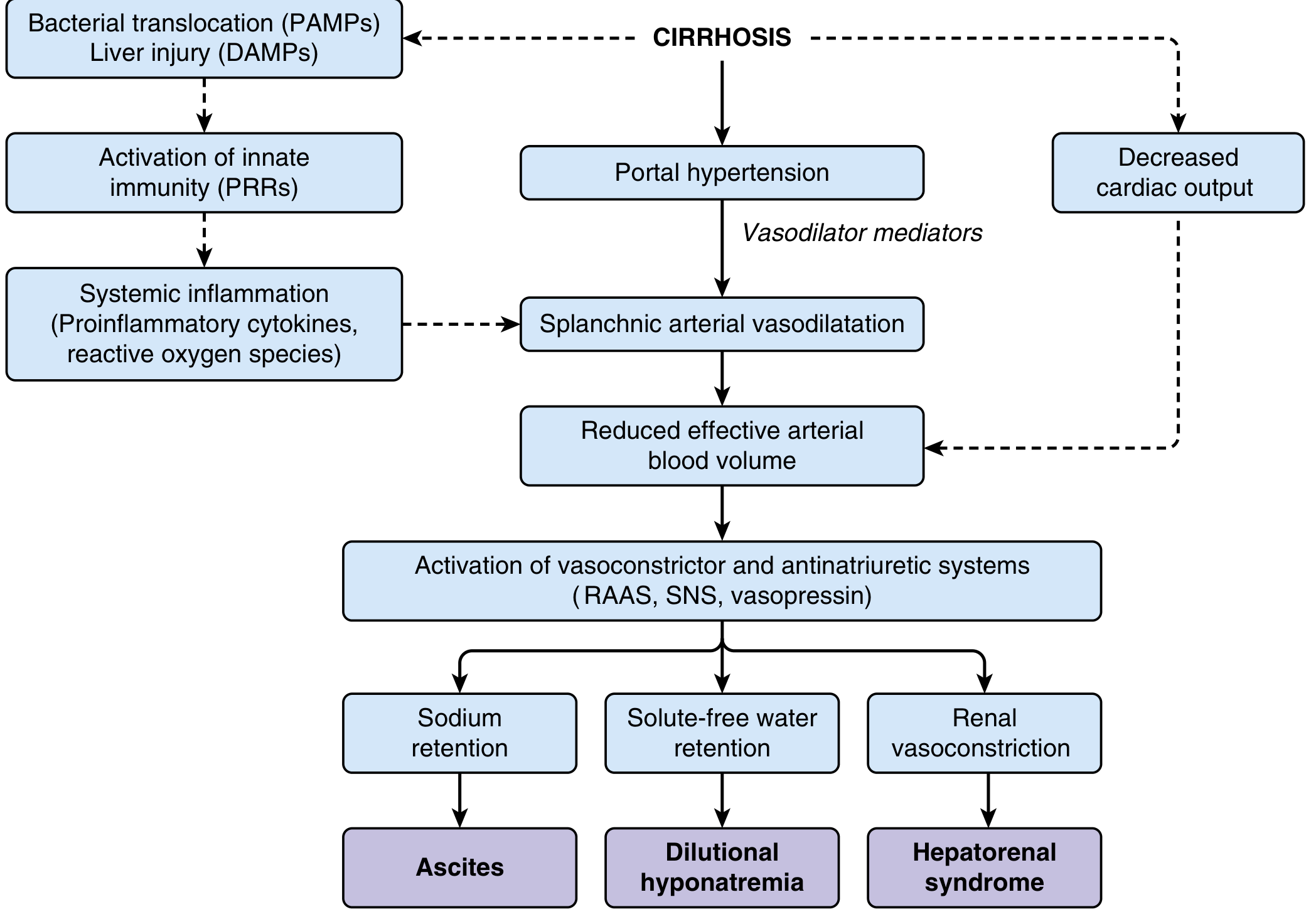
CAUSE 1: CIRRHOSIS OF THE LIVER (Most Common - ~80% of cases)
What Happens in Cirrhosis Step by Step:
Step 1: Portal Hypertension Develops
- Normal portal vein pressure = 5 mmHg
- Scar tissue in the liver blocks blood flow through it → blood backs up in the portal vein
- Portal pressure rises to 12+ mmHg = portal hypertension
Step 2: Splanchnic Vasodilation (The Key Event)
- High portal pressure → release of vasodilators (especially Nitric Oxide - NO) into the blood supply of the gut (splanchnic circulation)
- All the blood vessels around the gut relax and dilate (widen)
- Result: enormous amount of blood pools in the gut blood vessels
Step 3: "Effective Blood Volume" Falls (The Body Is Fooled)
- Even though total blood volume is normal or even high, most of it is trapped in dilated gut vessels
- The heart and kidneys "see" an empty circulation → the body thinks it is hypovolemic (blood loss)
- Analogy: Imagine a city with plenty of water, but all of it is stuck in flooded suburbs - the city center has no water. The body sees a "water shortage" and panics.
Step 4: The Kidney Panics and Retains Salt and Water
- The body activates emergency systems:
- RAAS (Renin-Angiotensin-Aldosterone System): Aldosterone is released → kidneys retain sodium
- SNS (Sympathetic Nervous System): Further sodium retention
- ADH/Vasopressin: Retains free water
- More and more sodium and water is retained → but it can't stay in the blood vessels (portal pressure too high) → it leaks out into the peritoneal cavity
Step 5: Low Albumin Makes It Worse
- The damaged liver also can't make albumin properly
- Low albumin = low oncotic pressure = even more fluid leaks out of vessels
- Result: Ascites + edema (ankle swelling)
- Spider nevi, palmar erythema, gynecomastia (signs of liver disease)
- Jaundice, caput medusae (dilated veins around the navel)
- Splenomegaly (spleen enlarged due to backed-up portal blood)
- Varices (dilated veins in esophagus/stomach - dangerous bleeding)
CAUSE 2: CONGESTIVE HEART FAILURE (Cardiac Ascites)
How:
- The failing heart cannot pump blood forward → blood backs up in the veins behind it
- Right heart failure → backs up into the inferior vena cava (IVC) → hepatic veins → liver → portal system
- Raised venous pressure in the liver → fluid leaks into the peritoneal cavity
- Additionally: poor cardiac output → kidneys retain salt and water (same RAAS activation as cirrhosis)
CAUSE 3: CONSTRICTIVE PERICARDITIS
How:
- The pericardium (sac around the heart) becomes thick and stiff (from old TB infection, radiation, or unknown)
- The stiff pericardium squeezes the heart → cannot fill properly → blood backs up into venous system
- Same mechanism as right heart failure → backs up into liver → ascites
- Classic sign: Kussmaul's sign (JVP rises on inspiration instead of falling)
- This is an important and frequently missed cause - always check JVP in ascites patients!
CAUSE 4: BUDD-CHIARI SYNDROME
How:
- The hepatic veins (which drain blood OUT of the liver) are blocked - by clot, tumor, or membrane
- Blood cannot exit the liver → gets trapped inside → liver becomes congested and swollen
- Raised pressure inside the liver → fluid leaks into the peritoneal cavity
- The liver is acutely engorged → RUQ pain + tender, enlarged liver + rapid-onset ascites
- Associated with: polycythemia vera, pregnancy, oral contraceptives, hypercoagulable states
CAUSE 5: NEPHROTIC SYNDROME
How:
- Kidney disease causes the glomeruli to leak albumin into the urine → severe hypoalbuminemia
- With very low albumin in the blood, the oncotic pressure (the "holding" force) drops dramatically
- Fluid cannot be retained inside blood vessels → leaks into tissues and peritoneal cavity
- Analogy: Without enough albumin "magnets," the water just pours out wherever it can
CAUSE 6: PERITONEAL CARCINOMATOSIS (Malignant Ascites)
How:
- Tumor cells seed the peritoneum (the lining of the belly) - most commonly from ovarian cancer, gastric cancer, colon cancer, pancreatic cancer
- Two mechanisms:
- Tumor implants on the peritoneum → secrete VEGF (vascular endothelial growth factor) → makes local blood vessels leaky → protein-rich fluid pours out
- Tumor implants block lymphatic drainage → lymph cannot be cleared → accumulates
- Result: rapidly accumulating, protein-rich ascitic fluid
- Cytology of ascitic fluid shows malignant cells (positive in 40-70%)
CAUSE 7: TUBERCULOUS PERITONITIS
How:
- Mycobacterium tuberculosis infects the peritoneum (either from gut TB or hematogenous spread)
- The peritoneum becomes inflamed with granulomas → rich in inflammatory cells and protein
- Inflamed peritoneum = leaky vessels = exudative, protein-rich ascites
- Also: TB blocks lymphatics → adds to fluid accumulation
CAUSE 8: PANCREATIC ASCITES
How:
- The pancreatic duct ruptures (from acute pancreatitis or chronic pancreatitis/pseudocyst)
- Pancreatic juice (rich in digestive enzymes) pours directly into the peritoneal cavity
- Amylase-rich, protein-rich ascitic fluid accumulates
- High amylase in ascitic fluid is the diagnostic clue
CAUSE 9: CHYLOUS ASCITES
How:
- The thoracic duct or mesenteric lymphatics are damaged/blocked (by lymphoma, lymph node metastases, surgery, filariasis, trauma)
- Lymph (chyle) - which is milky white from absorbed fat - leaks directly into the peritoneum
- Milky white, turbid ascitic fluid with very high triglycerides (>200 mg/dL)
PART 3: THE SAAG - THE MOST IMPORTANT DIAGNOSTIC TEST
What Is SAAG?
- Take blood → measure serum albumin
- Do paracentesis → measure albumin in ascitic fluid
- Subtract: SAAG = Serum albumin - Ascitic fluid albumin
Why Is SAAG So Useful?
The Simple Logic Behind SAAG:
SAAG ≥ 1.1 g/dL = HIGH SAAG = Portal Hypertension Related
Memory Trick: "HIGH SAAG = HIGH Pressure in the portal system"
| Cause | Why SAAG is HIGH |
|---|---|
| Cirrhosis | Portal hypertension (main cause) |
| Alcoholic hepatitis | Portal hypertension |
| Congestive heart failure | Raised hepatic venous pressure |
| Constrictive pericarditis | Raised venous pressure backing up |
| Budd-Chiari syndrome | Hepatic vein blockage → raised sinusoidal pressure |
| Portal vein thrombosis | Raised portal pressure |
| Fulminant liver failure | Raised sinusoidal pressure |
| Liver metastases (massive) | Obstructive portal hypertension |
| Myxedema (hypothyroidism) | Increased capillary permeability - rare cause |
- Cirrhosis
- Cardiac failure (CHF)
- Constrictive pericarditis
- Portal vein thrombosis
- Budd-Chiari syndrome
- Fulminant liver failure
SAAG < 1.1 g/dL = LOW SAAG = NOT Portal Hypertension
Memory Trick: "LOW SAAG = the fluid is coming from somewhere LOCAL (peritoneum) or from LOW albumin"
| Cause | Why SAAG is LOW |
|---|---|
| Peritoneal carcinomatosis | Tumor makes leaky vessels → protein-rich fluid, gradient narrows |
| TB peritonitis | Inflamed peritoneum → exudative, protein-rich fluid |
| Nephrotic syndrome | No portal hypertension; ascites purely from low oncotic pressure |
| Pancreatic ascites | Pancreatic juice in the belly |
| Chylous ascites | Lymph in the belly (high triglycerides) |
| Bile ascites | Bile duct rupture → bile in belly |
| Peritoneal mesothelioma | Peritoneal malignancy |
- Nephrotic syndrome
- TB peritonitis
- Pancreatic ascites
- Chylous ascites
- Peritoneal mesothelioma
- Malignancy (peritoneal carcinomatosis)
SAAG + Ascitic Protein: The Full 2x2 Table
| SAAG | Ascites Protein | Most Likely Cause |
|---|---|---|
| HIGH (≥1.1) | LOW (<2.5 g/dL) | Cirrhosis (commonest) |
| HIGH (≥1.1) | HIGH (≥2.5 g/dL) | Cardiac failure, Budd-Chiari, constrictive pericarditis |
| LOW (<1.1) | HIGH (≥2.5 g/dL) | TB peritonitis, Peritoneal carcinomatosis |
| LOW (<1.1) | LOW (<2.5 g/dL) | Nephrotic syndrome |
Why does cirrhosis have LOW ascites protein despite HIGH SAAG?
- In cirrhosis, the hepatic sinusoids become "capillarized" (lose their normal extreme leakiness) due to fibrosis
- They become relatively impermeable to large proteins like albumin
- So fluid leaks out (due to high portal pressure) but protein is left behind → low protein in ascites
- In heart failure, sinusoids are NORMAL and highly permeable → protein-rich fluid leaks → high ascites protein
PART 4: HISTORY TAKING FOR ASCITES
Chief Complaint Framework: Abdominal Distension + (any associated features)
Step 1: Onset and Duration
- How long has the belly been swelling?
- Gradual over months → cirrhosis, malignancy, TB
- Rapid over days/weeks → Budd-Chiari, malignancy, cardiac failure
Step 2: Associated Symptoms (The Most Important Step)
Liver/Portal Causes - Ask:
- Alcohol: How much? How long? Daily? (Alcoholic cirrhosis - commonest)
- Jaundice (yellow eyes/urine): Liver disease
- Blood in vomit or black stools: Variceal bleeding (portal hypertension)
- Confusion or drowsiness: Hepatic encephalopathy (ammonia affecting brain)
- Ankle swelling: Common with ascites in liver disease
- Itching all over the body: Cholestasis (bile salts deposit in skin)
Cardiac Causes - Ask:
- Breathlessness, orthopnea, PND: Heart failure
- Palpitations, chest pain: Cardiac disease
- Swelling of legs: Bilateral in heart failure
TB Causes - Ask:
- Fever, night sweats, weight loss: Classic TB constitutional symptoms
- Contact with TB patient
- Prior TB treatment
- HIV status (increases TB risk)
Malignancy Causes - Ask:
- Weight loss (involuntary): How much? Over how long?
- Loss of appetite, early satiety
- Known primary cancer: Ovarian, gastric, colon, pancreatic
- Hemoptysis, blood in stool, urinary symptoms: Points to primary
- Pelvic symptoms in women: Ovarian cancer (most common cause of malignant ascites in women)
Nephrotic Causes - Ask:
- Frothy urine (protein in urine looks frothy, like detergent)
- Swelling of face (periorbital, early morning) - nephrotic patients swell everywhere, including the face (unlike cirrhosis which mainly affects the legs and belly)
- Diabetes, autoimmune disease (causes of nephrotic syndrome)
Step 3: Past History
- History of liver disease (hepatitis B, C, NAFLD, alcohol abuse)
- History of heart disease, rheumatic fever, TB
- Known malignancy
- Prior episodes of ascites
- Vaccinations (hepatitis B)
Step 4: Drug History
- Alcohol (quantify carefully)
- Hepatotoxic drugs (methotrexate, amiodarone → liver fibrosis)
- NSAIDs (worsen renal function in cirrhosis)
- Diuretics (already on treatment?)
Step 5: Family History
- Liver disease, Wilson's disease, hemochromatosis (genetic liver diseases)
- Malignancy
Step 6: Social History
- Occupation: IV drug use (hepatitis B/C), alcohol
- Travel: Malaria-endemic areas, TB areas
- Sexual history (hepatitis B transmission)
PART 5: PHYSICAL EXAMINATION FINDINGS IN ASCITES
General Inspection
| Sign | Suggests |
|---|---|
| Jaundice, spider nevi, leukonychia, clubbing, caput medusae | Cirrhosis/liver disease |
| Raised JVP, pulsatile neck veins | Cardiac cause |
| Periorbital edema, anasarca | Nephrotic syndrome |
| Cachexia (extreme wasting), lymphadenopathy, hard abdominal mass | Malignancy |
| Low blood pressure with rising JVP on inspiration (Kussmaul) | Constrictive pericarditis |
Abdominal Examination
1. Inspection
- Distended abdomen, flanks full and bulging (fluid falls to sides by gravity)
- Everted umbilicus (fluid pushes umbilicus outwards)
- Caput medusae (dilated veins radiating from umbilicus = portal hypertension)
- Visible peristalsis (suggests bowel obstruction - differentiate from ascites)
2. Percussion - Two Key Signs
- Percuss from umbilicus to flank - fluid makes the flank dull (fluid is heavy, sinks to the flanks)
- Ask patient to lie on their side - the fluid shifts to the lower side → the previously dull side becomes resonant, and the previously resonant central area becomes dull
- Detects ascites of >500 mL
- Patient on all fours (like crawling position) for 5 minutes → fluid collects in the umbilical area
- Percuss at umbilicus - dull if ascites present
- Detects as little as 120 mL - most sensitive sign
3. Palpation
- Ask an assistant to place their hand on the midline (to block fat transmission)
- Flick one flank with a finger → the thrill/impulse is felt on the opposite flank
- Positive only in large amounts of ascites (tense ascites)
4. Grading of Ascites (WHO/Clinical)
| Grade | Clinical Finding |
|---|---|
| Grade 1 | Only detectable by ultrasound (not clinically) |
| Grade 2 | Moderate, detectable by shifting dullness |
| Grade 3 | Large, tense (drum-tight belly), obvious distension |
PART 6: INVESTIGATIONS AND DIAGNOSTIC APPROACH
Step 1: First Line Always
Ultrasound Abdomen (First investigation)
- Confirms ascites (detects even small amounts, >100 mL)
- Looks at: liver size/texture (cirrhosis = shrunken, nodular), splenomegaly (portal hypertension), portal vein diameter, masses, lymph nodes
- Guides paracentesis
Diagnostic Paracentesis (The Most Informative Test - MUST DO in every new case)
- Insert a needle into the left iliac fossa under sterile conditions → withdraw ascitic fluid
- Safe even in cirrhosis if done correctly (INR does not need to be corrected unless very high)
- Send fluid for:
| Test | Always | Sometimes |
|---|---|---|
| Cell count + differential | ✓ | |
| Albumin | ✓ | |
| Total protein | ✓ | |
| Gram stain + culture | ✓ | |
| Glucose, LDH | ✓ | |
| Amylase | Pancreatic ascites | |
| Triglycerides | Chylous ascites | |
| Bilirubin | Bile ascites | |
| Cytology | Malignancy suspected | |
| AFB culture + PCR | TB suspected | |
| ADA | TB suspected |
Ascitic Fluid Appearance:
| Appearance | Suggests |
|---|---|
| Clear, straw-colored | Cirrhosis, cardiac failure |
| Turbid/cloudy | Infection (SBP), high cell count |
| Milky white | Chylous ascites (triglycerides >200 mg/dL) |
| Blood-stained | Malignancy, trauma, TB (sometimes) |
| Dark brown/greenish | Bile ascites (bilirubin > serum bilirubin) |
Calculate SAAG (Same day as paracentesis):
- SAAG = Serum albumin - Ascites albumin
- ≥1.1 → portal hypertension related
- <1.1 → non-portal hypertension related
Step 2: Based on SAAG Result
If SAAG ≥ 1.1 (Portal Hypertension):
- LFTs, PT/INR, serum albumin → assess liver function (is this cirrhosis?)
- Hepatitis B surface antigen, Hepatitis C antibody → viral cirrhosis?
- USG liver with Doppler → hepatic vein flow (Budd-Chiari?), portal vein thrombosis?
- Echocardiogram → cardiac failure, constrictive pericarditis?
- CT abdomen → liver morphology, HCC (hepatocellular carcinoma) complicating cirrhosis?
- Upper GI endoscopy → esophageal varices (confirms portal hypertension)
- Liver biopsy (if etiology unclear) → establish cause of cirrhosis
If SAAG < 1.1 (Non-Portal Hypertension):
- Ascitic fluid cytology → malignant cells?
- AFP, CEA, CA-125, CA 19-9 → tumor markers (ovarian, colon, pancreatic cancer?)
- CT abdomen + pelvis → primary tumor, peritoneal deposits?
- Ascitic fluid ADA, AFB culture, PCR → TB peritonitis?
- Urine protein/creatinine ratio, renal biopsy → nephrotic syndrome?
- Laparoscopy + biopsy of peritoneum → if TB/malignancy not confirmed otherwise (gold standard for TB peritonitis)
Step 3: Important Complication to Always Rule Out - SBP
Spontaneous Bacterial Peritonitis (SBP)
- Bacteria from the gut translocate into the ascitic fluid → infection of the ascites
- No obvious source of infection (no perforation, no abscess)
- Common in: cirrhosis patients with ascites (lowered local immunity)
PART 7: QUICK CLINICAL REASONING - PUTTING IT ALL TOGETHER
The Diagnostic Algorithm
PATIENT PRESENTS WITH ABDOMINAL DISTENSION
            ↓
CONFIRM ASCITES (Ultrasound)
            ↓
DIAGNOSTIC PARACENTESIS → Calculate SAAG
            ↓
┌─────────────────────────┬─────────────────────────┐
│   SAAG ≥ 1.1            │   SAAG < 1.1            │
│ (Portal Hypertension)   │ (Non-Portal HTN)         │
├─────────────────────────┼─────────────────────────┤
│ Ascites protein LOW     │ Ascites protein HIGH     │
│ → CIRRHOSIS             │ → TB peritonitis         │
│                         │ → Malignancy             │
├─────────────────────────┼─────────────────────────┤
│ Ascites protein HIGH    │ Ascites protein LOW      │
│ → CHF                   │ → Nephrotic syndrome     │
│ → Budd-Chiari           │                         │
│ → Constrictive peric.   │                         │
└─────────────────────────┴─────────────────────────┘
Clinical Scenarios - Quick Matching
| Patient Scenario | Diagnosis |
|---|---|
| 45M, chronic alcoholic, jaundice, spider nevi, variceal history, SAAG 1.5, ascites protein 1.8 | Alcoholic Cirrhosis |
| 60F, breathless, orthopnea, raised JVP, pedal edema, SAAG 1.3, ascites protein 3.0 | Congestive Heart Failure |
| 30F, on OCP, rapid painful ascites, tender enlarged liver, Doppler shows no hepatic vein flow | Budd-Chiari Syndrome |
| 25F, frothy urine, periorbital puffiness, SAAG 0.7, ascites protein 1.5, massive proteinuria | Nephrotic Syndrome |
| 35M, fever + night sweats + weight loss, SAAG 0.6, high ADA in ascites, lymphocytic fluid | TB Peritonitis |
| 55F, known ovarian Ca, rapid ascites, cytology positive for malignant cells, SAAG 0.5 | Peritoneal Carcinomatosis |
| Cirrhotic patient admitted with sudden abdominal pain + fever, ascites PMN >250/mL | SBP (Spontaneous Bacterial Peritonitis) |
Give definition,indications,contraindications,technique as well as risks of therapeutic ascitic tap /large volune paracentesis
Large Volume Paracentesis (LVP) / Therapeutic Ascitic Tap
DEFINITION
| Type | Volume Drained | Purpose |
|---|---|---|
| Diagnostic paracentesis | 20-50 mL | To analyze fluid (SAAG, culture, cytology) - finding the cause |
| Therapeutic paracentesis / LVP | >5 liters (often 5-10 L or even total drainage) | To relieve symptoms caused by large/tense ascites |
WHY IS LVP NEEDED? (Rationale)
- Abdominal distension and discomfort - the belly is drum-tight and painful
- Early satiety - the stomach is compressed, patient can barely eat
- Shortness of breath - the diaphragm is pushed up by the fluid, limiting lung expansion
- Difficulty walking - the belly is so heavy and tense
INDICATIONS
Absolute / Clear Indications:
- The belly is massively distended, drum-tight
- Patient is in discomfort, breathless, or unable to eat
- Regardless of whether diuretics have been tried - immediate relief is the goal
- Ascites that does NOT respond to maximum doses of diuretics (spironolactone 400 mg/day + furosemide 160 mg/day)
- OR ascites that recurs rapidly despite diuretics
- LVP is the first-line intervention for refractory ascites while awaiting TIPS or transplant
- Massive ascites elevating the diaphragm → patient is hypoxic or severely breathless
- Emergency LVP needed to decompress
- Ascitic fluid migrates through small diaphragmatic defects into the pleural space
- Draining the ascites reduces the pleural fluid source
- In cancer patients where ascites repeatedly accumulates rapidly
- LVP for symptom relief (comfort/palliation)
- When symptomatic and not responding to conservative management
CONTRAINDICATIONS
Absolute Contraindications:
| Contraindication | Reason |
|---|---|
| Disseminated Intravascular Coagulation (DIC) | Uncontrolled bleeding risk at puncture site |
| Acute abdomen / Peritonitis requiring surgery | The underlying problem needs surgery, not drainage |
| No ascites confirmed | Nothing to drain - risk without benefit |
Relative Contraindications (Proceed with Caution):
| Contraindication | What To Do |
|---|---|
| Severe coagulopathy (INR > 2, Platelets < 50,000) | Traditionally a concern, but evidence shows paracentesis is SAFE even with abnormal clotting in cirrhosis. Fresh frozen plasma (FFP) or platelet transfusion is NOT routinely recommended. Use clinical judgment. |
| Pregnancy | Adjust needle site, use ultrasound guidance - not an absolute contraindication |
| Prior abdominal surgery / scars | Risk of bowel adhesions at the scar site - use ultrasound to find a safe pocket of fluid away from scars |
| Loculated (non-free) ascites | Fluid trapped in pockets, not freely flowing - ultrasound-guided approach mandatory |
| Active skin infection/cellulitis at the insertion site | Needle can carry skin bacteria into the peritoneum - find a different site |
| Bowel distension / obstruction | Risk of bowel puncture is higher |
| Abdominal wall hematoma | Avoid that site |
Important note: Coagulopathy (high INR, low platelets) in cirrhosis is NOT a reason to withhold paracentesis. Cirrhotic patients almost always have deranged clotting, yet paracentesis has a very low bleeding complication rate (<1%). The liver regenerates thrombin and other factors locally. Do NOT reflexively transfuse FFP before a tap in a cirrhotic patient.
TECHNIQUE - STEP BY STEP
Equipment Needed:
- Sterile gloves, drape, antiseptic solution (betadine/chlorhexidine)
- Local anaesthetic (lignocaine 2%)
- Paracentesis needle (14-18G) or cannula (trocar)
- 10 mL syringe (for anaesthetic) + 50 mL syringe (for aspiration)
- Drainage bag with tubing (for LVP)
- Albumin solution ready for IV infusion after the procedure
- Ultrasound machine (highly recommended)
Step 1: Patient Preparation
- Explain the procedure and get informed consent
- Patient empties the bladder (to avoid puncturing a full urinary bladder)
- Position: Patient lies supine (flat on back), or may be tilted slightly to the left (left lateral decubitus) to pool fluid on the left side
- Check: Blood pressure, pulse, fluid confirmed on clinical examination or ultrasound
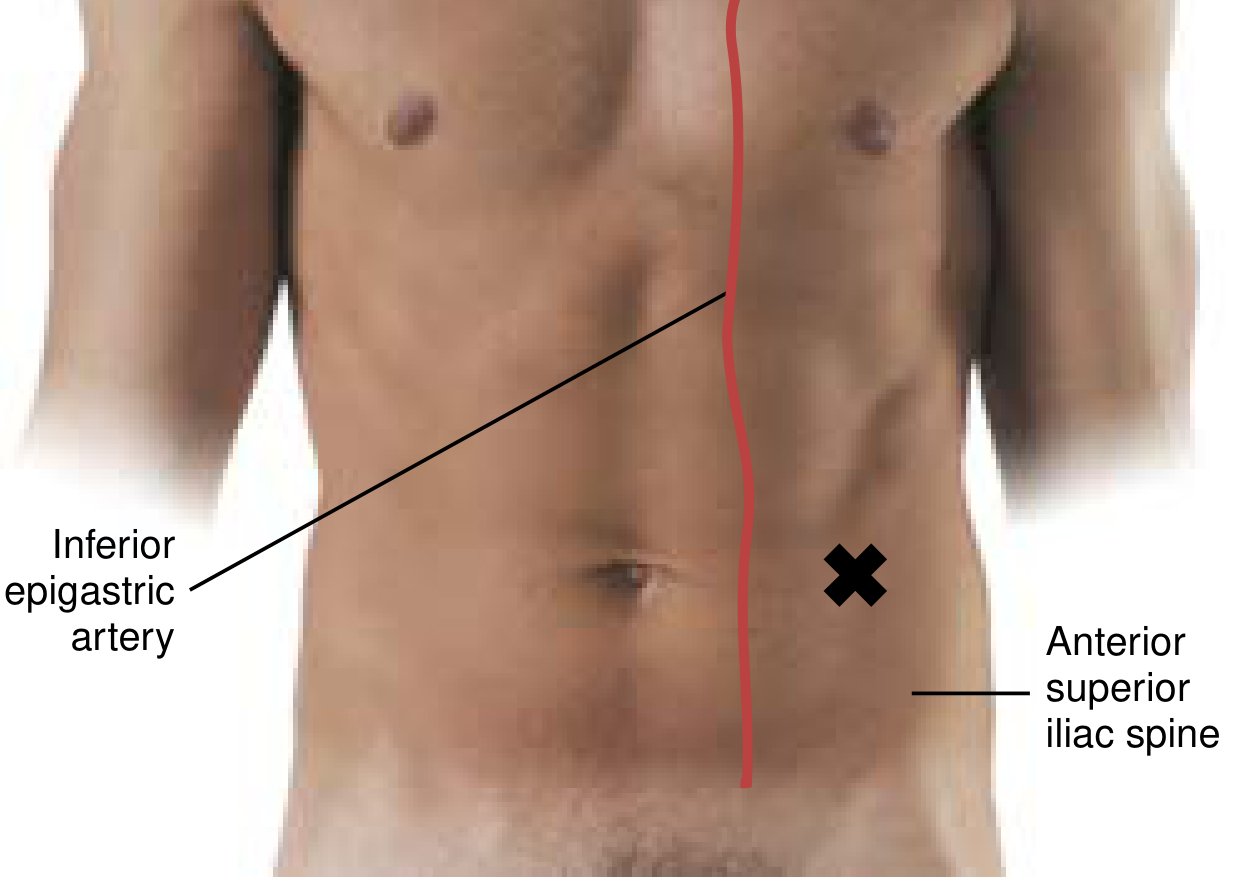
Step 2: Identify the Insertion Site
- 3 cm medial and 3 cm superior to the left anterior superior iliac spine (ASIS)
- This is the standard landmark-based site
- On the right side, the cecum may be distended (especially in cirrhotic patients who take lactulose - a common laxative given for hepatic encephalopathy). A distended cecum is more likely to be punctured on the right.
- The left iliac fossa has the sigmoid colon, which is smaller and more medial.
- Midline (linea alba) - 2-3 cm below the umbilicus - avascular, good when bilateral fluid is present. Avoid the inferior epigastric vessels (which run laterally).
- Right iliac fossa - acceptable but less preferred
- Previous scars (bowel adhesions underneath)
- Visible engorged subcutaneous vessels
- The inferior epigastric artery (runs laterally in the rectus sheath)
- Site of skin infection or hematoma
Step 3: Aseptic Preparation
- Clean the insertion site with antiseptic (betadine or chlorhexidine) in wide concentric circles
- Apply sterile drape
- Put on sterile gloves
Step 4: Local Anaesthesia
- Draw up lignocaine 2% (10-15 mL)
- First inject a small bleb into the skin (superficial) to numb the skin
- Then infiltrate deeper - through the subcutaneous fat and muscle layers, aspirating as you go (to confirm you are not in a vessel)
- When you reach the peritoneum, you may aspirate ascitic fluid into the syringe - confirming correct depth
- Give a generous amount - the peritoneum is sensitive
Step 5: Needle Insertion Technique
- Pull the skin downward (or to the side) by about 2 cm with your non-dominant hand, stretching it
- Insert the needle/cannula straight (perpendicular) through the skin while it is displaced
- When you release the skin, the skin track and the peritoneal puncture site no longer align - the Z-shape seals itself after removal
- Like threading a needle through displaced fabric - when you release, the holes don't line up
- Advantage: Significantly reduces post-procedure ascitic leakage
- Insert the needle at an angle (obliquely), so the skin entry point and peritoneal entry point are offset
- Achieves a similar "sealing" effect
- Advance the needle slowly through the skin, fat, muscle (you feel resistance) → then a "give" or pop as you enter the peritoneum
- Aspirate as you advance - when you freely aspirate clear/straw-colored ascitic fluid, you are in the correct position
Step 6: Drainage (for LVP)
- Attach drainage tubing and bag to the cannula
- Allow gravity drainage OR use a manual syringe technique with repeated aspiration
- For large volume drainage, attach a closed drainage system (vacuum bottle or gravity bag)
- Rate: Allow to drain freely - there is no evidence that slow drainage is safer than fast drainage in terms of PPCD (post-paracentesis circulatory dysfunction)
- Drain 5-10 litres (in refractory ascites, total drainage is acceptable)
- Limit each session to <8 litres if not giving proportionate albumin replacement
Step 7: Albumin Infusion (Critical Step - Must Not Be Skipped for LVP)
- As soon as >5 litres is drained (or at the end of the procedure):
- Give intravenous albumin 6-8 g per litre of ascites removed
- Example: If 6 litres removed → give 36-48 g of albumin IV (human albumin solution 20% or 25%)
- This is to prevent Post-Paracentesis Circulatory Dysfunction (PPCD) - explained in detail in the Risks section
For < 5 litres: Albumin is optional unless the patient has low BP (<90 mmHg systolic) or hyponatremia.
Step 8: Removal and Post-Procedure
- Remove the needle/cannula when drainage slows or stops
- Apply firm pressure to the site with sterile gauze for a few minutes
- If persistent leakage occurs → place a single suture or stitch to close the skin track, or apply an ostomy bag over the site
- Dress the wound with sterile dressing
- Observe patient for 1-2 hours: Check BP, pulse, abdominal signs
ULTRASOUND GUIDANCE - STRONGLY RECOMMENDED
- Confirms presence and size of the fluid pocket
- Identifies a safe area free of bowel, bladder, and major vessels
- Mandatory when: loculated ascites, obesity, previous surgery, small fluid volume, suspected abdominal wall hematoma
RISKS AND COMPLICATIONS
1. Post-Paracentesis Circulatory Dysfunction (PPCD) - Most Important
- After large volume drainage, the fluid that was in the belly disappears → the pressure inside the abdomen drops suddenly
- Blood vessels in the abdomen and gut (splanchnic circulation) expand rapidly to fill the space
- Blood pools in these dilated vessels → effective circulating blood volume suddenly drops
- The body panics → activates RAAS and sympathetic nervous system dramatically
- Result: kidney gets less blood → sodium retention → ascites rapidly recurs + kidney damage (hepatorenal syndrome)
- Happens 6-24 hours after LVP
- Detected by: rise in plasma renin activity, fall in blood pressure, hyponatremia, rising creatinine
- Patient may feel dizzy, develop hypotension
- Can progress to hepatorenal syndrome (kidneys shut down)
- IV Albumin 6-8 g/L of fluid removed → albumin stays in the circulation, maintains oncotic pressure, keeps fluid in the vessels, prevents the collapse of effective circulating volume
- Albumin is the ONLY proven agent to prevent PPCD and reduce mortality with LVP
- Alternative (less effective): Dextran-70 (6 g/L removed) or Gelofusine for taps <5 L in resource-limited settings
- Albumin molecules are large proteins - they stay in the bloodstream (don't leak out easily)
- They maintain oncotic pressure → keep fluid inside blood vessels
- They also bind inflammatory mediators produced during the post-drainage period
2. Local Complications at the Needle Site
| Complication | Frequency | Explanation |
|---|---|---|
| Persistent fluid leak | Common (5-10%) | Especially if Z-track technique not used. Treat with a suture or ostomy bag |
| Local hematoma | Uncommon | Usually minor bruising. Major if inferior epigastric artery is hit |
| Abdominal wall hematoma | Rare | From injury to a subcutaneous vessel |
| Wound site infection / cellulitis | Rare | Breach of aseptic technique |
3. Organ Injury (Rare with Proper Technique)
| Injury | When It Happens |
|---|---|
| Bowel perforation | If needle inserted into bowel (especially cecum on right side, or adhesions from prior surgery) |
| Bladder puncture | If patient did not empty bladder before the procedure |
| Hemorrhage | If inferior epigastric artery or other vessel is hit - rare but potentially serious |
| Introduction of infection | If aseptic technique is compromised → peritonitis |
4. Electrolyte and Metabolic Disturbances
| Disturbance | Mechanism |
|---|---|
| Hyponatremia | Rapid fluid shift + RAAS activation → water retention without sodium |
| Hypokalemia | Especially if diuretics are continued aggressively after LVP |
| Hypotension | PPCD - blood pressure drops due to reduced circulating volume |
5. Hepatic Encephalopathy (Worsening Confusion)
- LVP-induced PPCD → reduced perfusion to liver → ammonia clearance drops → patient becomes confused
- Also: PPCD activates RAAS → aldosterone rises → hypokalemia → ammonia production increases → encephalopathy
- Prevented by: albumin infusion + monitoring after procedure
6. Rapid Recurrence of Ascites
- Not a complication of the technique itself, but a clinical reality
- If the underlying cause (portal hypertension) is not addressed, ascites recurs within 2-4 weeks
- LVP is a temporizing measure - it treats the symptom, not the cause
- Definitive treatment: TIPS (Transjugular Intrahepatic Portosystemic Shunt) or Liver Transplantation
SUMMARY TABLE
| Feature | Detail |
|---|---|
| Definition | Draining >5 litres of ascitic fluid from the peritoneal cavity for symptom relief |
| Indications | Tense/grade 3 ascites, refractory ascites, respiratory distress from ascites, malignant ascites (palliation) |
| Absolute contraindications | DIC, acute abdomen needing surgery, no ascites |
| Relative contraindications | Coagulopathy (relative), pregnancy, scars, loculated fluid, skin infection at site |
| Preferred site | 3 cm medial + 3 cm superior to left ASIS |
| Technique | Z-track or angular technique under aseptic conditions, with lignocaine, ideally ultrasound-guided |
| Albumin dose | 6-8 g per litre of ascites removed, IV, given after procedure |
| Key complication | Post-paracentesis circulatory dysfunction (PPCD) → prevented by albumin |
| Other risks | Fluid leak, bleeding, bowel perforation, electrolyte imbalance, encephalopathy |
The most important thing to remember: LVP without albumin replacement is incomplete and potentially dangerous. Albumin after LVP >5 L is not optional - it is mandatory to prevent PPCD, hepatorenal syndrome, and death. Every textbook from Harrison's to Sleisenger agrees on this point.
Definition of ascites
- Normal: The peritoneal cavity contains up to ~50 mL of serous fluid - just enough to lubricate the bowel loops so they can move freely.
- Pathological: When fluid accumulates beyond this normal amount (detectable clinically at >500 mL, on ultrasound at >100 mL), it is called ascites.
- The fluid can be a transudate (low protein, from pressure/oncotic changes) or an exudate (high protein, from inflammation/malignancy), depending on the underlying cause.
- Ascites is a sign/symptom, not a diagnosis in itself - it always reflects an underlying disease (most commonly cirrhosis, heart failure, malignancy, TB, or nephrotic syndrome).
Hi, Aries. Can you please, uh, help me out? Uh, I have clinical postings for medicine for about, uh, four weeks and I need to complete my whole medicine in it as a clinician. So can you please give me, uh, like, sketch out schedule, like, in what-- for what day, what, uh, amount of things I have to cover up so that I am in a good state of giving a clinical viva and, uh, like, dodging the clinical classes? Um, the things must include, uh, the things must include the clinical things, uh, the symptomatology, the, uh, procedural skills which I need to learn as a final year MAB student. All the things that I need to learn as final year MAB students.
Mbbs final year studentt
4-Week Medicine Clinical Posting Schedule (Final Year MBBS)
How This Is Structured
WEEK 1: FOUNDATIONS - History Taking, General Examination, CVS + Respiratory
| Day | Morning (Ward) | Afternoon (Theory) | Evening (Revision) |
|---|---|---|---|
| Day 1 | Practice full history taking format (chief complaint → HPI → past/personal/family/drug history) on 2 patients | History taking framework + General Physical Examination (built, nourishment, pallor, icterus, cyanosis, clubbing, lymphadenopathy, edema) | List and rehearse all "general examination" findings and their causes |
| Day 2 | Vitals + general exam practice; learn to measure BP, pulse (character, volume, rhythm), JVP | Pulse examination in depth (all peripheral pulses, radio-femoral delay, collapsing pulse, pulsus paradoxus) + JVP interpretation | Quiz yourself: causes of each abnormal pulse/JVP pattern |
| Day 3 | CVS examination practice (inspection, palpation, percussion, auscultation) on 2-3 patients | CVS symptomatology: chest pain, palpitations, syncope, orthopnea, PND, pedal edema - causes + mechanisms | Auscultation areas + heart sounds + murmur grading (mnemonic practice) |
| Day 4 | Present a CVS case to your resident/PG (full case) | Common CVS diagnoses: Rheumatic heart disease (MS, MR, AS, AR), Heart failure, IHD, Congenital heart disease basics | Murmur differentiation table (timing, character, radiation) - self-test |
| Day 5 | Respiratory exam practice (inspection, palpation - chest expansion/vocal fremitus, percussion, auscultation) | Respiratory symptomatology: cough, expectoration, hemoptysis, dyspnea, chest pain, wheeze, stridor (mechanisms + causes) | Percussion note differentiation (resonant/dull/stony dull/hyperresonant) with causes |
| Day 6 | Present a respiratory case; practice identifying breath sounds (bronchial, vesicular, crackles, wheeze) | Common respiratory diagnoses: Pneumonia, COPD, Asthma, Bronchiectasis, Pleural effusion, Pneumothorax, TB | Compare CVS vs RS causes of dyspnea - clinical differentiation chart |
| Day 7 (Sunday) | Light ward rounds | Mock viva - Week 1: History + General exam + CVS + RS (get a peer or senior to grill you) | Note weak areas, re-read only those topics |
WEEK 2: Abdominal System + CNS Examination
| Day | Morning (Ward) | Afternoon (Theory) | Evening (Revision) |
|---|---|---|---|
| Day 8 | Abdominal exam practice (inspection, superficial/deep palpation, liver/spleen/kidney palpation) | Abdominal symptomatology: abdominal pain (visceral vs parietal), vomiting, diarrhea, constipation, jaundice, ascites (definition, causes, SAAG concept) | Revise ascites causes + SAAG table (portal HTN vs non-portal HTN) |
| Day 9 | Practice shifting dullness, fluid thrill, liver span measurement, splenomegaly grading | Hepatology: Cirrhosis, Portal hypertension, Viral hepatitis, Liver failure symptomatology (jaundice mechanism, encephalopathy grading) | Grading systems: Child-Pugh score, MELD score, hepatic encephalopathy grades |
| Day 10 | Present an abdominal/hepatology case fully | GI bleed (hematemesis vs melena), causes and mechanism; Peptic ulcer disease, IBD basics | Differentiate upper vs lower GI bleed causes and clinical clues |
| Day 11 | CNS exam practice - higher functions, cranial nerves (learn a systematic sequence for all 12) | Cranial nerve examination in detail with common lesions (e.g., 3rd nerve palsy, facial nerve palsy - UMN vs LMN) | Cranial nerve testing checklist - rehearse full sequence out loud |
| Day 12 | CNS motor exam (tone, power grading MRC 0-5, reflexes, clonus, plantar reflex) practice | Motor system: UMN vs LMN lesion differentiation table; Stroke syndromes, Hemiplegia localization | UMN vs LMN comparison chart - memorize cold |
| Day 13 | CNS sensory exam + cerebellar signs + gait examination practice | Sensory pathways basics, Cerebellar signs (DANISH mnemonic), Common gait abnormalities and their causes | Case scenario practice: given findings, localize the lesion |
| Day 14 (Sunday) | Ward rounds | Mock viva - Week 2: Abdomen + CNS full case presentation | Identify and re-read 2-3 weakest topics only |
WEEK 3: Renal, Endocrine, Hematology + Common Emergency Presentations
| Day | Morning (Ward) | Afternoon (Theory) | Evening (Revision) |
|---|---|---|---|
| Day 15 | Look for edema cases; correlate with renal/cardiac/hepatic causes | Renal symptomatology: Edema (mechanism - nephrotic vs nephritic vs cardiac vs hepatic), Oliguria/anuria, Hematuria | Nephrotic vs Nephritic syndrome comparison table |
| Day 16 | Practice examining for pitting edema, look for uremic signs if any inpatient available | AKI vs CKD, causes and staging; Dialysis basics (indications) | RIFLE/KDIGO staging - quick recall |
| Day 17 | Look for diabetic/thyroid patients on ward, practice thyroid examination | Endocrine symptomatology: Polyuria/polydipsia (DM), weight loss/gain, heat/cold intolerance (thyroid), Cushingoid features | DM complications list (acute + chronic) - self-test |
| Day 18 | Practice examining for pallor, lymphadenopathy, bleeding manifestations | Hematology: Anemia symptomatology and types (mechanism-based - hemolytic vs deficiency vs marrow failure), Bleeding disorders basics | Anemia workup algorithm (MCV-based classification) |
| Day 19 | Attend/observe any emergency admission if possible | High-yield emergencies: DKA, Hypoglycemia, Status epilepticus, Acute coronary syndrome, Acute severe asthma, Anaphylaxis - recognize + immediate steps | Emergency drug doses and first steps - flashcard style recall |
| Day 20 | Present any case with fever - practice fever history and workup | Fever symptomatology (patterns, mechanisms) revision; Common fevers - Typhoid, Malaria, Dengue, TB - differentiating clinical features | Fever pattern chart (continuous/remittent/intermittent/hectic) with causes |
| Day 21 (Sunday) | Ward rounds | Mock viva - Week 3: Renal + Endocrine + Hematology + one emergency scenario | Weak-topic revision only |
WEEK 4: Integration, Procedures, Case Presentation Mastery, and Full Mock Vivas
| Day | Morning (Ward) | Afternoon (Theory) | Evening (Revision) |
|---|---|---|---|
| Day 22 | Take 1 full long case (any system) independently, present to senior | Revise all system-wise "symptom → mechanism → cause" chains you've studied - pick 5 symptoms and explain out loud without notes | Self-test: pick a random symptom, explain mechanism + 5 causes |
| Day 23 | Practice 2 short cases (spot diagnosis based cases - clubbing, edema, murmur, skin lesion, goiter etc.) | Common short case topics: Anemia, Jaundice, Lymphadenopathy, Goiter, Ascites, Edema, Clubbing - quick recognition + causes | Flash through short case list rapid-fire |
| Day 24 | Revise and practice all procedural skills hands-on/simulation: Paracentesis, Pleural tap, Lumbar puncture (indications/technique), IV cannulation, ABG sampling, ECG lead placement | Read indications/contraindications/technique/complications for each of the above procedures (exactly like the ascites paracentesis breakdown you already have) | Procedure viva cards - definition, indications, contraindications, technique, complications for each |
| Day 25 | Present a full case start to finish (as if it's your actual exam) to a friend or senior, timed | Revise investigation interpretation: CBC, LFT, RFT, ABG, urine routine, CXR patterns, ECG patterns | Practice explaining "how to arrive at diagnosis" logically for 3 different cases |
| Day 26 | Repeat weak system's long case | Drug history + common drug side effects relevant to medicine (steroids, ACE inhibitors, anticoagulants, insulin) | Rapid recall: drug-induced conditions (e.g., ACE inhibitor cough, steroid-induced diabetes) |
| Day 27 | Full mock long case + short cases with feedback from a senior/resident | Review all differential diagnosis frameworks and "how to rule out" approaches across systems | Go through your own notes cover-to-cover, mark anything unclear |
| Day 28 (Sunday) | Final full mock viva - simulate actual exam (long case + 2 short cases + instruments/procedure questions) | Clear doubts from mock viva feedback | Light revision only - do not cram new material |
Daily Non-Negotiables (Applies to All 4 Weeks)
- See at least 2-3 patients per day actively (not passively standing in rounds) - take history and examine yourself before your senior does.
- Present at least 1 case out loud per day to anyone available (senior, peer, even to yourself in a mirror) - viva is about fluency, not just knowledge.
- Maintain a symptom-to-diagnosis logbook - every real patient you see, write down: chief complaint → your differential → actual diagnosis → what you'd have missed.
- Keep a "procedure card" file for: Paracentesis, Pleural tap, Lumbar puncture, Bone marrow aspiration (theory only), ABG sampling, IV cannulation, ECG recording, Blood transfusion protocol.
Core Topic Checklist (Tick Off As You Go)
A Note on Pace
Hi, can you please explain symptomatology in CNS? In this please try to tell causes but also try to explain how do these causes lead to the particular symptom elaboratively. What are important things to keep in mind while taking history? How to rule out the particular causes? How to arrive at diagnosis? Please explain in more understandable language giving examples and simplified wordings easy to understand and we can, what we can remember easily.
Hi, can you please explain symptomatology in CNS? In this please try to tell causes but also try to explain how do these causes lead to the particular symptom elaboratively. What are important things to keep in mind while taking history? How to rule out the particular causes? How to arrive at diagnosis? Please explain in more understandable language giving examples and simplified wordings easy to understand and we can, what we can remember easily.
CNS Symptomatology - Complete Student Guide
1. HEADACHE
What Is Headache and Why Does It Hurt?
- Blood vessels (arteries at the base of the brain, dural venous sinuses)
- The dura mater (the tough covering of the brain)
- Cranial nerves (especially the trigeminal nerve - CN V)
- Extracranial structures (scalp muscles, blood vessels, sinuses, teeth, eyes, neck muscles)
Mechanism-Based Causes of Headache
A. Vascular Mechanism - Migraine
- Trigeminovascular system gets activated - nerve fibers around blood vessels release inflammatory chemicals (CGRP - calcitonin gene-related peptide, substance P)
- This causes sterile neurogenic inflammation around the blood vessels in the meninges
- Blood vessels dilate and become hypersensitive → every pulse of blood becomes painful
- This explains the classic throbbing/pulsating quality of migraine pain
- Aura (visual flashes before headache) is due to a wave of neuronal suppression spreading across the cortex (cortical spreading depression)
B. Muscular Mechanism - Tension-Type Headache
- Sustained muscle contraction of the scalp, neck, and shoulder muscles (from stress, poor posture, anxiety)
- Contracted muscles compress local blood vessels and irritate nerve endings within the muscle itself
- Analogy: Like a tight rubber band wrapped around your head - constant dull pressure
C. Raised Intracranial Pressure (ICP) - Tumor, Hydrocephalus, Abscess
- A mass (tumor, blood, pus, excess CSF) takes up space inside the fixed, rigid skull
- Since the skull cannot expand, pressure rises inside → stretches the dura and blood vessels at the base of the brain
- This stretch is what generates the pain (not the tumor itself directly)
D. Meningeal Irritation - Meningitis, Subarachnoid Hemorrhage (SAH)
- Infection (meningitis) or blood (SAH) irritates the meninges directly
- The meninges are richly supplied with pain fibers → severe, diffuse pain
- Irritation also causes reflex spasm of the neck muscles → neck stiffness (meningismus)
E. Sinusitis
- Infected/inflamed sinus mucosa + trapped mucus/pus builds up pressure within the closed sinus cavity
- Pressure and inflammation irritate local nerve endings
- Clue: Pain over the specific sinus (frontal, maxillary), worse bending forward, associated with nasal discharge/congestion, tender over the sinus on palpation
F. Giant Cell (Temporal) Arteritis
- Autoimmune inflammation of the temporal artery wall → vessel wall swells and becomes tender
- Direct irritation of pain fibers in the inflamed vessel wall
- Clue: Elderly patient, unilateral scalp tenderness (combing hair hurts), jaw pain on chewing (jaw claudication), visual loss risk (ophthalmic artery involvement) - THIS IS AN EMERGENCY, treat with steroids immediately to prevent blindness
G. Cervicogenic Headache
- Pain referred from the neck (cervical spine arthritis, muscle spasm) via shared nerve pathways (upper cervical nerves converge with trigeminal nucleus)
- Clue: Neck stiffness, pain starts at the back of head/neck and radiates forward
RED FLAGS in Headache History (Always Screen For These)
| Red Flag | What It Suggests |
|---|---|
| Systemic symptoms (fever, weight loss) | Infection, malignancy |
| Neurological deficit (new weakness, confusion) | Stroke, tumor, bleed |
| Onset sudden/thunderclap | Subarachnoid hemorrhage |
| Older age (new-onset headache >50 yrs) | Temporal arteritis, tumor |
| Pattern change (progressively worsening, positional, "worst ever") | Raised ICP, mass lesion |
History Taking for Headache
- Onset: Sudden (SAH) vs gradual (tension, migraine, tumor)
- Site: Unilateral (migraine, cluster) vs bilateral (tension) vs specific sinus location
- Character: Throbbing (vascular) vs band-like (tension) vs severe/worst-ever (SAH)
- Duration and frequency: Episodic vs daily/constant
- Aggravating factors: Straining/coughing/bending (raised ICP), light/sound (migraine), chewing (jaw claudication - GCA)
- Relieving factors: Rest, dark room (migraine), analgesics
- Associated symptoms: Vomiting, visual disturbance, fever, neck stiffness, weakness, seizures
- Timing: Morning headache with vomiting (raised ICP) vs afternoon/evening (tension, eye strain)
- Family history: Migraine is often familial
2. SEIZURES
What Is a Seizure?
Mechanism
- Normally, excitation (glutamate) and inhibition (GABA) are balanced in the brain
- A seizure occurs when there is excessive excitation or reduced inhibition
- This can be due to: structural damage (scar tissue after stroke/trauma acting as an irritable focus), metabolic derangement (low sugar, low sodium, low calcium affecting neuronal membrane stability), genetic ion channel abnormalities, or withdrawal states (alcohol, benzodiazepine withdrawal removes the "brake" of GABA)
- The abnormal electrical discharge, depending on which part of the brain it starts and spreads to, produces different symptoms - motor jerking (motor cortex), sensory disturbance (sensory cortex), automatisms/lip-smacking (temporal lobe)
Causes and Mechanisms
- Inherited ion channel abnormalities → neurons are inherently more excitable
- Usually begins in childhood/adolescence
- Damaged brain tissue heals with gliosis (scar tissue)
- This scar tissue is electrically unstable and can act as an "irritable focus" that discharges abnormally
- Clue: Adult-onset seizure, especially with a focal onset (starts in one limb) → always look for a structural lesion with imaging
- These derange the electrical stability of neuronal membranes directly
- Low sodium/calcium alter membrane excitability threshold → neurons fire more easily
- Clue: Seizure in a known diabetic on insulin (hypoglycemia), or in renal failure (uremic encephalopathy)
- Direct inflammation/irritation of brain tissue → abnormal electrical activity
- Neurocysticercosis (pork tapeworm cysts in the brain) is a very common cause of adult-onset seizures in India
- Chronic alcohol use enhances GABA (inhibitory) system function
- Sudden withdrawal removes this inhibitory "brake" → neurons become hyperexcitable → seizure
- Clue: Seizure 24-48 hours after last drink in a chronic alcoholic
- Pregnancy-related hypertensive disorder → cerebral vasospasm and edema → seizures
- Clue: Pregnant woman, >20 weeks, hypertension + proteinuria + seizure
Types of Seizures (Simplified)
| Type | What Happens |
|---|---|
| Focal (Partial) | Starts in one specific area - jerking of one limb, or a strange sensation/smell, patient may stay conscious |
| Generalized Tonic-Clonic | Whole brain involved from the start - stiffening (tonic) then jerking (clonic) of all limbs, loss of consciousness |
| Absence | Brief (seconds) blank staring spells, common in children, no falling |
| Myoclonic | Sudden brief jerks, like an electric shock |
Seizure vs Syncope - The Most Important Differential
| Feature | Seizure | Syncope |
|---|---|---|
| Trigger | Often none, or sleep deprivation | Standing up, pain, emotional stress, heat |
| Warning | Aura (a specific sensation) may occur | Lightheadedness, sweating, tunnel vision before |
| Color change | Cyanosis (bluish) | Pallor (pale) |
| Duration of unconsciousness | Usually >1-2 minutes | Brief, seconds |
| Motor activity | Prolonged jerking (>15 sec), tongue biting | Brief myoclonic jerks may occur but short |
| Recovery | Slow, confused (postictal state), may take 30+ min | Rapid, alert almost immediately |
| Incontinence | Common | Uncommon (unreliable sign, can occur in either) |
| Muscle soreness after | Common | Uncommon |
History Taking for Seizures (CRITICAL - Get it From a Witness!)
- Before the event: Any warning/aura? What was the patient doing? (standing, exercising, sleep-deprived)
- During the event:
- Did the whole body jerk or just one part?
- Did the eyes deviate to one side?
- Was there tongue biting, frothing at mouth?
- Duration of the event?
- Color change (blue vs pale)?
- Incontinence (urine/stool)?
- After the event: How long to become normal again? Confusion? Weakness in one limb after (Todd's paralysis - suggests focal onset)?
- Past history: Previous seizures? Head injury? Birth history (birth asphyxia)? Developmental delay?
- Precipitating factors: Missed medications, alcohol, sleep deprivation, fever (in children)
- Family history: Epilepsy in family
3. WEAKNESS / PARALYSIS
The Most Important Concept: UMN vs LMN
The Pathway (Simplified)
- Upper Motor Neuron (UMN): Starts in the motor cortex (brain) → travels down through the corticospinal tract → synapses onto the LMN in the spinal cord anterior horn
- Lower Motor Neuron (LMN): Starts in the spinal cord anterior horn → travels via peripheral nerve → directly innervates the muscle
UMN vs LMN - Signs Comparison
| Feature | UMN Lesion | LMN Lesion |
|---|---|---|
| Weakness pattern | Pyramidal pattern (extensors weaker in arm, flexors weaker in leg) | Weakness in specific muscle/root/nerve distribution |
| Muscle bulk | Normal (or mild disuse atrophy later) | Wasting/atrophy (muscle has no nerve signal to maintain it) |
| Fasciculations | Absent | Present (irritated dying motor neurons twitch spontaneously) |
| Tone | Increased (spasticity) - after acute phase | Decreased (flaccid) |
| Reflexes | Exaggerated (hyperreflexia) | Diminished/absent (hyporeflexia) |
| Plantar reflex | Extensor (Babinski positive - toes go up) | Flexor (normal - toes go down) |
| Clonus | Present | Absent |
- The UMN normally sends both excitatory AND inhibitory signals to control movement smoothly
- When UMN is damaged, the descending inhibitory control is lost
- The LMN and spinal reflex arcs become "unchecked" - they fire excessively → spasticity and hyperreflexia
- Analogy: Like removing the brakes from a car - the engine (LMN) is fine, but without brakes (UMN inhibition) it revs out of control
- The muscle receives NO nerve signal at all
- Without nerve signals, the muscle atrophies from disuse (like a limb in a cast for months)
- The dying/irritated motor neuron axon fires spontaneously in an uncoordinated way → visible flickering under the skin = fasciculations
Causes and Mechanisms of Weakness
A. Stroke (UMN Cause)
- Blockage (ischemic) or rupture (hemorrhagic) of a blood vessel supplying the motor cortex or internal capsule
- Sudden loss of blood supply → neurons die within minutes (ischemic penumbra can be saved if treated fast - "time is brain")
- Produces contralateral hemiparesis (weakness on the opposite side of the body from the lesion, because the corticospinal tract crosses over/decussates in the medulla)
- Clue: Sudden onset weakness, often with facial droop and speech difficulty (FAST - Face, Arms, Speech, Time)
B. Spinal Cord Compression (UMN Cause, below the lesion)
- Tumor, disc prolapse, TB (Pott's spine), trauma compress the spinal cord
- Compresses the corticospinal tracts → weakness below the level of the lesion (paraparesis if thoracic/lumbar cord affected)
- Associated with sensory level and bladder/bowel involvement (autonomic fibers also run in the cord)
- Clue: Weakness in both legs with a "sensory level" on the trunk + urinary retention
C. Guillain-Barré Syndrome (LMN Cause)
- Autoimmune process (often post-infectious, e.g., after Campylobacter jejuni diarrhea) → antibodies attack the myelin sheath of peripheral nerves
- Demyelination → signal conduction along peripheral nerves fails
- Classic pattern: Ascending weakness - starts in the legs and moves upward, can involve respiratory muscles (needs ventilator support)
- Clue: Progressive ascending weakness over days, following a recent diarrheal or respiratory illness, areflexia (absent reflexes)
D. Peripheral Neuropathy (LMN Cause)
- Diabetes, vitamin B12 deficiency, alcohol, toxins damage peripheral nerves
- Longest nerves affected first (length-dependent) → glove and stocking pattern of weakness/numbness (starts in feet/hands)
- Clue: Diabetic patient with numbness/tingling in both feet, absent ankle reflexes
E. Myasthenia Gravis (Neuromuscular Junction Cause)
- Autoimmune antibodies attack acetylcholine receptors at the neuromuscular junction
- Signal from nerve cannot properly stimulate the muscle
- Classic feature: Fatigability - weakness worsens with repeated use and improves with rest
- Clue: Ptosis (drooping eyelids) and diplopia worse in the evening, improves with rest, weakness worsens with sustained activity
F. Myopathy (Muscle Disease)
- The muscle itself is diseased (inflammatory myositis, muscular dystrophy, steroid-induced)
- Weakness is typically proximal (shoulders, hips) rather than distal
- Clue: Difficulty climbing stairs, combing hair, rising from a chair (proximal muscle weakness) with normal sensation
History Taking for Weakness
- Onset: Sudden (stroke) vs progressive over days (GBS) vs chronic over months (myopathy, neuropathy)
- Distribution: One side of body (hemiparesis - stroke), both legs (paraparesis - cord lesion), ascending (GBS), distal (neuropathy) vs proximal (myopathy)
- Associated sensory symptoms: Numbness, tingling (suggests nerve involvement)
- Bladder/bowel involvement: Suggests spinal cord lesion
- Fatigability: Worse with activity, better with rest = myasthenia gravis
- Preceding illness: Diarrhea/URI before weakness = GBS
- Risk factors: Hypertension, diabetes, smoking (stroke risk factors)
- Family history: Muscular dystrophy, hereditary neuropathy
4. ALTERED SENSORIUM / COMA
What Is Altered Consciousness?
- Arousal (wakefulness) - controlled by the Reticular Activating System (RAS) in the brainstem
- Awareness (content of consciousness) - controlled by both cerebral hemispheres
- Both cerebral hemispheres are diffusely affected, OR
- The brainstem RAS (the "on switch" for the whole brain) is affected
Levels of Consciousness (Simple Scale)
| Level | Description |
|---|---|
| Alert | Fully awake, normal |
| Lethargic/Drowsy | Responds to verbal command, but drifts back to sleep |
| Obtunded | Reduced alertness, responds to louder stimuli |
| Stuporous | Responds only to painful stimuli, localizes to pain |
| Comatose | No response to any stimuli, eyes remain closed |
Glasgow Coma Scale (GCS) - The Standard Tool
| Component | Score Range | Best Response |
|---|---|---|
| Eye opening | 1-4 | 4 = spontaneous |
| Verbal response | 1-5 | 5 = oriented |
| Motor response | 1-6 | 6 = obeys commands |
Causes of Altered Consciousness - The "AEIOU TIPS" Mnemonic
| Letter | Cause |
|---|---|
| A | Alcohol / Acidosis |
| E | Epilepsy / Electrolytes / Encephalopathy |
| I | Insulin (hypo/hyperglycemia) |
| O | Overdose / Oxygen (hypoxia) |
| U | Uremia |
| T | Trauma / Temperature (hyper/hypothermia) |
| I | Infection (meningitis, sepsis) |
| P | Poisoning / Psychiatric |
| S | Stroke / Space-occupying lesion / Subarachnoid hemorrhage |
Mechanism Grouping (More Useful Clinically)
- Hypoglycemia: Brain runs almost exclusively on glucose - without it, neurons cannot generate ATP → widespread dysfunction
- Hepatic encephalopathy: Liver fails to clear ammonia → ammonia crosses blood-brain barrier → disrupts neurotransmission diffusely
- Uremic encephalopathy: Toxins normally cleared by kidney accumulate → diffuse neuronal dysfunction
- Hypoxia: Without oxygen, neurons cannot produce ATP → widespread cell dysfunction/death
- Key clue: NO focal neurological signs (both sides equally affected), pupils often normal and reactive
- Large stroke/hemorrhage with mass effect → pushes on the brainstem (herniation)
- Traumatic brain injury with hematoma
- Brain tumor with edema
- Key clue: Focal neurological signs (asymmetric pupils, one-sided weakness), signs of raised ICP
- Meningitis/Encephalitis - diffuse inflammation of brain/meninges
- Diffuse traumatic brain injury (diffuse axonal injury)
- Subarachnoid hemorrhage
History Taking for Altered Consciousness (From Attendants/Family - Patient Can't Give History!)
- Onset: Sudden (stroke, SAH) vs gradual (metabolic, infection, tumor)
- Preceding events: Head trauma? Fever? Seizure witnessed? Drug/alcohol use? Known diabetic on insulin?
- Associated symptoms before becoming unconscious: Headache, vomiting, weakness on one side, fever
- Past history: Diabetes, liver disease, kidney disease, epilepsy, hypertension, psychiatric illness
- Drug history: Sedatives, insulin, any new medications, possibility of overdose
- Rate of progression: Rapid deterioration suggests structural/vascular cause; slow suggests metabolic
5. VERTIGO / DIZZINESS
What Is Vertigo?
Mechanism
-
The brain determines your sense of balance by combining information from 3 sources:
- Vestibular system (inner ear) - detects head position/movement
- Vision - detects movement relative to surroundings
- Proprioception - joint/muscle position sense
-
Vertigo occurs when there is a mismatch or asymmetry between these inputs - most commonly, when one side's vestibular apparatus sends different signals than the other side
-
Analogy: Imagine two people (left ear and right ear balance organs) constantly reporting to the brain "we are moving this way." If one reports normally and the other malfunctions and reports something different, the brain gets confused signals and perceives false spinning motion
The Critical Distinction: Peripheral vs Central Vertigo
| Feature | Peripheral (Inner ear) | Central (Brainstem/Cerebellum) |
|---|---|---|
| Onset | Sudden, severe | Can be gradual |
| Severity | Intense | May be milder |
| Nausea/vomiting | Severe | Variable |
| Hearing loss/tinnitus | Often present | Usually absent |
| Nystagmus | Horizontal, fatigable, suppressed by visual fixation | Vertical or direction-changing, NOT suppressed by fixation |
| Associated neuro signs | Absent | Present (weakness, slurred speech, ataxia) |
| Danger | Usually benign | Can indicate stroke - DANGEROUS |
Causes
- Tiny calcium carbonate crystals (otoconia) that normally sit in the utricle become dislodged and float into the semicircular canals
- Head movement causes these crystals to move abnormally within the canal → sends a false signal of rotation to the brain
- Clue: Brief vertigo (seconds) triggered specifically by head position change - rolling in bed, looking up
- Viral infection inflames the vestibular nerve or labyrinth → sudden unilateral loss of vestibular function
- Sudden imbalance in signals between the two ears → severe vertigo
- Clue: Sudden severe vertigo lasting days, often after a viral illness, no hearing loss (neuritis) or with hearing loss (labyrinthitis)
- Excess fluid (endolymph) builds up in the inner ear → distends the labyrinth
- Causes episodic dysfunction of the vestibular and cochlear apparatus
- Clue: Recurrent episodes of vertigo + hearing loss + tinnitus + ear fullness, lasting hours
- Blockage of the vertebrobasilar arterial system → ischemia to the brainstem/cerebellum (which house central vestibular pathways)
- Clue: Sudden vertigo + other brainstem signs (double vision, slurred speech, limb weakness, ataxia) - "5 D's": Dizziness, Diplopia, Dysarthria, Dysphagia, Ataxia (Dystaxia)
History Taking for Vertigo
- True vertigo (spinning) or vague dizziness/lightheadedness? - clarify this first
- Duration: Seconds (BPPV) vs hours (Ménière's, migraine) vs days (neuritis) vs continuous
- Triggers: Positional change (BPPV) vs spontaneous
- Associated hearing symptoms: Hearing loss, tinnitus, ear fullness (suggests peripheral/inner ear cause)
- Associated neurological symptoms: Double vision, slurred speech, weakness, difficulty swallowing (suggests DANGEROUS central cause - needs urgent imaging)
- Cardiovascular history: Risk factors for stroke (hypertension, diabetes, smoking, atrial fibrillation)
6. OTHER IMPORTANT CNS SYMPTOMS
A. Speech Disturbance
- Dysarthria = difficulty articulating words (muscles of speech affected) - slurred speech, but the content/language is normal. Causes: cerebellar disease, bulbar palsy, myasthenia
- Aphasia = difficulty with the language content itself (can't find words, can't understand, or both) - due to damage to language areas in the DOMINANT hemisphere (usually left). Broca's aphasia (can't produce fluent speech, understanding intact) vs Wernicke's aphasia (fluent but nonsensical speech, poor comprehension)
B. Sensory Disturbance (Numbness, Tingling, Paresthesia)
- Damage anywhere along the sensory pathway (peripheral nerve → spinal cord → thalamus → sensory cortex) can cause abnormal sensation
- Glove-and-stocking pattern = peripheral neuropathy (length-dependent nerve damage)
- Dermatomal pattern = nerve root compression (e.g., disc prolapse)
- Hemisensory loss (one side of body) = lesion in the contralateral thalamus or cortex
C. Involuntary Movements (Tremor, Chorea, Dystonia)
- Resting tremor = Parkinson's disease (loss of dopaminergic neurons in the substantia nigra → imbalance between dopamine and acetylcholine in basal ganglia circuits)
- Chorea (irregular dance-like movements) = Huntington's disease, Sydenham's chorea (post-streptococcal, basal ganglia affected by cross-reacting antibodies)
- Action tremor = essential tremor, hyperthyroidism, anxiety
D. Gait Disturbance
- Spastic/scissoring gait = UMN lesion (cerebral palsy, stroke) - stiff legs crossing over each other
- Steppage gait = foot drop from peripheral neuropathy/LMN lesion - high lift of foot to avoid dragging toes
- Ataxic/broad-based gait = cerebellar disease - unsteady, wide stance, like a drunk person walking
- Shuffling/festinant gait = Parkinson's disease - small steps, stooped posture, difficulty initiating movement
7. RULING OUT CAUSES AND ARRIVING AT DIAGNOSIS - GENERAL APPROACH
Step 1: Localize the Lesion First (Before Naming the Disease)
| Clinical Clue | Localization |
|---|---|
| Weakness + face involved on same side | Cerebral hemisphere/internal capsule |
| Weakness both legs + sensory level + bladder involvement | Spinal cord |
| Weakness distal, glove-stocking sensory loss | Peripheral nerve |
| Weakness proximal, normal sensation | Muscle (myopathy) |
| Weakness worse with activity, ocular symptoms | Neuromuscular junction |
| Ataxia, intention tremor, nystagmus | Cerebellum |
| Vertigo + diplopia + dysarthria + weakness | Brainstem |
Step 2: Examination - Key Findings to Document
- GCS/Sensorium
- Pupils (size, symmetry, reaction to light) - asymmetric pupils = warning sign of herniation
- Cranial nerves (all 12, systematically)
- Motor system: Tone, power (MRC grading 0-5), reflexes, plantar response
- Sensory system: Pain, touch, vibration, proprioception - map out the pattern
- Cerebellar signs: Finger-nose test, heel-shin test, rapid alternating movements, gait
- Meningeal signs: Neck stiffness, Kernig's sign, Brudzinski's sign
- Signs of raised ICP: Papilledema on fundoscopy, bradycardia + hypertension (Cushing's triad - late sign)
Step 3: Investigations
First Line:
- Blood glucose (bedside) - ALWAYS check first in any altered consciousness/seizure - hypoglycemia is rapidly reversible and easily missed
- CBC, electrolytes, renal/liver function - rule out metabolic causes
- CT Head (non-contrast) - first imaging in emergency setting (bleed, mass effect, midline shift)
Second Line:
- MRI Brain - better for stroke (especially early ischemic changes), demyelination, tumors, infection
- Lumbar puncture (CSF analysis) - for suspected meningitis/encephalitis/SAH (only after ruling out raised ICP with imaging, to avoid herniation risk)
- EEG - for suspected seizures, to characterize epilepsy type
- Nerve conduction studies/EMG - for suspected peripheral neuropathy, myopathy, neuromuscular junction disease
- MRI Spine - for suspected cord compression
- Doppler/CT angiography of neck vessels - for suspected stroke etiology (carotid stenosis)
- Autoimmune/infectious workup - ANA, anti-AChR antibodies (myasthenia), VDRL, HIV as indicated
Step 4: Putting It Together - Example
- Blood glucose (rule out hypoglycemia mimicking stroke)
- CT Head immediately (rule out hemorrhage - determines if thrombolysis is safe)
- If ischemic and within window → thrombolysis/thrombectomy
- ECG (rule out AF as embolic source)
- Carotid Doppler, Echo (find the source of stroke)
Quick Reference Table - Symptom to Diagnosis
SUDDEN THUNDERCLAP HEADACHE + NECK STIFFNESS
→ SUBARACHNOID HEMORRHAGE

MORNING HEADACHE + VOMITING + PAPILLEDEMA
→ RAISED INTRACRANIAL PRESSURE (Tumor/Hydrocephalus)

ASCENDING WEAKNESS + AREFLEXIA + RECENT DIARRHEA
→ GUILLAIN-BARRE SYNDROME

SUDDEN HEMIPARESIS + FACIAL DROOP + SLURRED SPEECH
→ STROKE

FATIGABLE WEAKNESS + PTOSIS + WORSE IN EVENING
→ MYASTHENIA GRAVIS

VERTIGO + HEARING LOSS + TINNITUS (EPISODIC)
→ MENIERE'S DISEASE

VERTIGO + DIPLOPIA + DYSARTHRIA + LIMB WEAKNESS
→ POSTERIOR CIRCULATION STROKE (Central, dangerous)

FEVER + HEADACHE + NECK STIFFNESS + PHOTOPHOBIA
→ MENINGITIS

RESTING TREMOR + RIGIDITY + SHUFFLING GAIT + BRADYKINESIA
→ PARKINSON'S DISEASE

CONFUSION + ASTERIXIS + KNOWN LIVER DISEASE
→ HEPATIC ENCEPHALOPATHY
Hii can u tell me the weightage list unit wise for medicine in neet pg snd inicet ?? After having proper research
NEET PG Medicine subject unit wise weightage topics high yield
INICET Medicine unit wise weightage topic distribution
Medicine subject topic wise weightage NEET PG cardiology CNS GI endocrine nephrology number of questions
https://neetpgai.com/neet-pg-study-material/ini-cet-subject-…
1. Medicine's Overall Share (Subject-level, for context)
| Exam | Medicine Weightage (incl. Derm/Psych) | Approx. Questions (out of 200) |
|---|---|---|
| NEET PG | 15-22% | 30-45 |
| INI-CET | 18-22% | 36-44 |
2. Medicine UNIT-WISE Breakdown (within Medicine itself)
| Unit/System | Relative Weightage in Medicine | High-Yield Topics |
|---|---|---|
| Cardiology | Highest (~20-25% of Medicine questions) | ECG interpretation (arrhythmias, blocks, ischemia patterns), Heart failure (HFrEF vs HFpEF classification/management), ACS, valvular disease, antiarrhythmics |
| Endocrinology | High | Diabetes management (drug selection, targets, complications), thyroid disorders, Cushing's vs Addison's, MEN syndromes, pituitary tumors |
| Nephrology & Acid-Base | High | CKD staging, glomerulonephritis types, RTA, RPGN, dialysis indications, electrolyte/acid-base vignettes |
| Neurology (CNS) | High | Stroke management, seizure classification, movement disorders, demyelinating disease, CSF findings |
| Infectious Disease | High | TB/HIV, fever workup, first-line antimicrobial choice, resistance-aware management |
| Pulmonology/Respiratory | Moderate-High | COPD vs asthma management, pleural effusion analysis, ILDs, pulmonary hypertension, CXR pattern recognition |
| Gastroenterology | Moderate | Liver cirrhosis complications, IBD, pancreatitis scoring systems |
| Rheumatology | Moderate | SLE criteria, RA vs other arthritis, vasculitis classification |
| Hematology (as part of Medicine) | Moderate | Anemia workup, bleeding disorders (often overlaps with Pathology) |
| Geriatrics/Poisoning/Community Medicine crossover | Lower but recurring | Toxicology antidotes, geriatric syndromes |
3. Cross-check: Full NEET PG Subject-wise table (for context on Medicine's rank)
| Subject | Weightage |
|---|---|
| Medicine (incl. Derm/Psych) | 15-22% (highest single clinical subject) |
| Surgery (incl. Ortho/Anesthesia/Radio) | 14-18% |
| Obs & Gynae | 10-15% |
| Pathology | 6-13% |
| Pharmacology | 7-10% |
| Microbiology | 6-7% |
| PSM/Community Medicine | 4-8% |
| Anatomy/Physiology/Biochemistry | 5-8% each |
| Pediatrics | 2.5-5% |
| Remaining short subjects (ENT, Ophtho, Derm, Psych, Anesthesia, Radiology, FMT) | 2-4% each |
4. INI-CET Subject-wise (Medicine's context)
| Subject | Weightage | Priority |
|---|---|---|
| Medicine | 18-22% | P1 |
| Surgery | 14-18% | P1 |
| Obs & Gynae | 12-15% | P1 |
| Pathology | 10-13% | P2 |
| Pharmacology | 8-10% | P2 |
| Anatomy | 6-8% | P2 |
| Physiology | 5-7% | P2 |
| Microbiology | 4-6% | P2 |
| Community Medicine | 4-6% | P3 |
| Rest (Ped, FMT, Ophtho, ENT, Ortho, Derm, Psych, Anesthesia, Radio, Biochem) | 1-5% each | P3 |
My honest take
Hi, can you please explain symptomatology in liver disease Eith blood in vomiting? In this please try to tell causes but also try to explain how do these causes lead to the particular symptom elaboratively. What are important things to keep in mind while taking history? How to rule out the particular causes? How to arrive at diagnosis? Please explain in more understandable language giving examples and simplified wordings easy to understand and we can, what we can remember easily.