
Osteo arthritis
Reading File
Now let me search the medical library for osteoarthritis content in parallel:
Finding Sources
Searching PubMed
"osteoarthritis"[MeSH Terms] AND "management"[MeSH Terms]
Good - I have several strong textbook sources. Let me read the most authoritative ones in parallel - Robbins Pathology, Goldman-Cecil Medicine, and Firestein Rheumatology:
Reading File
Reading File
Reading File
Good material from Robbins. Now let me get the rest of the Robbins pathology section (pathogenesis + morphology) and the Goldman-Cecil management section:
Reading File
Reading File
Excellent content. Let me also fetch the key diagrams and get the management details from Goldman-Cecil's dedicated OA chapter:
Loading Image
Loading Image
Loading Image
Finding Sources
I now have rich, comprehensive content from Robbins Pathology and Goldman-Cecil Medicine, plus three excellent diagrams. Let me also quickly read the pathogenesis section I truncated:
Reading File
Searching PubMed
"osteoarthritis"[MeSH Terms] AND "therapy"[MeSH Terms]
I now have everything I need to write a comprehensive answer. Here it is:
Osteoarthritis (OA)
Definition
Osteoarthritis is characterized by failed repair of synovial joint tissues after intra-articular stress. The resulting breakdown of cartilage and bone leads to pain, stiffness, and functional disability. Structural changes on imaging often correlate poorly with patient-reported symptoms. - Goldman-Cecil Medicine
Although the term implies inflammation, OA is primarily a degenerative disorder of articular cartilage, with inflammation acting as a secondary contributor. - Robbins & Kumar Basic Pathology
Epidemiology
- The most common disease of joints worldwide, affecting >50 million people
- Women are disproportionately affected
- Prevalence increases exponentially after age 50; ~40% of people older than 70 are affected
- Primary (idiopathic) OA: appears without apparent cause as an aging phenomenon; typically oligoarticular, affecting weight-bearing joints
- Secondary OA (~5% of cases): younger individuals with a predisposing condition - joint deformity, prior injury, obesity, or diabetes
Pathogenesis
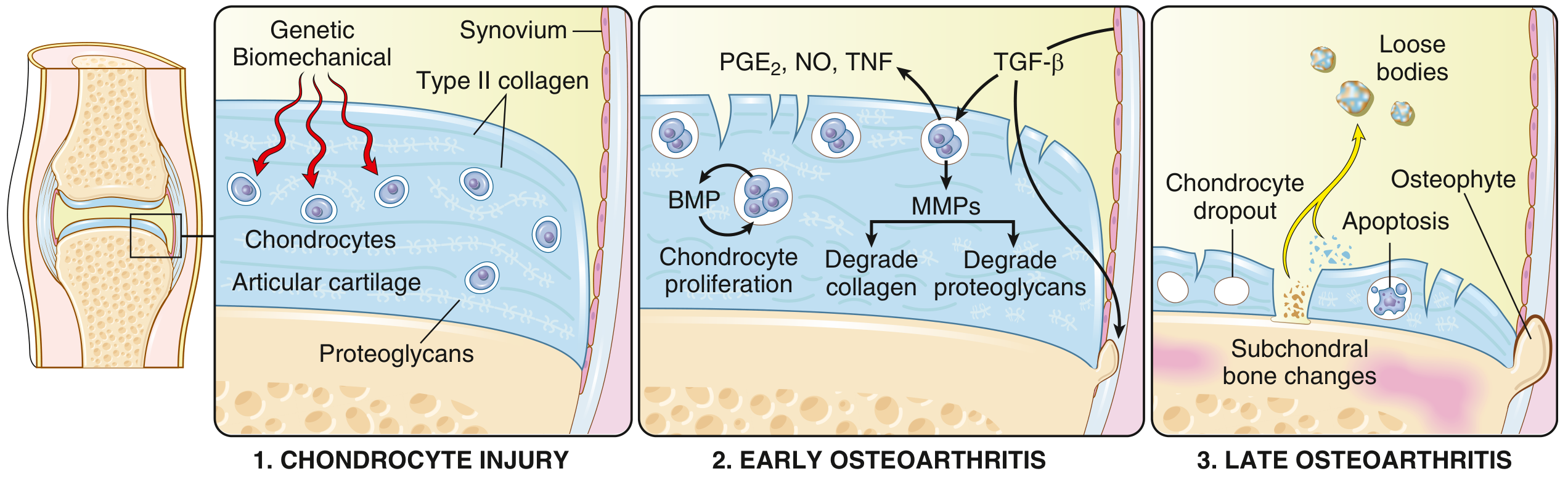
Biomechanical stress is the principal pathogenic mechanism, but genetic polymorphisms in matrix and signaling molecules predispose to chondrocyte injury. The sequence proceeds:
- Chondrocyte injury (genetic + biomechanical triggers) → early matrix changes
- Early OA: Chondrocytes proliferate and secrete matrix metalloproteinases (MMPs) that degrade type II collagen. Water content of matrix increases; proteoglycan concentration decreases. Collagen fibers are cleaved, creating surface fissures (fibrillation)
- Cytokines (TGF-β, IL-1, IL-6), prostaglandins (PGE2), and nitric oxide (NO) from chondrocytes, synovial cells, and macrophages amplify damage
- BMPs and TGF-β drive osteophyte formation at articular margins
- Late OA: Chondrocyte loss, full-thickness sloughing, loose bodies ("joint mice"), subchondral bone exposure and eburnation
Morphology (Pathological Changes)
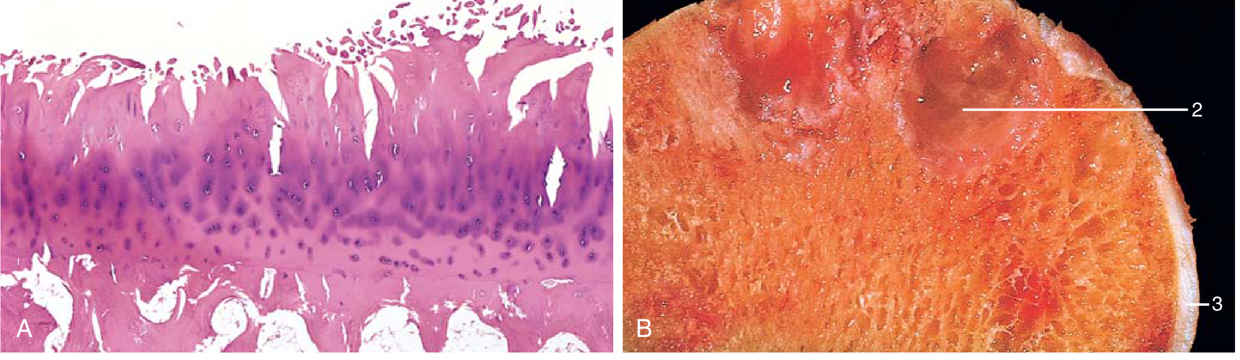
| Feature | Description |
|---|---|
| Cartilage fibrillation | Surface cracking and clefting; earliest histologic sign |
| Cartilage loss | Full-thickness sloughing in advanced disease |
| Loose bodies ("joint mice") | Dislodged fragments of cartilage and subchondral bone |
| Bone eburnation | Exposed subchondral bone polished to ivory-like appearance by friction |
| Subchondral cysts | Formed by synovial fluid forced into fracture gaps (ball-valve mechanism) |
| Osteophytes | Bony outgrowths at articular margins capped by fibrocartilage, then ossify |
| Synovium | Mildly congested and fibrotic; only scattered chronic inflammatory cells (unlike RA) |
OA vs. Rheumatoid Arthritis - Key Differences
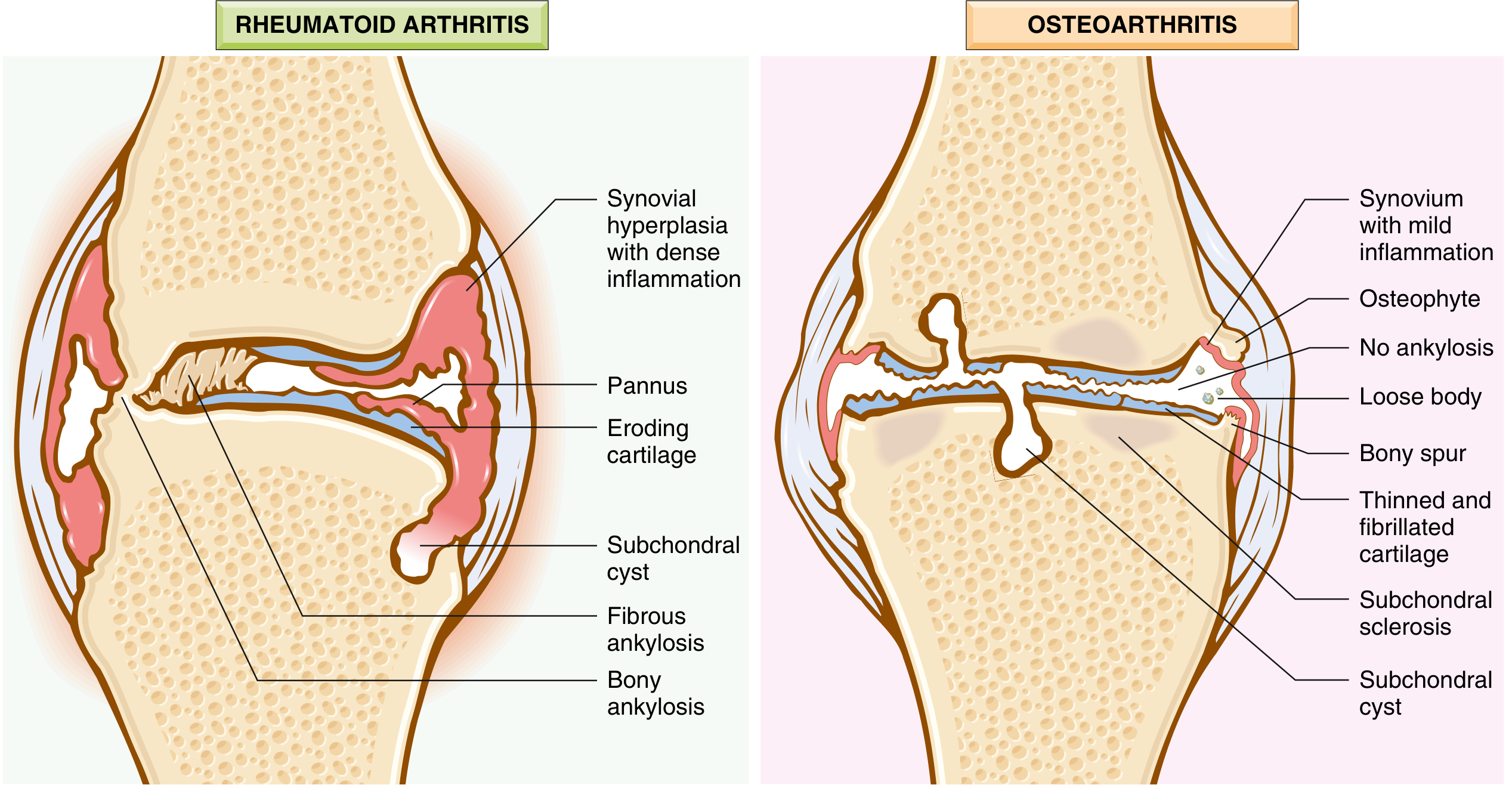
| Feature | Osteoarthritis | Rheumatoid Arthritis |
|---|---|---|
| Primary mechanism | Mechanical injury to cartilage | Autoimmunity |
| Inflammation | Secondary; low-grade | Primary driver |
| Joints involved | Weight-bearing (knees, hips, spine) | Small joints of fingers first; then multiple joints |
| Pathology | Cartilage degeneration, osteophytes, subchondral cysts | Inflammatory pannus, joint erosion, ankylosis |
| Serum antibodies | None | ACPA, rheumatoid factor |
| Other organs | No | Yes (lungs, heart, etc.) |
| Joint fusion | Does NOT occur | Occurs (ankylosis) |
Clinical Features
- Primary OA typically presents after age 50
- Joint pain that worsens with use (activity-related pain)
- Morning stiffness (typically brief, <30 minutes - unlike RA where it lasts >1 hour)
- Crepitus on movement
- Limited range of motion
- Joint deformity over time, but no fusion
Commonly involved joints:
- Knees and hips (weight-bearing)
- Lower lumbar and cervical vertebrae
- Proximal interphalangeal (PIP) and distal interphalangeal (DIP) joints of fingers
- First carpometacarpal joints
- First tarsometatarsal joints
Heberden nodes - osteophytes at the DIP joints; more common in women
Bouchard nodes - osteophytes at the PIP joints
Spinal OA: Osteophytes impinge on spinal foramina causing nerve root compression, radicular pain, muscle spasms, atrophy, and neurologic deficits.
Important: Radiographic severity does not correlate well with pain and disability.
Management
There are no treatments to prevent or halt OA progression. Therapy is directed at symptom control and functional restoration. - Robbins Pathology; Goldman-Cecil Medicine
Non-pharmacologic:
- Physical and occupational therapy to restore functional status (a critical component of care)
- Weight loss (particularly for knee/hip OA)
- Activity modification
- Assistive devices
Pharmacologic:
- NSAIDs - reduce inflammation and pain; first-line pharmacologic agents
- Intra-articular corticosteroids - for acute flares
- Analgesics (acetaminophen, topical agents)
- Pain management strategies
Surgical:
- Joint replacement is indicated when functional status interferes with activities of daily living and supportive care has failed; highly effective for end-stage hip and knee OA
Genetics (Key Points)
The largest OA GWAS (>800,000 samples from 9 populations) identified >52 OA risk loci, demonstrating different genetic risk variants for different joint sites. Key genes include:
-
GDF5 (chromosome 20) - codes for growth factor protein important in chondrogenesis
-
RUNX2, SMAD3, PTHLH - important in skeletal/bone development
-
BMI has a causative effect on OA risk (demonstrated via Mendelian randomization)
-
Firestein & Kelley's Textbook of Rheumatology
Sources: Robbins & Kumar Basic Pathology; Goldman-Cecil Medicine International Edition; Firestein & Kelley's Textbook of Rheumatology
This is a shared conversation. Sign in to Orris to start your own chat.