
Lab Investigations of transfusion reaction and mismatched blood transfusion
transfusion reaction hemolysis laboratory workup direct antiglobulin test
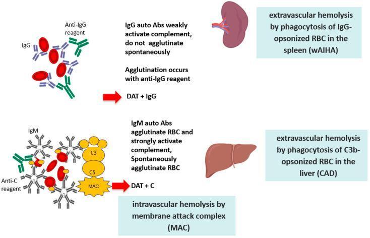
This pathophysiology diagram illustrates the two primary mechanisms of red blood cell (RBC) destruction in Autoimmune Hemolytic Anemia (AIHA). The top section depicts Warm AIHA (wAIHA), characterized by IgG autoantibodies. These monomeric antibodies weakly activate the complement system and do not cause spontaneous RBC agglutination; instead, agglutination occurs only with an anti-IgG reagent, resulting in a positive Direct Antiglobulin Test (DAT + IgG). This pathway leads to extravascular hemolysis via phagocytosis of IgG-opsonized RBCs primarily in the spleen. The bottom section depicts Cold Agglutinin Disease (CAD), mediated by pentameric IgM autoantibodies. These antibodies cause spontaneous RBC agglutination and strongly activate the complement cascade. The diagram shows the binding of C3, C5, and the formation of the Membrane Attack Complex (MAC), leading to intravascular hemolysis. Additionally, it highlights extravascular hemolysis occurring via phagocytosis of C3b-opsonized RBCs in the liver, with a positive Direct Antiglobulin Test for complement (DAT + C). Key anatomical landmarks include the spleen for IgG-mediated clearance and the liver for C3b-mediated clearance.

Summary : This image shows two process steps related to allergy management in the context of DAT (Direct Antiglobulin Test) administration, each presented as a separate node with directional connectors. flowchart: Nodes : • "Perform allergy testing, and (if necessary) desensitisation before DAT is given" (text node) • "Give DAT and manage subsequent allergy and reactions as they arise*" (text node) Connectors : • Each node has a diagonal arrow pointing away from it, suggesting a sequence or branching, but the destination of the arrows is not visible in the crop. Layout : • Two horizontally aligned nodes, each with an outgoing diagonal arrow. Analysis : • The diagram outlines two alternative or sequential steps in allergy management for DAT: pre-administration testing/desensitisation, and post-administration management of allergic reactions. The arrows indicate process flow, but the next steps are not shown in this crop.
This diagnostic clinical image displays a close-up, grayscale view of the bottom portion of a laboratory test tube containing a liquid sample. The image illustrates a state of incomplete dissolution or insolubility, characterized by clear stratification of the contents. A distinct, highly opaque, and dark region is concentrated at the base of the tube, indicating the presence of a heavy precipitate or sedimented particulate matter. The upper portion of the liquid appears relatively lighter and less dense, though it remains non-homogeneous. This visual is used in medical laboratory science to teach the identification of insoluble samples, often in the context of hemolysis detection or serum/plasma quality assessment. The educational focus is on recognizing physical properties such as precipitation and density-based layering, which are critical for determining specimen integrity and the suitability of a sample for diagnostic testing.
<table> <tr> <td>Aggregate Evidence Quality</td> <td>C</td> </tr> <tr> <td>Benefits</td> <td>Use of IVIG during escalation of care may reduce hemolysis and thereby stabilize or reduce TSB concentrations, preventing the need for exchange transfusion.</td> </tr> <tr> <td>Risk, harm, and cost</td> <td>The effect of IVIG for immune-mediated hemolytic disease has been understudied with conflicting evidence supporting a reduction in exchange transfusions. Recent investigations using routine single early dose prophylactic IVIG do not demonstrate benefit in reducing the need for exchange transfusion and the routine use of prophylactic IVIG in DAT + neonates should be discouraged. However, targeted dosing may be more effective. Although observational studies suggest that IVIG may be associated with necrotizing enterocolitis, the risk of necrotizing enterocolitis with exchange transfusion is well documented so the benefits of IVIG may outweigh this potential harm when exchange thresholds are approached.</td> </tr> <tr> <td>Benefit-harm assessment</td> <td>The benefits of IVIG are not clear, and there is a small risk of harm. Treatment with IVIG may be more strongly considered if there is a poor response to phototherapy and there is difficulty in obtaining an exchange transfusion.</td> </tr> <tr> <td>Intentional vagueness</td> <td>None</td> </tr> <tr> <td>Role of patient preferences</td> <td>Some families may want to avoid IVIG treatment given limited evidence for its effectiveness, especially for Rh+ infants and the potential risk of necrotizing enterocolitis.</td> </tr> <tr> <td>Exclusions</td> <td>None</td> </tr> <tr> <td>Strength</td> <td>Option</td> </tr> <tr> <td>Key references</td> <td>Technical report and 66–75</td> </tr> </table>
Lab Investigations of Transfusion Reactions and Mismatched Blood Transfusion
Overview: Immediate Action First
Step-by-Step Laboratory Workup
1. Clerical Check (Pre-analytical)
- Check all paperwork: patient ID band, blood bag label, compatibility label, and request form
- Confirm patient name, ID number, ABO/Rh group on the unit vs. the patient
2. Visual Inspection for Hemolysis
| Test | What to Look For |
|---|---|
| Patient plasma/serum | Pink or red discoloration = hemoglobinemia (free Hb) |
| Urine dipstick | Positive for blood = hemoglobinuria (no RBCs on microscopy in intravascular hemolysis) |
| Urine microscopy | Confirms hemoglobinuria vs. hematuria vs. myoglobinuria |
- Blood drawn >8 hours after the event may be visually clear but icteric; check serum bilirubin (elevated up to 24-36 hours)
- Free serum myoglobin can give a false-positive visual result - in that case, look for a clinical history of trauma
- Hemoglobinuria without hematuria = pathognomonic of intravascular hemolytic reaction
3. Repeat ABO/Rh Grouping
- Retype the patient (post-transfusion sample)
- Retype the donor unit (from the blood bag)
- Compare both against the original pre-transfusion sample
- Any discrepancy confirms ABO mismatch
4. Direct Antiglobulin Test (DAT / Coombs Test)
| Result | Interpretation |
|---|---|
| Positive DAT | Antibody or complement coated on transfused RBCs - immune-mediated hemolysis |
| Negative DAT | If severe hemolysis, may be negative due to rapid destruction of all coated cells; does not exclude hemolysis |
| Positive DAT + positive elution | Confirms immune-mediated AHTR |
| Negative serology + confirmed physical cause | Non-immune hemolysis (e.g., thermal injury, mechanical) |
- Always compare to the pre-transfusion DAT sample
- An elution study is performed on DAT-positive cells to identify the offending antibody
5. Hemolysis Panel - Core Lab Tests
| Investigation | Finding in Hemolytic Reaction | Notes |
|---|---|---|
| Serum haptoglobin | Decreased / absent | Haptoglobin binds free Hb; saturated and cleared rapidly in intravascular hemolysis |
| Serum LDH | Elevated | Released from lysed RBCs |
| Serum bilirubin (indirect/unconjugated) | Elevated | Product of Hb catabolism; elevated up to 24-36 h |
| Plasma hemoglobin (hemoglobinemia) | Elevated | Pink/red serum; direct evidence of intravascular lysis |
| Urine hemoglobin (hemoglobinuria) | Positive | Indicates heavy intravascular hemolysis |
| Peripheral blood smear | Spherocytes, fragmented RBCs | Extravascular hemolysis produces spherocytes |
| Serum potassium | Elevated | Released from lysed RBCs; risk of arrhythmia |
| Fibrinogen | Decreased | Indicates developing DIC |
| Hemoglobin (CBC) | Falling Hb | Inadequate post-transfusion rise or active fall |
Tietz diagnostic criterion: ≥2 of these hemolysis findings are required to confirm AHTR
6. Coagulation Screen (for DIC)
- PT / INR - prolonged
- aPTT - prolonged
- Fibrinogen - decreased
- D-dimer / FDPs - elevated
- Platelet count - falling
- Blood film - schistocytes
7. Renal Function Tests
- Serum creatinine and urea - rising values indicate acute kidney injury
- Urine output - oliguria/anuria is a danger sign
- Urine Hb - hemoglobin precipitates in renal tubules causing tubular necrosis
- Maintain urine output >1 mL/kg/hour with IV fluids + diuretics (furosemide)
8. Antibody Identification Panel
- Antibody screen on post-transfusion sample
- Antibody identification panel (panel cells) to determine the specificity of the alloantibody (e.g., anti-K, anti-Jka, anti-E)
- Crossmatch between donor unit and patient serum (repeat)
- Future transfusions: use antigen-negative, crossmatch-compatible blood
9. Blood Cultures
- From patient and donor unit (if septic transfusion reaction is suspected)
- Gram stain of the donor unit
- Bacterial contamination is a leading cause of transfusion fatality, especially with platelet units
Summary Table by Reaction Type
| Reaction Type | Key Lab Findings | Specific Tests |
|---|---|---|
| Acute Hemolytic (AHTR) - ABO mismatch | Hemoglobinemia, hemoglobinuria, ↓haptoglobin, ↑LDH, ↑bilirubin, spherocytes, + DAT | DAT + elution, repeat ABO, antibody panel, DIC screen |
| Delayed Hemolytic (DHTR) | Positive DAT 24h-28 days post-transfusion, new alloantibody, spherocytes, inadequate Hb rise | DAT, elution, antibody screen |
| Delayed Serologic (DSTR) | New alloantibody, positive DAT - NO clinical hemolysis | Antibody screen |
| Febrile Non-hemolytic (FNHTR) | Fever ≥38°C, ≥1°C rise from baseline within 4h - NO hemolysis | Rule out AHTR; blood cultures if severe |
| Allergic / Anaphylactic | No hemolysis; eosinophilia; low serum IgA (if anti-IgA mediated) | Serum IgA levels, anti-IgA antibodies |
| TRALI | Bilateral infiltrates on CXR; hypoxemia (PaO2/FiO2 <300); donor HLA/neutrophil antibodies | CXR, ABG, donor HLA antibody testing |
| TACO | Elevated BNP/NT-proBNP, CXR pulmonary edema | BNP, echocardiography |
| Septic | Positive blood cultures (patient + unit), DIC, high fever | Blood cultures, Gram stain |
Diagnostic Criteria for AHTR (NHSN/Tietz Standard)
-
Any key symptom during or within 24 hours of transfusion:
- Back/flank pain, chills/rigors, DIC, epistaxis, fever, hematuria, hypotension, oliguria/anuria, pain/oozing at IV site, renal failure
-
≥2 hemolysis laboratory findings from:
- Decreased fibrinogen, decreased haptoglobin, increased bilirubin, increased LDH, hemoglobinemia, hemoglobinuria, plasma discoloration, spherocytes on smear
-
Either:
- Positive DAT AND positive elution (immune-mediated), OR
- Negative serologic testing AND confirmed physical cause (non-immune)
Pathophysiology of AIHA/Hemolysis Mechanisms (Visual)
Key Points to Remember
- The most common cause of fatal hemolytic reactions is clerical/identification error, not laboratory failure
- ABO incompatibility (due to naturally occurring IgM isoagglutinins) causes intravascular hemolysis - the most severe type
- Non-ABO antigen incompatible reactions (e.g., anti-K, anti-Jka) are more frequent but less severe, causing extravascular hemolysis (IgG-mediated)
- A negative DAT does not exclude hemolysis if all coated cells have been rapidly destroyed
- Approximately 50% of ABO-incompatible transfusions have no adverse effect, but 5% are fatal
- All transfusion fatalities must be reported to the FDA (written report within 7 days in the US)
Description of indivial cell in "positive for malignancy, ascitic fluid cytology"
malignant cells ascitic fluid cytology adenocarcinoma peritoneal
malignant cells body fluid cytology nuclear enlargement hyperchromasia
?
Histopathology description of well differentiated Adenocarcinoma - Esophagus