
Perianal absses
perianal abscess anatomy classification
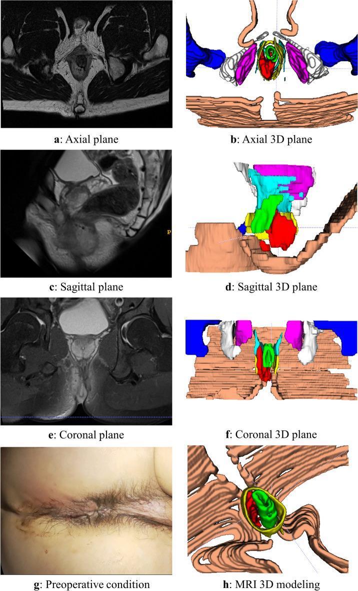
This composite educational image illustrates a case of an intersphincteric anorectal abscess through multi-modal diagnostic views. The panel includes original MRI scans and corresponding color-coded 3D reconstructions across axial (a, b), sagittal (c, d), and coronal (e, f) planes. The MRI images demonstrate the complex anatomy of the perianal region with the abscess appearing as a localized, high-signal intensity lesion on T2-weighted imaging. In the 3D models, the red structure represents the abscess, highlighting its spatial relationship to the anal canal, internal and external sphincters (represented by concentric green and yellow layers), and the levator ani muscle. Panel (g) provides a clinical photograph of the preoperative perianal region, and panel (h) displays a detailed 3D modeling view. This visual aid is designed to teach colorectal surgeons and radiologists the volumetric assessment and anatomical mapping of perianal fistulas and abscesses, emphasizing the value of 3D reconstruction in clarifying tissue demarcations that may be obscured in standard 2D grayscale radiology.
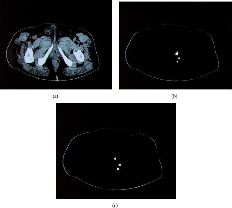
This composite diagnostic image demonstrates computerized tomography (CT) segmentation techniques for identifying a perianal abscess. Image (a) is an axial (transverse) CT slice of the pelvic and upper thigh region, showing high-density (white) femoral and pelvic bone structures, lower-density soft tissues, and the anal canal area where a perianal lesion is located. Images (b) and (c) present the results of automated lesion extraction using different machine learning models. Subfigure (b) shows the segmentation output of a standard Convolutional Neural Network (CNN), which identifies the abscess as scattered, less-defined white pixels against a masked dark background. Subfigure (c) demonstrates the result of a Deep Learning Fusion Convolutional Neural Network (DLFCNN) algorithm, showing a more precise and localized segmentation of the abscess tissue with reduced noise and clearer borders compared to the standard CNN. This comparison illustrates the clinical application of deep learning in radiology for the accurate classification and boundary delineation of soft tissue infections in the perianal region.
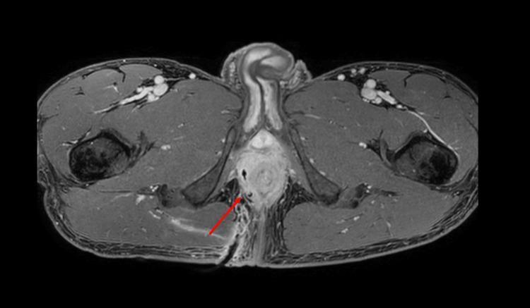
This diagnostic image is an axial MRI of the male pelvic region, showing the anatomy at the level of the anal canal and ischioanal fossa. The scan reveals a post-procedural state following the drainage of a complex perianal abscess. A red arrow points to a hypointense, linear tubular structure representing a percutaneous drain coursing through the right ischioanal fossa toward the central perianal area. The surrounding tissue shows a reduction in high-signal inflammatory fluid compared to a pre-drainage state, indicating abscess involution. Key visible structures include the central anal canal, the prostate gland located anteriorly, the external genitalia, and the bilateral gluteus maximus muscles and ischioanal fat. The image demonstrates the clinical application of interventional radiology in managing pelvic infections and the use of MRI for monitoring treatment response in complex fistulating disease.
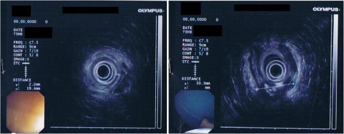
This diagnostic image displays two panels of transrectal endoscopic ultrasonography (TRUS) alongside corresponding endoscopic views. The TRUS images provide a cross-sectional view of the perianal and rectal anatomy. Key features include the internal anal sphincter (IAS), which is visualized as a continuous circular band with a measured thickness of approximately 2.2 mm. In the right posterior-lateral quadrant, a well-defined pathological lesion measuring approximately 30 mm x 15 mm is evident. This structure is predominantly hypoechoic, indicating fluid or soft tissue, and contains internal hyperechoic foci suggestive of gas bubbles—features characteristic of a perianal abscess with gas-forming components. The adjacent rectal mucosa appears normal in the integrated endoscopic inset. This imaging is clinically significant for evaluating the depth, extent, and specific echogenic characteristics of perianal inflammatory conditions or abscesses relative to the sphincteric complex.
Perianal Abscess
Definition & Pathogenesis
Classification by Anatomical Location
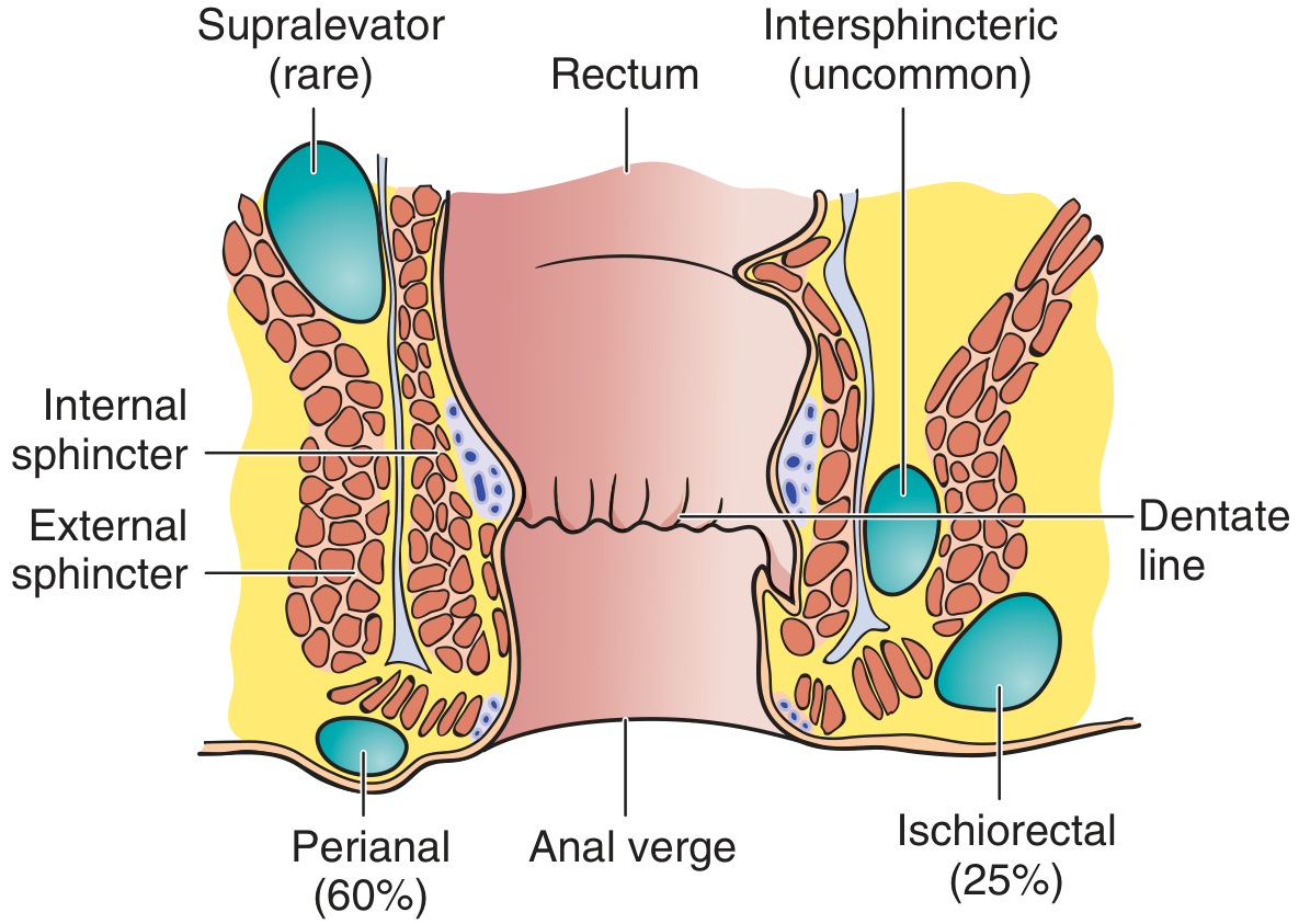
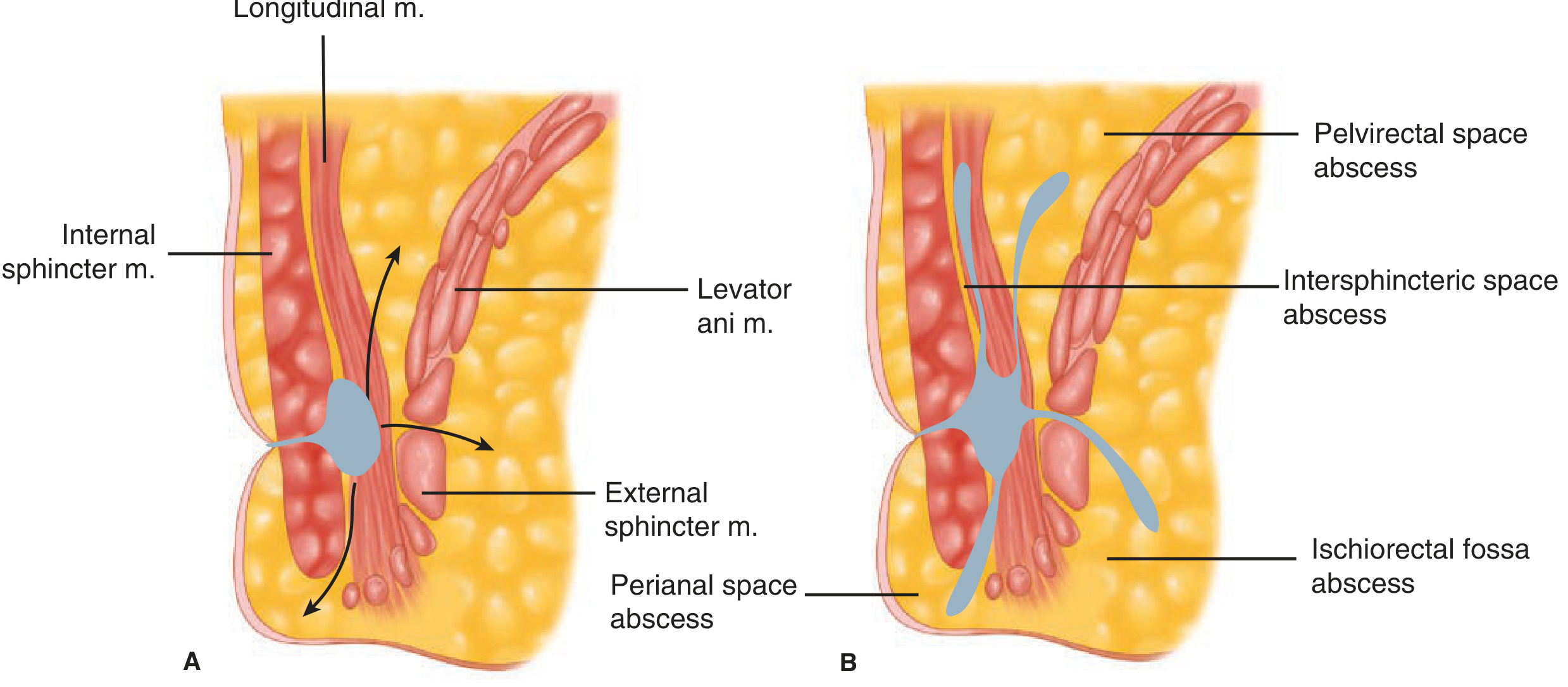
| Type | Incidence | Location | Clinical Features |
|---|---|---|---|
| Perianal | 60% | Adjacent to anal verge, in perianal tissues | Most common; easily visible and palpable externally |
| Ischiorectal | 25% | Ischiorectal fossa, outside the sphincters, 2-3 cm from anal verge | Diffuse swelling; may form horseshoe abscess |
| Intersphincteric | Uncommon | Between internal & external sphincters | NOT externally visible; deep "up inside" pain worsened by coughing/sneezing; diagnosed by digital rectal exam |
| Supraelevator (supralevator) | Rare | Above levator ani; perirectal rather than perianal | Must rule out intra-abdominal/pelvic source (appendicitis, diverticulitis, PID) |
- Pfenninger & Fowler's Procedures for Primary Care; Schwartz's Principles of Surgery, 11th ed.
Clinical Presentation
- Perianal pain and swelling - cardinal features
- Fever and marked leukocytosis (depending on severity)
- Erythema, tenderness, and fluctuation on exam (may be absent with intersphincteric, supraelevator, or deep ischiorectal abscesses)
- A history of increasing pain + fever + focal perianal tenderness should raise suspicion even without external signs
Diagnosis
- Clinical in most cases (visible fluctuant perianal swelling)
- Examination under anesthesia (EUA) when the cause cannot be determined in the office
- CT scan, MRI, or endorectal ultrasound - useful in occult, recurrent, or complex disease (e.g., supralevator, horseshoe abscesses)
- Anoscopy before I&D - if gently pressing the abscess expresses pus from an internal opening at the dentate line, this confirms an associated anal fistula
Treatment
- Necrotizing fasciitis
- Perineal sepsis (classic triad: pain + fever + inability to void) - a life-threatening emergency
I&D Technique (Pfenninger & Fowler's)
- Anesthesia: 2% lidocaine with epinephrine (27-30 gauge needle). Local anesthesia is often only marginally effective due to the acid environment of infected tissue - spinal or general anesthesia may be needed for larger abscesses.
- Incision: Radial to the anal canal circumference over the fluctuant area. This limits damage to underlying sphincter muscles.
- Excise a disk of skin to prevent premature closure - no packing is necessary for simple perianal abscesses. Sitz baths begin the next day.
- Break down loculations with a hemostat after entering the abscess.
- Drain or pack with iodiform gauze if needed; suction for large cavities.
Special Situations
- Ischiorectal abscess: Simple drainage through skin incision. Horseshoe abscesses require drainage of the deep postanal space + counterincisions over one or both ischiorectal spaces.
- Intersphincteric abscess: Requires EUA; internal sphincterotomy may be needed.
- Supraelevator abscess: Must work up for intra-abdominal source; requires more complex surgical drainage.
- Horseshoe abscess: Drainage of the deep postanal space with counterincisions bilaterally.
Antibiotics
- Not the primary treatment; used as adjuncts when there is:
- Significant surrounding cellulitis
- Systemic sepsis
- Immunocompromised patient, diabetes, or hematologic malignancy (leukemia, lymphoma, granulocytopenia) - in these patients, conservative antibiotic therapy +/- local radiation may be considered, though some advocate aggressive surgical drainage
Associated Anal Fistula
- ~50% of perianal abscesses have an associated anal fistula (some texts report up to 50% or higher)
- The fistula represents the chronic form - a persistent tract from the infected anal gland to the external skin
- Identifying the fistula at the time of I&D (via anoscopy) is important; unrecognized fistula is the most common cause of recurrence
Complications
| Complication | Notes |
|---|---|
| Recurrence | Most common complication; usually due to an unrecognized anal fistula |
| Anal fistula | Develops in ~50% if fistula not addressed at initial drainage |
| Necrotizing fasciitis | Life-threatening; if I&D is delayed - especially in immunocompromised patients |
| Perineal sepsis (Fournier's gangrene) | Medical emergency: pain + fever + inability to void |
| Bleeding / excess pain | Rare post-procedure complications |
Post-Procedure Care
- Sitz baths 10-15 min, 2-4 times per day
- Wound must heal from the inside out - prevent premature skin edge closure
- Daily wound irrigations if no gauze packing used
- Follow-up to rule out anal fistula or Crohn's disease if abscess recurs
Special Populations
- Immunocompromised / steroids / diabetes: Require prompt, aggressive I&D without delay; at high risk for rapid progression to necrotizing fasciitis
- Hematologic malignancy (leukemia, lymphoma): May harbor unusual organisms; individualized management (conservative antibiotics vs. surgical drainage)
- Crohn's disease: Always consider as an underlying cause, especially with recurrent perianal abscess; management in context of IBD is complex and often multidisciplinary