


25 year old male patient complaint of burning sensation since 4-5 days no other symptoms on other body parth cause and full line of treatment
- Image 1: Perioral area with glistening/greasy skin (possibly topical applied), mild hyperpigmentation, dry/wrinkled upper lip vermilion
- Image 2: Lower lip with asymmetric swelling/edema, multiple vertical fissures, dry/chapped appearance, no vesicles or crusting
cheilitis dry cracked swollen lips fissures
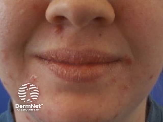
This frontal, close-up clinical photograph depicts the perioral region with multiple reddish papules surrounding the mouth, crusted fissures at the commissures, and mild desquamation of the lips. The vermilion border shows dry, cracked skin with papular lesions at the perioral gingivo-labial junction. There is bilateral involvement around the lips, with a pattern consistent with perioral dermatitis or angular cheilitis. No obvious swelling or mucosal involvement beyond the perioral skin. The skin tone shows mild erythema, dryness, and scaling. The image is captured in standard clinical photography settings with frontal orientation; there is no magnification evidence of dermoscopy. The observed lesions are non-ulcerative, non-vesicular, and present as clustered papules with crusts. The differential includes perioral dermatitis, angular cheilitis, irritant contact dermatitis, seborrheic dermatitis of the perioral region, candidal infection, and less likely herpes simplex infection which would show grouped vesicles. The clinical significance lies in distinguishing inflammatory, infectious, and irritant etiologies to guide therapy: topical antibiotics or antifungals, barrier repair, avoidance of irritants, and consideration of topical steroids in selective cases. This image is appropriate for dermatology education, clinical case discussions, and image-based training. Clinical relevance includes monitoring treatment response progress.
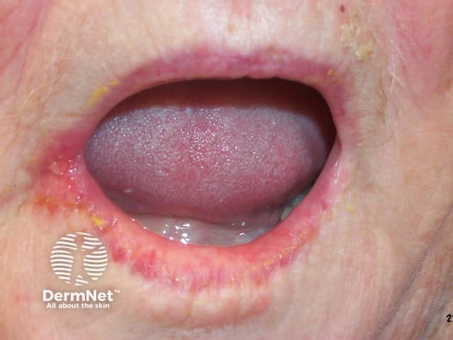
Clinical photography of a mucocutaneous lesion at the oral commissures illustrating angular cheilitis. The image shows erythematous, cracked corners of the mouth with superficial fissures, mild edema of perioral skin, and crust formation along the oral angles. The lips and adjacent skin are moist, and there is a subtle erythema extending onto the perioral mucosa. This presentation is characteristic of inflammatory cheilitis at the labial commissures and may reflect secondary fungal colonization or bacterial superinfection. The visual pattern—bilateral, symmetric fissuring with crusting—helps distinguish angular cheilitis from other perioral dermatoses such as irritant dermatitis, morsicatio labiorum, or herpetic stomatitis. Clinically, the finding signals possible underlying contributing factors including denture-mediated trauma, xerostomia, nutritional deficiencies (iron, zinc, B vitamins), or immune compromise. Diagnostic significance lies in recognizing the fissured commissures as a common, treatable condition; appropriate management includes barrier protection (emollients, ointments), antifungal therapy if candidiasis is suspected, and addressing causative factors. This image is valuable for dermatology and oral medicine education, differential diagnosis practice, and guiding holistic patient care in perioral inflammation. It supports case-based learning, standardizes descriptor terminology for commissural lesions, and can be paired with history, exam findings, and tests (iron studies, zinc, B12) to refine diagnosis and treatment planning.
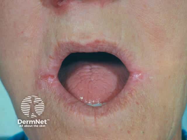
Clinical photograph of the perioral region in a denture wearer showing angular cheilitis at the oral commissures. This frontal close‑up captures bilateral fissuring and inflammatory erythema at the corners of the mouth, with occasional desquamation and crusting along the commissural folds. The lips appear dry and mildly macerated adjacent to the fissures, consistent with chronic irritation from ill‑fitting dentures, moisture accumulation, and repetitive lip movement. Superimposed candidal infection is a common comorbidity in denture wearers and may contribute to persistent cracking and maceration; yeast colonization can manifest as red, moist plaques or white pseudomembranes at the commissures, though not always evident in this image. The appearance supports a diagnosis of angular cheilitis (perleches angularis) secondary to denture‑related trauma, chronic salivary pooling, and local mucocutaneous barrier disruption. Differential considerations include irritant dermatitis, candidal angular cheilitis, bacterial superinfection (Staphylococcus), nutritional deficiencies (riboflavin, iron), and denture stomatitis with secondary involvement of the commissures. Clinically, management includes optimizing denture fit and hygiene, barrier protection with emollients, antifungal therapy if yeast is suspected, and addressing systemic risk factors. This image is valuable for education, dermatology and dentistry training, and multimodal retrieval of perioral inflammatory dermatoses. High-resolution capture aids pattern recognition and comparative diagnosis worldwide today.
Clinical Assessment
Diagnosis: Cheilitis (Exfoliative/Contact/Angular Type)
- Swelling and edema of the lower lip (right side predominant)
- Multiple vertical fissures across the lower lip vermilion
- Dry, chapped appearance with prominent mucosal creases
- Perioral skin with a glistening/applied-product appearance
- No vesicles, pustules, or hemorrhagic crusting (ruling out herpes labialis as primary)
- Burning sensation x 4-5 days, isolated to lips, no systemic involvement
Causes (Etiology)
| Category | Specific Cause |
|---|---|
| Habits | Lip licking, lip biting (most common in young males) |
| Nutritional deficiency | Vitamin B2 (riboflavin), B6 (pyridoxine), Iron deficiency |
| Irritant/Allergic contact | Toothpaste, mouthwash, lip balm, flavored foods, fragrances, lanolin, balsam of Peru |
| Atopic / Seborrheic | Atopic dermatitis, seborrheic dermatitis |
| Infectious | Secondary Candida albicans, Staphylococcus aureus (can complicate angular cheilitis) |
| Dry weather / environmental | Dehydration, cold/dry climate |
| Drug-related | Retinoid therapy (isotretinoin), protease inhibitors |
Investigations to Order
- CBC - rule out iron deficiency anemia
- Serum ferritin, iron studies
- Vitamin B2 (riboflavin) and B12 levels
- Random blood glucose / HbA1c - Candidal cheilitis is more common in diabetics
- KOH swab from lip fissures - if Candida suspected
- HIV screening - angular cheilitis is a WHO Clinical Stage 2 feature of HIV
- Patch testing - if allergic contact cheilitis is suspected (fragrances, toothpaste, balm ingredients)
Full Line of Treatment
Step 1 - Remove the Cause
- Stop all potential irritants: flavored/medicated toothpaste, fragrances, lip balms with fragrance/lanolin, hot spicy food, mouthwashes with alcohol
- Stop lip licking (most common habit in young men; saliva dries and worsens chapping)
- Keep lips well hydrated with plain water
Step 2 - Barrier Repair (Topical Emollients)
- White soft paraffin (petroleum jelly / Vaseline) - applied liberally 4-6 times/day; heals fissures effectively
- Zinc oxide ointment - applied to fissures, promotes healing
- Avoid commercial flavored lip balms during active inflammation
Step 3 - Topical Anti-inflammatory
- Mild topical corticosteroid (e.g., Hydrocortisone 1% ointment OR Mometasone 0.1% ointment) applied twice daily x 1-2 weeks
- Topical tacrolimus 0.1% ointment (Protopic) OR pimecrolimus 1% cream (Elidel) - second-line calcineurin inhibitors, especially if corticosteroids are not suitable; useful for chronic/recurrent cases
- As documented in Andrews' Diseases of the Skin, these are usually helpful for exfoliative cheilitis
Step 4 - Treat Infection (if present)
- If Candida suspected (white coating, commissure involvement): Clotrimazole 1% cream or Miconazole gel applied to lips TID x 2 weeks
- Or combined preparation: Nystatin + hydrocortisone ointment
- If Staphylococcal secondary infection (yellow crusting, painful): Mupirocin 2% ointment topically
Step 5 - Nutritional Supplementation (empirically in young patients)
- Vitamin B-complex tablet (especially riboflavin B2, pyridoxine B6, B12) once daily x 4-6 weeks
- Iron supplement (Ferrous sulfate 200mg OD) if ferritin low
- Zinc supplementation if dietary deficiency suspected
Step 6 - Systemic (if severe/unresponsive)
- Oral antihistamine (e.g., Cetirizine 10mg OD or Hydroxyzine 25mg HS) for pruritus/burning relief
- For significant inflammation not responding to topicals: short course oral prednisolone 0.5 mg/kg/day x 5-7 days (rarely needed)
Prognosis & Follow-up
- Acute exfoliative/contact cheilitis typically resolves in 2-4 weeks with proper topical treatment and trigger removal
- Review at 2 weeks to assess healing
- If not improving, consider patch testing, nutritional workup, or KOH culture
- In a 25-year-old male, rule out lip-licking habit and contact allergen first - these are the most common causes at this age
Clinical note: This is an educational overview. A proper diagnosis requires direct clinical examination. If the patient has risk factors for HIV (given angular cheilitis is a WHO Stage 2 feature), testing is strongly recommended. If the lesion does not respond in 3-4 weeks, biopsy should be considered to rule out rare causes such as plasma cell cheilitis or granulomatous cheilitis.