
today lets learn about cataract surgery from basic to advanced level, how does it work, it mechanism, why is it done, why in that plane, what are its pros and cons. Since this is the basic every ophthalmologist should know, explain in detail each and every line and also i have my thesis on this topic as well 'pain during cataract surgery using peribulbar vs topical anesthesia'. the viva questions that can be asked or anything; make me thorough about this topic
Reading File
Finding Sources
Finding Sources
Finding Sources
Searching PubMed
peribulbar anesthesia AND topical anesthesia AND cataract surgery pain
Reading File
Reading File
Reading Article
PMID: 22365066
Finding Sources
Reading File
Reading File
Reading File
Finding Sources
Reading File
Reading File
Reading File
Reading File
Reading File
Finding Sources
Loading Image
Reading File
Loading Image
Here is your complete, thorough guide to cataract surgery - from first principles to thesis-level depth.
CATARACT SURGERY: A COMPLETE GUIDE
From Basics to Advanced - Including Peribulbar vs Topical Anesthesia
PART 1: THE LENS - ANATOMY & PATHOPHYSIOLOGY
Normal Lens Structure
The crystalline lens is a transparent biconvex avascular structure suspended behind the iris by the zonular fibres (zonules of Zinn), which connect it to the ciliary body. Its layers from outside to inside:
- Lens capsule - the outermost layer; a thin, elastic, PAS-positive basement membrane that surrounds the entire lens. It is thickest anteriorly (14 µm) and thinnest posteriorly (4 µm). This is why the anterior capsule is easier to manipulate - and why the posterior capsule is more prone to rupture.
- Lens epithelium - a single layer of cells beneath the anterior capsule only. These cells are metabolically active and divide at the equator to form new lens fibers throughout life.
- Lens cortex - the outer softer layer made of recently formed lens fibers. These contain more water and are more easily aspirated.
- Epinucleus - a transitional zone between cortex and nucleus.
- Nucleus - the central, oldest, densest layer. The embryonic nucleus is formed in utero; fetal, infantile, and adult nuclei are added throughout life. With age the nucleus hardens (nuclear sclerosis) and turns yellow-brown (brunescent).
Why this anatomy matters for surgery: The surgeon exploits the distinct planes between nucleus, epinucleus, cortex, and capsule. Hydrodissection separates these planes deliberately before phacoemulsification begins.
What is a Cataract?
A cataract is any opacification of the crystalline lens, whether involving the nucleus, cortex, or subcapsular region. The lens doubles in volume between birth and age 70 as new lens fiber cells are laid down beneath the capsule.
The lens receives nutrients by diffusion from aqueous humor (it has no blood supply). When this diffusion is disrupted - by aging, oxidative stress, UV radiation, diabetes, steroids, trauma, or congenital causes - protein aggregation and water clefts form, causing opacification.
Types of Cataracts
| Type | Location | Key Features | Common Cause |
|---|---|---|---|
| Nuclear sclerosis | Nucleus | Yellow-brown discoloration, myopic shift | Aging |
| Posterior subcapsular (PSC) | Posterior capsule | Worst in bright light/reading, early visual loss | Steroids, diabetes, radiation |
| Anterior subcapsular | Anterior capsule | Less common, often due to uveitis | Trauma, inflammation |
| Cortical | Cortex, spoke-like | Water clefts, vacuoles, spokes | Diabetes, UV |
| Mature | Entire lens | White opaque lens, no red reflex | Advanced aging |
| Hypermature | Entire lens | Shrunken wrinkled anterior capsule, Morgagnian type | Neglected |
| Morgagnian | Liquefied cortex | Nucleus sinks inferiorly in liquid cortex | Very advanced |
Source: Kanski's Clinical Ophthalmology, 10th ed.
PART 2: INDICATIONS FOR SURGERY
Why Operate?
- Visual rehabilitation - the primary indication. When visual acuity impairs daily activities (driving, reading, work), the patient and surgeon together decide timing.
- Medical/complications:
- Phacolytic glaucoma - mature cataract leaks lens proteins through intact capsule, macrophages clog trabecular meshwork
- Phacomorphic glaucoma - intumescent lens causes pupil block or closes the angle
- Phacoanaphylaxis - lens protein acts as antigen; uveitis with possible hypotony
- Lens-particle glaucoma - post-traumatic lens cortex obstructs drainage
- To permit fundal examination - in patients where cataract prevents proper assessment of diabetic retinopathy, glaucoma, etc.
- Congenital cataracts - must be removed urgently to prevent amblyopia
Contraindications / Delay
From Miller's Anesthesia, 10th ed.:
- MI within 30 days (uncomplicated) or 60 days (complicated)
- PCI within 14 days (no stent) or 30 days (with stent)
- Decompensated heart failure
- Malignant hypertension
- Active infections, pulmonary embolus within 3 months
- Patient cannot lie flat
Important note: Elevated BP or hyperglycemia on the day of surgery does NOT automatically warrant cancellation. Antiplatelet/anticoagulation therapy does NOT need to be stopped for cataract surgery.
PART 3: PREOPERATIVE ASSESSMENT
- Biometry - measurement of axial length (A-scan ultrasound or optical biometry e.g. IOLMaster) and keratometry to calculate IOL power using formulae (SRK/T, Haigis, Barrett Universal II, etc.)
- Corneal topography if astigmatism management is planned
- Specular microscopy - endothelial cell count. If low (<1000 cells/mm²), higher risk of corneal decompensation
- Pupil dilation - mydriatics (tropicamide 1%, phenylephrine 2.5%)
- Povidone-iodine instillation - the single most important intervention to reduce endophthalmitis risk
- Routine medical tests are NOT required unless an acute comorbidity exists (per CMS/Society of Ambulatory Anesthesia guidelines)
PART 4: ANESTHESIA IN CATARACT SURGERY
This is the central topic of your thesis - let's go deep.
Overview of Options
| Method | Needle | Akinesia | Pain Relief | Complications |
|---|---|---|---|---|
| Topical | No | None | Cornea/conjunctiva only | Minimal |
| Sub-Tenon | Blunt cannula | Variable | Good | Chemosis, subconj. hemorrhage |
| Peribulbar | 25mm needle, outside cone | Good | Good-excellent | Globe perf (rare), chemosis, periorbital hematoma |
| Retrobulbar | Needle inside muscle cone | Excellent | Excellent | Globe perf, RB hemorrhage, brainstem anesthesia |
| General | - | Complete | Complete | GA risks |
A. TOPICAL ANESTHESIA
What it is: Local anesthetic applied as drops or gel to the ocular surface, blocking corneal and conjunctival sensation without any injection.
Agents used:
- Proxymetacaine (proparacaine) 0.5%
- Tetracaine (amethocaine) 0.5-1%
- Lidocaine 2% gel
Intracameral augmentation: Preservative-free lidocaine 0.2-1% is injected into the anterior chamber during hydrodissection. This significantly reduces intraoperative pain by anesthetizing the iris and ciliary body from inside. Combined Mydrane (tropicamide 0.02% + phenylephrine 0.31% + lidocaine 1%) is commonly used for simultaneous dilation and anesthesia.
Mechanism of action: Reversibly blocks sodium channels in nerve membranes, preventing depolarization. Blocks unmyelinated C-fibers and thin Aδ-fibers carrying pain. Does NOT block proprioception from extraocular muscles (hence no akinesia).
Pros:
- No injection - avoids injection-related complications
- No akinesia needed in experienced hands with cooperative patients
- No risk of globe perforation, retrobulbar hemorrhage, or brainstem anesthesia
- Rapid onset, no effect on IOP
- Patient preferred (no needle fear) - patients significantly prefer topical (Zhao et al., 2012, Ophthalmology [PMID 22365066])
- Day-case friendly; no pressure patching required
Cons:
- NO akinesia - patient eye movement is a significant risk; not suitable for uncooperative patients
- Intraoperative pain is significantly higher than with regional blocks (p<0.05, Zhao meta-analysis)
- Iris and ciliary body not adequately anesthetized by topical drops alone (require intracameral supplement)
- Not suitable for longer or more complex procedures
- Not suitable for patients with high pre-operative anxiety or high initial blood pressure
- Greater need for supplementary intraoperative anesthesia (p=0.03)
B. PERIBULBAR (PERICONAL) BLOCK
History: First described by Davis and Mandel in 1986 as a safer alternative to retrobulbar block.
Anatomical basis: The needle is placed outside the muscle cone (extra-conal space), in the peribulbar fat. Local anesthetic diffuses across the thin orbital septum into the retrobulbar space and muscle cone, eventually blocking the ciliary ganglion and all orbital nerves.
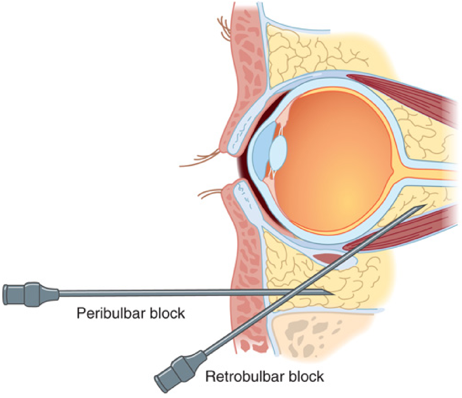
Why outside the cone? The classic retrobulbar block puts the needle INSIDE the muscle cone - behind the globe, in close proximity to the optic nerve and ophthalmic artery. This risks:
- Optic nerve injury
- Globe perforation
- Retrobulbar hemorrhage (1% incidence)
- Brainstem spread of anesthetic via optic nerve sheath
The peribulbar block deliberately stays outside this cone, making it significantly safer.
Technique (Classic):
- Classic: two injections - inferotemporal AND superonasal
- Modified single-injection: 3-cm, 23-gauge Atkinson needle at the junction of the middle and lateral thirds of the lower eyelid, just above the inferior orbital rim
- Direction: strictly parallel to the floor of the orbit (NOT angled superiorly - this risks globe perforation)
- Insertion depth: < 25 mm (this is critical - most globes are 24-25mm in length)
- If bone is contacted, redirect slightly upward
- Volume: 5-10 mL of local anesthetic
Why the plane matters:
- The orbital floor is flat; directing the needle parallel to it means it travels in the peribulbar fat without entering the cone
- Angling superiorly risks hitting the globe (which occupies ~80% of the orbital volume anteriorly)
- Depth <25mm ensures the needle does not pass behind the equator into the retrobulbar space
Agents used:
- Lidocaine 2% with hyaluronidase (hyaluronidase aids diffusion)
- Bupivacaine 0.5-0.75% (for longer duration)
- Mixture of lignocaine + bupivacaine + hyaluronidase is common
- Adrenaline (epinephrine) may be added for vasoconstriction (prolongs block)
Onset: Slower than retrobulbar (10-15 minutes vs 5 minutes) due to the need for the anesthetic to diffuse across the orbital septum.
Akinesia: Good but not complete - the superior oblique (CN IV), which lies outside the cone, may not be fully blocked (causing some intorsion on downgaze). Facial nerve block (van Lint, O'Brien, Atkinson, Nadbath) can be added if orbicularis akinesia is needed.
Pros:
- Superior pain relief vs topical anesthesia
- Complete akinesia - patient cannot move the eye, much safer for less-experienced surgeons
- Much safer than retrobulbar - no risk of retrobulbar hemorrhage
- Suitable for anxious patients, longer cases, high-volume cataracts
- Excellent surgical conditions (immobile eye + hypotony)
Cons:
- Injection itself is painful (needle injection)
- Higher incidence of chemosis, periorbital hematoma, subconjunctival hemorrhage vs topical (Zhao et al. 2012)
- Risk of globe perforation (rare, especially in long eyes >25mm axial length)
- Requires pressure on the eye post-injection (ocular massage/Honan balloon) to soften the eye and distribute anesthetic - adds time
- Requires facial nerve block if complete orbicularis akinesia needed
- Post-operative patching may be required until akinesia resolves
PART 5: THE SURGICAL PROCEDURE - STEP BY STEP
Historical Evolution
- Couching (ancient) - lens pushed posteriorly with a sharp instrument. No longer practiced.
- Intracapsular cataract extraction (ICCE) - entire lens + capsule removed through a large limbal incision. Needs cryoprobe. No posterior capsule remains, so IOL is placed in anterior chamber or sutured. High complication rate (vitreous loss, retinal detachment). Now obsolete.
- Extracapsular cataract extraction (ECCE) - anterior capsule opened, nucleus expressed manually, cortex aspirated. Posterior capsule left intact. Large incision (10-12mm), requires sutures. Still used in resource-limited settings and for very hard nuclei.
- Manual Small-Incision Cataract Surgery (MSICS) - modified ECCE via a scleral tunnel incision (6-7mm). No phaco machine needed. Widely used in developing world - fast, cheap, excellent results.
- Phacoemulsification (Phaco) - gold standard. Lens broken up ultrasonically, aspirated through a 2.2-2.8mm clear corneal incision. Foldable IOL inserted. Self-sealing wound.
- Femtosecond laser-assisted cataract surgery (FLACS) - laser performs incisions, capsulotomy, and lens fragmentation; then phaco completes extraction.
The Plane of Incision - Why Clear Cornea?
Modern phacoemulsification uses a clear corneal incision (CCI) in the corneal stroma, just anterior to the limbus. It is a 3-plane, self-sealing tunnel incision.
Why this plane:
- Self-sealing - the tunnel architecture (corneal bevel, pocket, and internal opening) means IOP closes the wound; no sutures needed
- Avascular - cornea has no blood vessels, so no bleeding
- Small (2.2-2.8mm) - induces minimal astigmatism, allows rapid healing
- Temporal location is preferred because:
- Better surgical access (the phaco tip can be angled more comfortably)
- Temporal incisions induce less surgically-induced astigmatism than superior incisions (which are closer to the steep vertical meridian)
- BUT: temporal incisions may carry slightly higher endophthalmitis risk than limbal incisions (greater exposure to lid flora)
Scleral tunnel (in MSICS): Made ~1-2mm behind the limbus through the sclera, into the scleral stroma, then forward into the anterior chamber. The conjunctival flap covers the scleral tunnel. This is self-sealing and less prone to endophthalmitis than clear corneal incisions.
Step-by-Step Phacoemulsification
1. Preparation
- Povidone-iodine 5% drops + eyelid cleaning (minimum 3 minutes contact time)
- Draping to exclude lashes from field
- Speculum insertion
2. Side-port incision
- ~1mm paracentesis, ~60° from the main incision (left of main incision for a right-handed surgeon)
- Used for the second instrument (manipulator/chopper)
- May do two side ports 180° apart
3. Viscoelastic (OVD) injection
- Injected into the anterior chamber (AC) through the side port
- Maintains AC depth, protects corneal endothelium, and stabilizes the iris
Cohesive OVDs (e.g. Healon, Provisc): high MW, maintain space well. Better for capsulotomy and IOL insertion. Can cause IOP spike postoperatively.
Dispersive OVDs (e.g. Viscoat, Ocucoat): adhere to surfaces, protect endothelium. Used when cornea is at risk (Fuchs' dystrophy). Harder to remove. Less IOP spike.
Soft-shell technique: Dispersive OVD placed first (protects endothelium), then cohesive OVD on top (to maintain AC). Used in high-risk corneas.
4. Main corneal incision
- 2.2-2.8mm clear corneal incision
- Three planes: initial bevel entry, corneal pocket, and internal opening (slightly larger to create valve)
5. Continuous Curvilinear Capsulorhexis (CCC)
- The most critical step
- A circular opening is made in the anterior capsule using a cystotome (bent 27G needle) or capsule forceps
- Why circular and continuous? A circular tear distributes stress evenly around the entire opening; a radial tear runs to the equator and beyond, making IOL implantation impossible
- Size: ideally 5.5-6mm - slightly smaller than a standard 6mm IOL optic, so the IOL is held within the capsular bag with the anterior capsule overlapping the optic edge (reduces posterior capsule opacification)
- Trypan blue (0.1%) can be used to stain the anterior capsule in dense white mature cataracts to make it visible when there is no red reflex
- Why is CCR difficult in hard cataracts? In hypermature cataracts with raised intralenticular pressure, the anterior capsule is under tension and the capsulotomy tends to "run out" peripherally. Dispersive OVD is injected first to lower intraocular pressure.
6. Hydrodissection
- A blunt cannula is inserted beneath the edge of the capsulorhexis and BSS (balanced salt solution) is gently injected
- A hydrodissection wave (fluid wave) should be visible beneath the capsule, separating the lens contents from the capsule
- The lens is then gently rotated to confirm free mobility
- Why: Without hydrodissection, the cortex remains adherent to the capsule; attempts to rotate or emulsify will tear the posterior capsule
- Also: hydrodelineation separates the epinucleus from the nucleus, leaving a soft shell around the nucleus that protects the posterior capsule
7. Phacoemulsification - Nucleus Emulsification
The phaco handpiece has a titanium hollow needle tip surrounded by a fluid-cooling sleeve. It operates at 28,000-40,000 Hz ultrasonic frequency. The tip moves back and forth (longitudinal phaco) or rotates (torsional phaco).
Three mechanisms of action:
- Jackhammer effect - mechanical microimpacts of the tip directly fragment the lens
- Cavitation - ultrasonic energy creates microscopic vapor bubbles that implode, releasing energy that breaks down lens material
- Acoustic streaming - fluid movement generated by ultrasound aids emulsification
Fluidics:
- Irrigation - BSS flows in through the sleeve around the tip, maintaining AC depth and cooling the tip
- Aspiration flow rate (AFR) - volume of fluid removed per minute. Higher AFR = faster lens attraction to tip but more surge risk.
- Vacuum - negative pressure that holds lens material against the tip during occlusion. Determines grip strength.
- Post-occlusion surge - when an occluded tip suddenly breaks contact with a lens fragment, the pent-up vacuum causes a sudden rush of fluid inward, potentially collapsing the AC and rupturing the posterior capsule. Modern phaco machines suppress surge.
Nuclear disassembly techniques:
Divide and conquer (safe, learner-friendly):
- Two perpendicular grooves sculpted into the nucleus (cross)
- The phaco tip and a second instrument are engaged in opposite walls of the groove
- Force applied in opposite directions to crack the nucleus into quadrants (the cracking step - nuclear material has cleavage planes)
- Each quadrant is then emulsified and aspirated
- Why effective: hard nucleus is broken into small pieces, each manageable
Phaco chop:
- A sharp-tipped chopper is embedded into the hard nucleus at the equator
- The chopper and phaco tip are pulled apart horizontally, splitting the nucleus
- Faster and uses less total ultrasound energy (less heat, better for corneal endothelium)
- Requires more skill - the chopper must reach under the capsulotomy to the equator
Stop and chop: Hybrid - initial groove then chop technique.
8. Epinucleus and Cortex Removal
- After the central nucleus is removed, the softer epinuclear shell is aspirated
- Cortical aspiration: Cortical spokes are engaged with an irrigation-aspiration (I/A) handpiece or bimanual system; the cortex is peeled toward the center (centripetal stripping) and aspirated
- Critical step: thorough cortex removal reduces posterior capsule opacification (PCO); retained cortex is the primary cause of postoperative inflammation and PCO
9. Posterior Capsule Polishing (optional)
- The posterior capsule is polished with a silicone-tipped cannula to remove remaining epithelial cells
- Reduces PCO
10. IOL Implantation
- Capsular bag is inflated with cohesive OVD
- Foldable IOL (acrylic or silicone) loaded into an injector cartridge
- Inserted through the main incision in folded form; unfolds within the capsular bag
- Haptics position themselves in the capsular bag equator, centering the optic
- Posterior capsule is left intact - this is the key difference from ICCE; it supports the IOL and prevents vitreous prolapse
11. Wound Hydration
- BSS injected into the wound stroma causes it to swell, sealing the self-sealing valve
- Wound integrity tested by pressing next to the wound (Seidel test)
- No sutures required for a well-constructed CCI <3mm
12. Subconjunctival or Intracameral Antibiotics
- Intracameral cefuroxime 1mg in 0.1mL is the evidence-based gold standard for endophthalmitis prophylaxis (ESCRS trial)
PART 6: INTRAOCULAR LENS (IOL)
Design Elements
Optic: The refracting element, typically 5.5-6.5mm diameter.
Haptics: Arms that stabilize the IOL in the capsular bag.
Materials: Hydrophobic acrylic (most common), hydrophilic acrylic, PMMA (rigid, ECCE), silicone.
Square-edge optic
Sharp 360° posterior edge creates a physical barrier to lens epithelial cell migration onto the posterior capsule, significantly reducing PCO. This is the most important design feature for reducing PCO.
Types of IOL
| Type | Function | Notes |
|---|---|---|
| Monofocal | Single focal point | Most common; patient needs glasses for near or distance |
| Toric | Corrects astigmatism | Must be aligned with steep meridian; rotation is main complication |
| Bifocal/Multifocal | Multiple focal points | Diffractive or refractive zones; halos/glare are common side effects |
| EDOF (Extended Depth of Focus) | Elongated focal range | Less halos than multifocal |
| Accommodative | Attempts to flex with ciliary body | Limited clinical accommodation in practice |
| Light-adjustable | UV post-operative power adjustment | Premium option, high precision |
Power Calculation
The IOL power is calculated using the lens formula relating corneal power (keratometry), axial length (biometry), and desired refraction. Modern formulae:
- SRK/T - widely used, regression-based
- Haigis - uses A-constant and anterior chamber depth
- Barrett Universal II - currently best for normal eyes
- Hill-RBF, Kane formula - AI-based, increasingly popular for extreme axial lengths
PART 7: COMPLICATIONS
Intraoperative
| Complication | Cause | Management |
|---|---|---|
| Posterior capsule rupture (PCR) | Excessive phaco energy, poor technique, weak capsule | Dispersive OVD to push vitreous back; anterior vitrectomy if needed; IOL in sulcus or ACIOL |
| Vitreous loss | PCR with vitreous prolapse | Vitrectomy; endophthalmitis risk |
| Zonular dialysis | Weak zonules (PXF, trauma, Marfan) | Capsular tension ring (CTR) |
| Corneal burn | Phaco tip occlusion, inadequate cooling | Stop phaco; increase irrigation |
| Iris prolapse | Shallow AC, high IOP | Viscoelastic to push iris back |
| Dropped nucleus | PCR with nucleus falling posteriorly | Stop, close, refer for pars plana vitrectomy |
| Expulsive hemorrhage | Rupture of choroidal vessels | Immediate wound closure; emergent canthotomy |
Signs of PCR:
- Sudden deepening or shallowing of the AC
- Momentary pupillary dilation
- Nucleus falls away from the phaco tip
- Vitreous enters phaco tip (slows aspiration)
- Torn capsule or vitreous visible
Postoperative
| Complication | Timing | Notes |
|---|---|---|
| Endophthalmitis | 1-7 days (acute bacterial) or weeks-months (chronic) | Emergency vitreous tap and intravitreal antibiotics; intracameral cefuroxime prophylaxis reduces risk by 5x |
| CMO (Irvine-Gass syndrome) | 4-12 weeks | Most common cause of reduced BCVA after uncomplicated phaco; NSAIDs + steroids |
| PCO | Months-years | Nd:YAG laser capsulotomy |
| Corneal decompensation (Bullous keratopathy) | Variable | Pre-existing low endothelial count + surgical trauma → DSEK/DMEK |
| Refractive surprise | Early | Biometry error; IOL exchange if significant |
| Uveitis | Early | Retained cortex, toxic OVD, infection |
| Raised IOP | Early | Retained OVD (treated with acetazolamide, topical anti-glaucoma drops) |
| Retinal detachment | Late | More common post-ICCE; young myopes at risk |
| Dysphotopsia | Any time | Positive (glare, halos - multifocal IOL) or negative (temporal arc) |
PART 8: FEMTOSECOND LASER-ASSISTED CATARACT SURGERY (FLACS)
The femtosecond laser (infrared, 1053nm) delivers ultrashort pulses that cause photodisruption of tissue at precise depths.
What the laser does:
- Corneal incisions - precise, reproducible
- Capsulotomy - more circular, more precisely centred, consistent size
- Lens fragmentation - pre-softens the nucleus (reduces phaco energy needed)
- Astigmatic keratotomy - limbal relaxing incisions
Advantages: Greater precision, reduced phaco energy (protects endothelium), potentially better refractive outcomes.
Disadvantages: Substantially higher cost, longer total operating time, technically challenging in small pupils, miosis during docking, suction ring-related subconjunctival hemorrhage, and no definitive evidence of better visual outcomes in uncomplicated cases.
PART 9: YOUR THESIS TOPIC - PERIBULBAR vs TOPICAL ANESTHESIA: PAIN IN CATARACT SURGERY
This is where you must be absolutely solid.
Background / Rationale
Both methods are widely used for phacoemulsification cataract surgery. The core clinical question is: does the superior pain relief of peribulbar block justify its additional injection-related risks and invasiveness compared to the simpler, safer topical anesthetic technique?
Key Evidence
Zhao LQ et al. (2012) Ophthalmology [PMID 22365066] - the landmark meta-analysis:
- 15 RCTs; 1084 eyes (topical) vs 1121 eyes (peribulbar/retrobulbar)
- All undergoing phacoemulsification
Results (critical for viva):
- Intraoperative pain: Significantly HIGHER in topical group (p<0.05)
- Postoperative pain: Significantly HIGHER in topical group (p<0.05)
- Inadvertent eye movement: More frequent in topical group (p<0.05)
- Need for supplementary anesthesia: Greater in topical group (p=0.03)
- Surgical difficulty (surgeon-assessed): No significant difference
- Patient preference: Patients significantly preferred topical (p<0.00001) - because they feared the injection
- Anesthesia-related complications (chemosis, periorbital hematoma, subconjunctival hemorrhage): More in peribulbar group (p<0.05)
- Surgical complications: No significant difference between groups
Conclusion from evidence: Regional (peribulbar) anesthesia provides better pain control; topical anesthesia achieves similar surgical outcomes but with more patient movement and more need for supplementary anesthesia. Patient preference for topical is driven by avoiding needle fear. Topical is not ideal for patients with high BP or high pain perception.
Pain Mechanisms During Cataract Surgery
With topical anesthesia, where does pain come from?
- Iris/ciliary body traction - the iris is the most pain-sensitive intraocular structure; manipulation during capsulorhexis, nucleus rotation, and IOL implantation causes ciliary nerve stimulation
- Light sensitivity - bright operating microscope light causes discomfort (retinal stimulation)
- Pressure changes - phaco energy transmission causes pressure waves inside the eye
- Conjunctival and corneal sensation - topical agents block this well, but NOT the deeper iris/ciliary body
- Oculocardiac reflex - traction on ocular structures can trigger bradycardia via the trigemino-vagal arc; peribulbar block partially prevents this (Dandekar et al., 2021 Indian J Ophthalmol [PMID 33727460])
With peribulbar anesthesia, pain is blocked because:
- The ciliary ganglion and short ciliary nerves are bathed in anesthetic as it diffuses into the cone
- Sensation from the iris, ciliary body, choroid, and cornea is completely blocked
- The oculomotor, trochlear, and abducens nerves are also blocked (akinesia)
Pain Assessment Tools Used in Research
- Visual Analogue Scale (VAS) - 0-10 horizontal line; simplest, most widely used
- Numerical Rating Scale (NRS) - 0-10 verbal
- Verbal Rating Scale (VRS) - none/mild/moderate/severe
- Your thesis should specify which scale you used, when pain was assessed (intraoperative vs postoperative), and who assessed it (patient or surgeon).
Confounders to Address in Your Thesis
This is gold for viva questions:
- Surgeon experience - inexperienced surgeons take longer; longer surgery = more pain in topical group
- Nucleus hardness (LOCS grading) - harder nucleus requires more phaco energy/time; more pain
- Patient anxiety - anxious patients perceive more pain; pre-operative counseling matters
- Intracameral lidocaine - topical + intracameral is significantly better than topical alone; your study must specify whether intracameral was used
- Sedation - midazolam, dexmedetomidine affect pain perception; must be standardized
- Pain during injection - the peribulbar injection itself is painful; this must be included in total pain assessment
- Intraoperative complications - PCR, vitreous loss cause more pain in topical group
- Patient selection bias - anxious/uncooperative patients are excluded from topical groups in practice
PART 10: VIVA QUESTIONS AND MODEL ANSWERS
Basic Questions
Q: Define cataract.
A: Any opacity of the crystalline lens, whether nuclear, cortical, or subcapsular, resulting in impaired vision.
Q: What is the most common type of cataract?
A: Age-related (senile) nuclear sclerosis - yellowing and hardening of the central nucleus due to progressive protein aggregation and loss of water content with aging.
Q: Why does nuclear sclerosis cause a myopic shift?
A: The hardened, denser nucleus has an increased refractive index, effectively increasing the power of the lens - causing index myopia. Patients may temporarily read without glasses ("second sight of the aged").
Q: What is the difference between ECCE and phacoemulsification?
A: ECCE uses a large incision (10-12mm) and expresses the nucleus manually, requiring sutures. Phacoemulsification uses ultrasound to emulsify the nucleus through a small (2.2-2.8mm) self-sealing corneal incision, requiring no sutures and allowing faster visual rehabilitation.
Q: Why is the posterior capsule preserved in modern surgery?
A: It supports the IOL in the capsular bag, prevents vitreous prolapse into the anterior segment, reduces CMO risk, and enables implantation of posterior chamber IOLs.
Q: What is capsulorhexis and why must it be circular and continuous?
A: Capsulorhexis is the creation of a smooth, circular opening in the anterior lens capsule. It must be circular and continuous because a circular tear distributes stress uniformly - any radial extension runs to the equator and beyond, preventing safe nucleus manipulation and IOL implantation within the capsular bag.
Q: What is hydrodissection? Why is it done?
A: BSS is injected beneath the anterior capsule edge to separate the lens cortex and epinucleus from the capsule. This allows safe nuclear rotation, reduces stress on the zonules during manipulation, and permits cortical stripping during aspiration.
Intermediate Questions
Q: Explain the mechanism of phacoemulsification.
A: The titanium phaco tip oscillates at 28,000-40,000 Hz. It works via: (1) jackhammer mechanical fragmentation, (2) cavitation - implosion of microscopic vapor bubbles releasing energy, and (3) acoustic streaming - fluid movements that aid emulsification. Simultaneously, irrigation maintains AC volume and temperature, while aspiration removes emulsified material.
Q: What is post-occlusion surge and how is it prevented?
A: When the phaco tip is occluded by lens material and occlusion suddenly breaks, the stored vacuum energy causes a rapid rush of fluid into the tip - surge - which can collapse the AC and rupture the posterior capsule. Modern machines use software algorithms, compliance chamber systems, and reduced tubing to dampen surge.
Q: What is the role of OVDs in cataract surgery?
A: OVDs (viscoelastics) maintain AC depth and protect the corneal endothelium from phaco energy. Cohesive OVDs maintain space; dispersive OVDs protect surfaces. The soft-shell technique (dispersive first, then cohesive) provides both benefits. All OVDs must be completely removed at the end to prevent postoperative IOP spike.
Q: What is the significance of the square optic edge on IOLs?
A: A sharp 360° posterior edge creates a physical barrier that prevents lens epithelial cells from migrating posteriorly onto the posterior capsule, significantly reducing PCO incidence compared to round-edged designs.
Q: What are the signs of posterior capsule rupture intraoperatively?
A: Sudden change in AC depth (deepening or shallowing), momentary pupillary dilation as the zonulo-hyaloid diaphragm shifts, nucleus falling away from the phaco tip, markedly slowed aspiration as vitreous enters the tip, and direct visualization of torn capsule or vitreous.
Anesthesia Questions (Thesis-Specific)
Q: What are the anatomical boundaries of the peribulbar space?
A: The peribulbar (extraconal) space is bounded anteriorly by the orbital septum, posteriorly by the annulus of Zinn, medially and laterally by the extraocular muscles forming the muscle cone. The peribulbar block deposits anesthetic outside this cone.
Q: Why does peribulbar block have a slower onset than retrobulbar?
A: Because the anesthetic is deposited outside the muscle cone and must diffuse across the intramuscular septum into the retrobulbar space and cone to reach the ciliary ganglion and optic nerve sheath. Retrobulbar block deposits the anesthetic directly inside the cone, adjacent to the ciliary ganglion. Hyaluronidase is added to the peribulbar injection to facilitate this diffusion.
Q: What is the risk of globe perforation with peribulbar block and how is it minimized?
A: Globe perforation is rare (<1 in 1000) but serious, leading to retinal detachment, vitreous hemorrhage, and blindness. It is minimized by: using a 25mm needle (not longer), directing it parallel to the orbital floor (not angled superiorly), limiting insertion depth to <25mm, avoiding highly myopic eyes (axial length >26mm) where globe diameter is enlarged, and using blunt-tipped needles in some protocols.
Q: Why do patients prefer topical anesthesia despite more intraoperative pain?
A: Multiple studies show patients prefer topical primarily because it involves no injection - they fear the needle more than the intraoperative discomfort. Patient satisfaction surveys consistently show higher preference for topical even when pain scores during surgery are higher.
Q: What is intracameral anesthesia and how does it improve topical anesthesia?
A: Preservative-free lidocaine 0.2-1% injected directly into the anterior chamber during surgery provides anesthesia to the iris and ciliary body - structures that topical drops cannot reach. Studies show significantly less pain with topical + intracameral compared to topical drops alone. Intracameral Mydrane (lidocaine + tropicamide + phenylephrine) additionally provides dilation.
Q: What is the oculocardiac reflex and how does choice of anesthesia affect it?
A: The OCR is a trigeminovagal reflex where traction on ocular structures (particularly the extraocular muscles) triggers the afferent trigeminal arc, leading to vagally-mediated bradycardia, arrhythmias, and occasionally cardiac arrest. Peribulbar block reduces OCR incidence by blocking the afferent trigeminal arc from intraocular structures. A 2021 RCT (Dandekar et al., PMID 33727460) in Indian J Ophthalmol found OCR was more frequent with topical than peribulbar anesthesia during phacoemulsification.
Q: What is the role of facial nerve block in cataract surgery?
A: Cataract surgery requires an immobile orbit, but also requires the patient not to forcefully squeeze the eyelids (which raises IOP and can extrude intraocular contents). Topical anesthesia provides no orbicularis akinesia. Facial nerve blocks (van Lint - at orbital rim; O'Brien - at condyle; Atkinson - at zygoma; Nadbath - at mastoid/mandible) block branches of CN VII supplying the orbicularis oculi. Van Lint is most commonly used.
Q: What are the contraindications to topical anesthesia for cataract surgery?
A: Uncooperative patient, dementia, cognitive impairment, head tremor, excessive anxiety, high pre-operative blood pressure, communication difficulties, long/complex surgery anticipated, surgeon inexperience, and patient with very low pain threshold.
Q: How would you measure pain in your thesis study, and at what time points?
A: Pain is best measured using the Visual Analogue Scale (VAS) 0-10 cm. Time points should include: (1) during anesthesia administration (injection pain for peribulbar; drop instillation for topical), (2) intraoperatively at defined steps (capsulorhexis, phaco, cortex aspiration, IOL insertion), (3) immediately postoperative (30 minutes), and (4) at 24 hours. Both patient and observer (surgeon) ratings should ideally be captured.
Advanced Questions
Q: What are the advantages of FLACS over conventional phacoemulsification?
A: Greater precision of capsulotomy (more circular, better centred), reduced phaco energy (lower endothelial cell loss), automated corneal incisions, and integration with astigmatic correction. However, evidence does not show better visual outcomes in uncomplicated cases, and the cost is substantially higher.
Q: What is the Irvine-Gass syndrome?
A: Cystoid macular oedema (CMO) occurring after cataract surgery, classically 4-12 weeks postoperatively, due to prostaglandin-mediated disruption of the blood-retinal barrier. It is the most common cause of unexplained reduced visual acuity after an apparently successful phacoemulsification. Risk factors include vitreous loss, uveitis, and use of prostaglandin analogue glaucoma drops. Treated with topical NSAIDs and steroids.
Q: A patient has axial length 28mm. What precautions should you take during anesthesia?
A: Avoid peribulbar/retrobulbar block if possible - the enlarged globe occupies more orbital volume, increasing risk of globe perforation. Topical or sub-Tenon block is preferred. If peribulbar is necessary, use a shorter needle (<23mm), ensure it is directed strictly parallel to the orbital floor, and limit depth to <25mm.
Q: What is pseudoexfoliation and how does it affect cataract surgery?
A: Pseudoexfoliation (PXF) is a systemic condition where abnormal fibrillar extracellular material deposits on the lens capsule, ciliary body, iris, and trabecular meshwork. It weakens the zonular fibers, leading to zonular laxity or dialysis during phacoemulsification. Management includes: preoperative iris dilation (PXF eyes dilate poorly), use of a capsular tension ring (CTR) to stabilize the capsular bag, gentle phaco technique, and awareness that the nucleus may be very hard.
Q: What is posterior capsule opacification (PCO) and how is it treated?
A: PCO (after-cataract) occurs when residual lens epithelial cells at the equator undergo fibrous metaplasia and migrate onto the posterior capsule, causing it to opacify and reducing vision. It is the most common long-term complication of cataract surgery. Treatment is Nd:YAG laser posterior capsulotomy - the laser creates a central opening in the opacified capsule. Prevention includes: thorough cortex removal, square-edge IOL design, and small capsulorhexis that overlaps the optic.
Summary Comparison Table: Peribulbar vs Topical
| Parameter | Peribulbar | Topical |
|---|---|---|
| Injection | Yes (needle) | No |
| Akinesia | Good (complete in most) | None |
| Pain relief (intraop) | Superior | Less effective alone |
| Need for supplementary | Less | More |
| Patient preference | Less preferred | Preferred (no needle) |
| Onset | 10-15 min | Immediate |
| Duration | 2-3 hours | 20-30 min |
| Volume | 5-10 mL | 2-4 drops |
| Chemosis/hematoma | More common | Rare |
| Globe perforation | Rare (0.1%) | None |
| Brainstem spread | Possible (rare) | None |
| Suitability | Anxious, complex cases | Cooperative, experienced surgeon |
| OCR prevention | Better | Less effective |
| Postop patch | Sometimes needed | Not needed |
Key References:
- Zhao LQ et al. (2012), topical vs regional anesthesia meta-analysis - 15 RCTs, 2205 eyes; landmark paper for your thesis.
- Dandekar P et al. (2021), oculocardiac reflex peribulbar vs topical - Indian J Ophthalmol RCT.
- Kanski's Clinical Ophthalmology, 10th ed. - Chapter 10 (The Lens), pp. 333-342.
- Miller's Anesthesia, 10th ed. - Chapter 65 (Ophthalmic Anesthesia), pp. 9724-9740.
This guide covers everything from the first principles of why a cataract forms, through every surgical step and why each is done in that plane and sequence, to a full thesis-level analysis of the pain comparison between peribulbar and topical anesthesia with the supporting evidence. Use the viva questions to test yourself systematically - examiners often push from basic anatomy into intraoperative reasoning, and then pivot to complications and anesthesia choice logic. Good luck with your thesis!
what about the phacodynamics and other techniques extracapsular and intracapsular
Your credits are exhausted. You can purchase a top-up or subscribe to a plan to continue using Orris. If you're already subscribed, your daily credits will reset tomorrow.
This is a shared conversation. Sign in to Orris to start your own chat.