
anatomy of eye including defination structures and their functions
Reading File
Now let me search the medical library for eye anatomy content in parallel:
Finding Sources
Finding Sources
Reading File
Reading File
Reading File
Reading File
Reading File
Loading Image
I now have rich content from multiple authoritative textbooks. Let me fetch one more diagram for the cross-section and compile the full answer.
Loading Image
Loading Image
Excellent - I have all the content and diagrams needed for a comprehensive answer. Here it is:
Anatomy of the Eye
Definition
The eye (Latin: oculus; Greek: ophthalmos) is the paired sensory organ of vision. It is a roughly spherical structure approximately 24 mm in diameter, housed within the bony orbit of the skull. Its function is to convert light energy in the visible spectrum (397-723 nm) into electrical impulses that travel via the optic nerve to the visual cortex, where they produce the sensation of sight.
The globe has a unique double-sphere shape - the cornea at the front has a steeper radius of curvature than the sclera behind it, which is what allows it to begin refracting light immediately on entry.
Overview Diagram
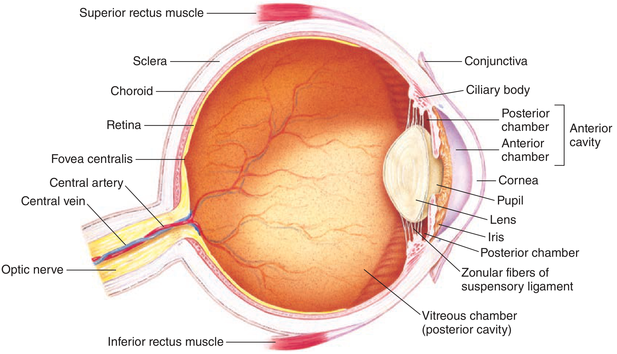
Cross-sectional anatomy of the human eye - Ganong's Review of Medical Physiology
Three Structural Layers (Tunics) of the Eye
The eye wall is composed of three concentric layers:
| Layer | Components |
|---|---|
| Outer fibrous layer | Sclera + Cornea |
| Middle vascular layer (Uvea) | Choroid + Ciliary body + Iris |
| Inner neural layer | Retina |
1. Outer Layer (Fibrous Tunic)
Sclera
- The tough, white, opaque outer coat of the eye
- Composed of dense fibrous connective tissue
- Function: Provides structural rigidity and maintains the shape of the eyeball; protects internal structures; serves as attachment for the six extraocular muscles
- No light passes through it
Cornea
- The transparent, avascular, dome-shaped anterior continuation of the sclera
- Has a steeper radius of curvature than the sclera
- Function: The primary refracting surface of the eye (accounts for ~70% of total refractive power). Its avascular nature is maintained by nourishment from the aqueous humor behind it and tears in front
- The lateral margin of the cornea is continuous with the conjunctiva
2. Middle Layer (Uveal Tract / Vascular Tunic)
The uvea consists of three parts: choroid, ciliary body, and iris.
Choroid
- A vascular layer lining the inner surface of the sclera, covering the posterior two-thirds of the globe
- Contains large vessels, small vessels, and a network of capillaries (choriocapillaris)
- Function: Supplies oxygen and nutrients to the outer layers of the retina; its dark pigment absorbs stray light to prevent internal reflection
Ciliary Body
- A thickened ring of tissue located between the choroid and the iris
- Contains the ciliary muscle (smooth muscle) and produces aqueous humor
- Functions:
- Ciliary muscle contraction/relaxation changes tension on the zonule fibers, altering lens shape for accommodation (focusing at different distances)
- Actively secretes aqueous humor (via carbonic anhydrase and cytochrome oxidase systems at the ciliary epithelium) at ~2 µL/min
- The iris dilator muscle is sympathetically innervated; the iris sphincter and ciliary muscle have parasympathetic innervation
Iris
- The pigmented, opaque, circular diaphragm in front of the lens
- Contains two sets of muscles:
- Sphincter pupillae (circular fibers) - constricts the pupil (miosis); parasympathetic innervation
- Dilator pupillae (radial fibers) - dilates the pupil (mydriasis); sympathetic innervation
- Function: Controls the amount of light entering the eye. Variations in pupil diameter can produce up to a 16-fold change in the amount of light reaching the retina
Pupil
- The central aperture/opening in the iris
- Appears dark because of the light-absorbing pigments in the retina
- Function: Regulates light entry
3. Inner Layer (Neural Tunic)
Retina
- A neurosensory membrane lining the posterior two-thirds of the choroid
- Composed of 10 histological layers that convert light impulses into neural impulses
- Contains two types of photoreceptors:
- Rods - for dim light / peripheral vision / motion detection (~120 million)
- Cones - for color vision and fine detail / high-acuity vision (~6 million; concentrated in the fovea)
Key retinal landmarks:
| Structure | Location | Function |
|---|---|---|
| Macula lutea | Center of the posterior retina | Area of highest cone density; responsible for central, high-acuity color vision |
| Fovea centralis | Center of the macula | Contains only cones; point of maximum visual acuity |
| Optic disc (optic disk) | Nasal to the fovea, where optic nerve exits | No photoreceptors here = the "blind spot" |
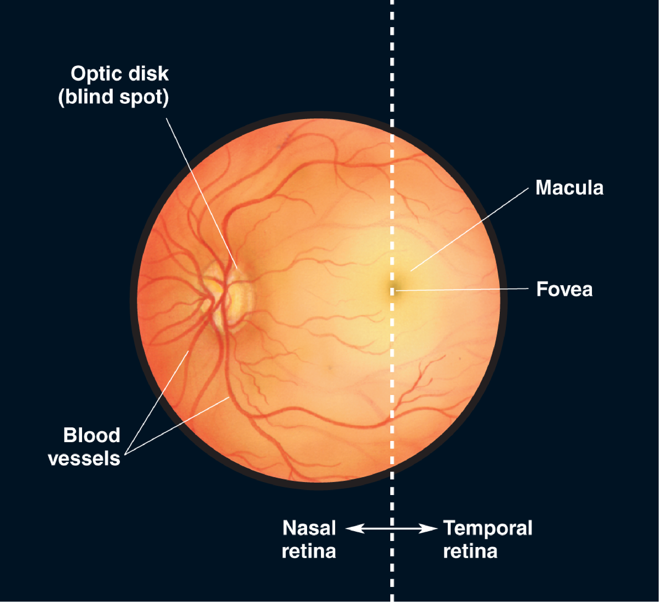
Retina as seen through an ophthalmoscope - Neuroscience: Exploring the Brain, 5th Ed.
Internal Structures
Crystalline Lens
- A transparent, biconvex, elastic structure located posterior to the pupil and iris
- Held in place by the zonule fibers (suspensory ligaments) attached to the ciliary body
- Function: Fine-tunes refraction to focus images on the retina; changes shape (accommodation) to focus near and far objects
- When the ciliary muscle contracts, tension on zonule fibers decreases and the lens becomes more rounded (increases refractive power for near vision)
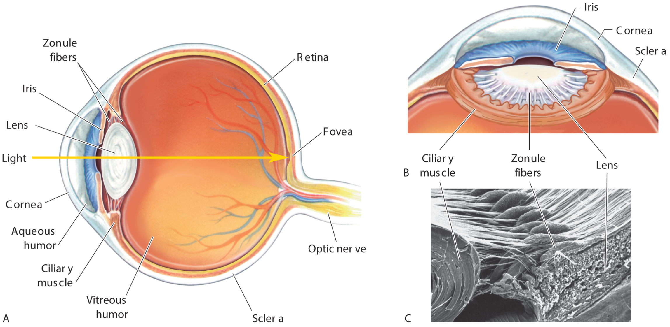
Lens and its supporting structures - Neuroscience: Exploring the Brain, 5th Ed.
Aqueous Humor
- A clear, protein-free fluid filling the anterior chamber (between cornea and iris) and posterior chamber (narrow space between iris, zonule, and lens)
- Produced in the ciliary body, flows through the pupil into the anterior chamber, and drains via the trabecular meshwork into the Canal of Schlemm
- Function: Nourishes the avascular cornea and lens; maintains intraocular pressure (IOP). Obstruction of drainage raises IOP - a key risk factor for glaucoma
Vitreous Humor
- A clear, gelatinous material filling the large vitreous chamber (posterior cavity) between the lens and the retina
- Function: Keeps the eyeball spherical; maintains the retina pressed against the choroid; transmits light
Accessory / Protective Structures
Conjunctiva
- A clear mucous membrane covering the anterior sclera and lining the inner surface of the eyelids
- Function: Protects the eye; serves as a site for drug absorption (popular route for ophthalmic drug administration)
Eyelids
- Composed of four layers: conjunctiva, cartilaginous tarsal plate, muscle layer (orbicularis oculi + levator palpebrae superioris), and skin
- Function: Protect the eye from foreign objects; spreading of the tear film during blinking keeps the cornea moist
Lacrimal System
- Lacrimal gland - located in the superior temporal orbit; produces the tear film
- Lacrimal drainage system - composed of the puncta, canaliculi, lacrimal sac, and nasolacrimal duct, draining into the nasal cavity
- Function: Maintain a moist anterior ocular surface; protect the cornea
Orbit
- A bony pyramidal cavity in the skull formed by seven bones: frontal, zygomatic, greater wing of sphenoid, maxilla, palatine, lacrimal, and ethmoid
- Function: Protects the globe and its associated structures from mechanical injury
- The optic foramen at the orbital apex transmits the optic nerve and ophthalmic artery
Extraocular Muscles (6 muscles)
| Muscle | Action | Nerve |
|---|---|---|
| Superior rectus | Elevation, adduction, intorsion | CN III (Oculomotor) |
| Inferior rectus | Depression, adduction, extorsion | CN III |
| Medial rectus | Adduction | CN III |
| Lateral rectus | Abduction | CN VI (Abducens) |
| Superior oblique | Depression, abduction, intorsion | CN IV (Trochlear) |
| Inferior oblique | Elevation, abduction, extorsion | CN III |
Innervation Summary
- Optic nerve (CN II) - carries visual impulses from the retina to the brain
- Oculomotor nerve (CN III) - motor to 4 extraocular muscles + levator palpebrae; parasympathetic to iris sphincter and ciliary muscle (via ciliary ganglion)
- Trochlear nerve (CN IV) - motor to superior oblique muscle
- Abducens nerve (CN VI) - motor to lateral rectus muscle
- Trigeminal nerve (CN V1 - ophthalmic branch) - sensory to cornea, iris, ciliary body
- Sympathetic fibers (from carotid plexus, through superior orbital fissure) - dilator pupillae and lid retraction
Blood Supply
- Arterial supply: Branches of the ophthalmic artery (from the internal carotid artery), including the central retinal artery
- Venous drainage: Superior and inferior ophthalmic veins + central retinal vein, all draining into the cavernous sinus
Sources: Ganong's Review of Medical Physiology, 26th Ed. | Barash, Cullen & Stoelting's Clinical Anesthesia, 9th Ed. | Neuroscience: Exploring the Brain, 5th Ed.
This is a shared conversation. Sign in to Orris to start your own chat.