
Help me in making a ppt of subject ophthalmology for medical and the topic is describe the surgical anatomy and the metabolism of lens Make the ppt in 10 slides from the textbook named AK Khurana and the content should be similar to that textbook and add pictures very relatable to the context 
lens anatomy surgical cross section crystalline lens capsule cortex nucleus
crystalline lens structure layers nucleus cortex capsule ophthalmology diagram
lens epithelium fiber cells zonule accommodation eye
cataract surgery phacoemulsification lens extraction eye surgery
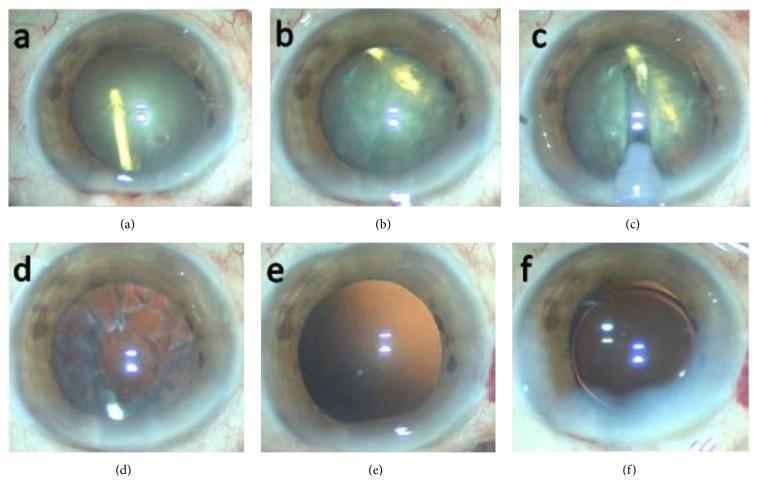
A series of six clinical intraoperative photographs (a–f) documenting a phacoemulsification cataract surgery complicated by an accidental intralenticular Ozurdex (dexamethasone) implant. (a) Displays a completed anterior continuous curvilinear capsulorrhexis (CCC). (b) Shows the intralenticular dexamethasone implant, visible as a rod-shaped semi-opaque structure, rotating within the opacified crystalline lens nucleus. (c) Illustrates the phacoemulsification probe creating a central groove in the lens nucleus to facilitate fragmentation. (d) Shows the eye after removal of the nucleus and implant, with remaining cortical remnants visible against the red reflex. (e) Demonstrates an intact and clear posterior capsule following complete irrigation and aspiration of cortical material. (f) Final stage showing the successful implantation of a three-piece hydrophobic acrylic intraocular lens (IOL) within the capsular bag. This series highlights the surgical management of lens trauma and iatrogenic implant displacement during cataract extraction.
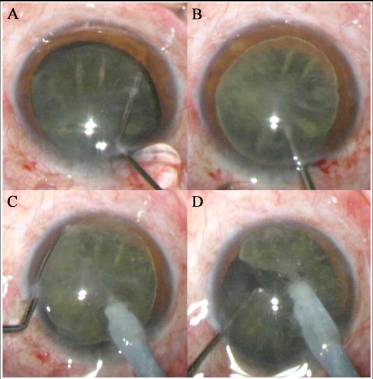
A series of four intraoperative clinical photographs (A-D) demonstrating critical stages of phacoemulsification cataract surgery. Frame A shows hydrodissection where fluid is injected into the eye using a thin cannula to separate the lens nucleus from the capsule. Frame B depicts nucleus rotation, confirming successful mobilization. Frames C and D illustrate the bimanual 'stop and chop' or 'phaco chop' technique: a large-bore phacoemulsification tip provides vacuum to hold the lens nucleus firmly in the center of the pupillary field, while a metallic second instrument (chopper) is hooked around the equator of the nucleus and moved centripetally to fragment the crystalline lens. The images highlight the anatomical relationship between the dilated pupil, the anterior capsule, and the surgical instrumentation within the anterior chamber. This series serves as an educational resource for ophthalmic surgical training, focusing on nucleus manipulation and fragmentation strategies during cataract extraction.
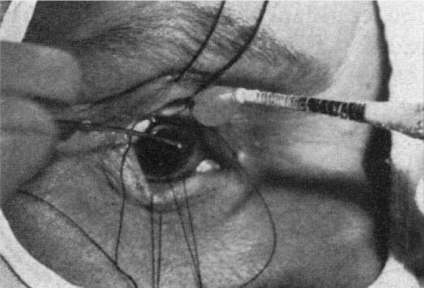
This monochrome clinical photograph captures an intraoperative view of a cryoextraction procedure for lens removal, a historical technique for intracapsular cataract extraction (ICCE). The image shows a close-up of the human eye with the surgical field prepared. A specialized cryoprobe, characterized by its cylindrical shape and alternating dark and light banded markings, is positioned horizontally from the lateral aspect, with its tip in direct contact with the superior portion of the crystalline lens. Multiple traction sutures are visible, anchored to the episclera or limbal area to retract the globe and provide surgical exposure. The anatomical structures depicted include the dilated pupil revealing the intumescent cataract, the limbus, and partially retracted eyelids. The educational focus of this image is to demonstrate the application of low-temperature instruments to adhere to and extract the lens capsule and its contents intact, primarily used in ophthalmic surgery before the advent of modern phacoemulsification.
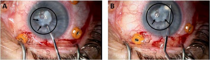
This image consists of two clinical photographs (A and B) showing the intraoperative removal of a grade 3 cataract from a human eye. The photographs display a high-magnification view of the anterior segment during surgery. In both frames, the central lens is visibly opacified, exhibiting a cloudy, white, and non-transparent appearance characteristic of advanced lenticular sclerosis. A black circle and black arrows highlight the primary area of dense opacification. Fine surgical instruments, including a slender metal probe or phacoemulsification tip, are shown entering the anterior chamber to manipulate the lens material. Small air bubbles are visible on the lens surface and within the surgical field. The surrounding anatomy shows a dilated pupil, conjunctival injection with visible surgical bleeding, and several orange-colored trocars or fixation devices positioned around the sclera to stabilize the eye or provide ports for vitrectomy instruments. This clinical visual demonstrates the morphology of high-grade cataracts and the micro-surgical environment required for extraction.
intraocular lens IOL implant capsular bag pseudophakia
mkdir -p /tmp/workspace/ophthalmology-lens-ppt
~/ophthalmology-lens-ppt/build_ppt.py
import json
import subprocess
import base64
from io import BytesIO
from pptx import Presentation
from pptx.util import Inches, Pt, Emu
from pptx.dml.color import RGBColor
from pptx.enum.text import PP_ALIGN, MSO_ANCHOR
from pptx.util import Inches, Pt

# ── Colours ──────────────────────────────────────────────────────────────────
DARK_BLUE  = RGBColor(0x0D, 0x2B, 0x55)   # deep navy
MID_BLUE   = RGBColor(0x1A, 0x57, 0x8C)   # medium blue
LIGHT_BLUE = RGBColor(0xD6, 0xE8, 0xF7)   # very light blue bg
ACCENT     = RGBColor(0x00, 0x8B, 0xD1)   # bright accent
WHITE      = RGBColor(0xFF, 0xFF, 0xFF)
GOLD       = RGBColor(0xF5, 0xA6, 0x23)
DARK_TEXT  = RGBColor(0x1A, 0x1A, 0x2E)
GREY_BG    = RGBColor(0xF0, 0xF4, 0xF8)

def hex_to_rgb(hex_str):
    h = hex_str.lstrip('#')
    return RGBColor(int(h[0:2],16), int(h[2:4],16), int(h[4:6],16))

# ── Presentation setup ────────────────────────────────────────────────────────
prs = Presentation()
prs.slide_width  = Inches(13.333)
prs.slide_height = Inches(7.5)
blank = prs.slide_layouts[6]   # fully blank layout

def add_rect(slide, x, y, w, h, fill_color, alpha=None):
    shape = slide.shapes.add_shape(1, Inches(x), Inches(y), Inches(w), Inches(h))
    shape.line.fill.background()
    shape.fill.solid()
    shape.fill.fore_color.rgb = fill_color
    return shape

def add_textbox(slide, x, y, w, h, text, font_size=18, bold=False,
                color=DARK_TEXT, align=PP_ALIGN.LEFT, italic=False, wrap=True):
    tb = slide.shapes.add_textbox(Inches(x), Inches(y), Inches(w), Inches(h))
    tf = tb.text_frame
    tf.word_wrap = wrap
    tf.margin_left  = Inches(0.05)
    tf.margin_right = Inches(0.05)
    tf.margin_top   = Inches(0.03)
    tf.margin_bottom= Inches(0.03)
    p = tf.paragraphs[0]
    p.alignment = align
    r = p.add_run()
    r.text = text
    r.font.size   = Pt(font_size)
    r.font.bold   = bold
    r.font.italic = italic
    r.font.color.rgb = color
    r.font.name = "Calibri"
    return tf

def add_bullet_textbox(slide, x, y, w, h, bullets, font_size=16,
                       title=None, title_size=20, title_color=MID_BLUE,
                       bullet_color=DARK_TEXT, bg_color=None):
    if bg_color:
        add_rect(slide, x, y, w, h, bg_color)
    tb = slide.shapes.add_textbox(Inches(x), Inches(y), Inches(w), Inches(h))
    tf = tb.text_frame
    tf.word_wrap = True
    tf.margin_left  = Inches(0.12)
    tf.margin_right = Inches(0.08)
    tf.margin_top   = Inches(0.08)
    tf.margin_bottom= Inches(0.05)

    if title:
        p = tf.paragraphs[0]
        p.alignment = PP_ALIGN.LEFT
        r = p.add_run()
        r.text = title
        r.font.size  = Pt(title_size)
        r.font.bold  = True
        r.font.color.rgb = title_color
        r.font.name  = "Calibri"

    for bullet in bullets:
        p = tf.add_paragraph()
        p.alignment = PP_ALIGN.LEFT
        r = p.add_run()
        r.text = f"• {bullet}"
        r.font.size  = Pt(font_size)
        r.font.color.rgb = bullet_color
        r.font.name  = "Calibri"
        p.space_before = Pt(4)
    return tf

def fetch_img(url):
    try:
        result = json.loads(subprocess.check_output(
            ["python", "/tmp/skills/shared/scripts/fetch_images.py", url],
            timeout=30
        ))
        if result and result[0]["base64"]:
            data = result[0]["base64"]
            if "," in data:
                data = data.split(",",1)[1]
            return BytesIO(base64.b64decode(data))
    except Exception as e:
        print(f"Image fetch failed: {e}")
    return None

def add_image(slide, url, x, y, w, h):
    img_io = fetch_img(url)
    if img_io:
        slide.shapes.add_picture(img_io, Inches(x), Inches(y), Inches(w), Inches(h))
        return True
    return False

# ═══════════════════════════════════════════════════════════════════════════════
# SLIDE 1 — Title Slide
# ═══════════════════════════════════════════════════════════════════════════════
slide = prs.slides.add_slide(blank)

# Full background
add_rect(slide, 0, 0, 13.333, 7.5, DARK_BLUE)
# Accent band
add_rect(slide, 0, 5.8, 13.333, 0.06, GOLD)
# Bottom strip
add_rect(slide, 0, 5.86, 13.333, 1.64, MID_BLUE)

# Eye/lens decorative circle (simple)
add_rect(slide, 10.5, 0.5, 2.2, 4.8, MID_BLUE)

# Title
add_textbox(slide, 0.6, 1.2, 10, 1.0,
            "SURGICAL ANATOMY & METABOLISM OF THE LENS",
            font_size=34, bold=True, color=WHITE, align=PP_ALIGN.LEFT)

# Subtitle
add_textbox(slide, 0.6, 2.4, 9, 0.6,
            "Comprehensive Ophthalmology  |  AK Khurana",
            font_size=20, bold=False, color=GOLD, align=PP_ALIGN.LEFT, italic=True)

add_textbox(slide, 0.6, 3.1, 9, 0.5,
            "Diseases of the Lens  •  Chapter Overview",
            font_size=16, color=LIGHT_BLUE, align=PP_ALIGN.LEFT)

add_textbox(slide, 0.6, 6.1, 9, 0.6,
            "Department of Ophthalmology  |  Medical Education",
            font_size=14, color=WHITE, align=PP_ALIGN.LEFT)

# ═══════════════════════════════════════════════════════════════════════════════
# SLIDE 2 — Introduction & Overview
# ═══════════════════════════════════════════════════════════════════════════════
slide = prs.slides.add_slide(blank)
add_rect(slide, 0, 0, 13.333, 7.5, GREY_BG)
add_rect(slide, 0, 0, 13.333, 1.1, DARK_BLUE)
add_rect(slide, 0, 1.1, 0.08, 6.4, GOLD)

add_textbox(slide, 0.3, 0.15, 12, 0.8,
            "INTRODUCTION TO THE CRYSTALLINE LENS",
            font_size=26, bold=True, color=WHITE)

bullets_left = [
    "Biconvex, transparent, avascular structure",
    "Located behind iris and in front of vitreous",
    "Held in position by zonular fibres (suspensory ligament of Zinn)",
    "Accounts for approximately 1/3 of total refracting power of the eye (~+16 to +20 D)",
    "Contributes to accommodation by changing shape (ciliary muscle)",
    "Completely enclosed within the lens capsule",
    "Has no blood supply, nerves, or lymphatics in adult life",
]
add_bullet_textbox(slide, 0.3, 1.25, 6.3, 5.9, bullets_left, font_size=15,
                   title="Key Features", title_color=DARK_BLUE, bg_color=WHITE)

bullets_right = [
    "Embryological origin: surface ectoderm",
    "Lens vesicle forms at ~5th week of gestation",
    "Primary lens fibres fill the vesicle lumen",
    "Lens grows throughout life (new secondary fibres added)",
    "Weight: ~65–255 mg (increases with age)",
    "Diameter: ~9–10 mm (equatorial)",
    "Axial thickness: ~3.5 mm (rest) → ~4.5 mm (accommodation)",
]
add_bullet_textbox(slide, 6.8, 1.25, 6.2, 5.9, bullets_right, font_size=15,
                   title="Embryology & Dimensions", title_color=MID_BLUE, bg_color=WHITE)

# ═══════════════════════════════════════════════════════════════════════════════
# SLIDE 3 — Surgical Anatomy: Gross Structure
# ═══════════════════════════════════════════════════════════════════════════════
slide = prs.slides.add_slide(blank)
add_rect(slide, 0, 0, 13.333, 7.5, GREY_BG)
add_rect(slide, 0, 0, 13.333, 1.1, MID_BLUE)
add_rect(slide, 0, 1.1, 0.08, 6.4, GOLD)

add_textbox(slide, 0.3, 0.15, 12, 0.8,
            "SURGICAL ANATOMY — GROSS STRUCTURE",
            font_size=26, bold=True, color=WHITE)

# Left column
parts = [
    ("LENS CAPSULE",
     ["Basement membrane of lens epithelium",
      "Thickest at pre-equatorial region (anterior: 14 µm; posterior: 4 µm)",
      "Anterior capsule thicker than posterior",
      "Elastic — important in phacoemulsification CCC",
      "Composed of type IV collagen & laminin"]),
    ("LENS EPITHELIUM",
     ["Single layer beneath anterior & equatorial capsule",
      "Only nucleated cells of the lens",
      "Central zone: low mitotic activity",
      "Pre-equatorial (germinative) zone: active mitosis → new fibres",
      "Absent from posterior surface"]),
]
y = 1.25
for title, buls in parts:
    add_rect(slide, 0.3, y, 6.0, 0.38, MID_BLUE)
    add_textbox(slide, 0.35, y+0.02, 5.9, 0.36, title, font_size=14, bold=True, color=WHITE)
    add_bullet_textbox(slide, 0.3, y+0.38, 6.0, len(buls)*0.38+0.1, buls,
                       font_size=13, bg_color=WHITE)
    y += len(buls)*0.38 + 0.65

# Right column
parts2 = [
    ("LENS SUBSTANCE (CORTEX & NUCLEUS)",
     ["Made up of lens fibres (secondary lens fibres)",
      "Nucleus: oldest fibres, centrally located, harder",
      "Embryonic nucleus (innermost) → fetal → adult nucleus",
      "Cortex: newer, softer peripheral fibres",
      "Fibres contain crystallins (α, β, γ) — maintain transparency",
      "Y-shaped anterior & inverted Y posterior sutures"]),
    ("ZONULAR FIBRES",
     ["Arise from ciliary processes (non-pigmented epithelium)",
      "Attach to lens capsule at equator",
      "Three groups: anterior, equatorial, posterior",
      "Tensed by ciliary muscle relaxation → distant vision",
      "Relaxed by ciliary muscle contraction → near vision (accommodation)"]),
]
y = 1.25
for title, buls in parts2:
    add_rect(slide, 6.6, y, 6.5, 0.38, DARK_BLUE)
    add_textbox(slide, 6.65, y+0.02, 6.4, 0.36, title, font_size=14, bold=True, color=WHITE)
    add_bullet_textbox(slide, 6.6, y+0.38, 6.5, len(buls)*0.38+0.1, buls,
                       font_size=13, bg_color=WHITE)
    y += len(buls)*0.38 + 0.65

# ═══════════════════════════════════════════════════════════════════════════════
# SLIDE 4 — Lens Capsule in Detail
# ═══════════════════════════════════════════════════════════════════════════════
slide = prs.slides.add_slide(blank)
add_rect(slide, 0, 0, 13.333, 7.5, GREY_BG)
add_rect(slide, 0, 0, 13.333, 1.1, DARK_BLUE)
add_rect(slide, 0, 1.1, 0.08, 6.4, GOLD)

add_textbox(slide, 0.3, 0.15, 12, 0.8,
            "LENS CAPSULE — SURGICAL SIGNIFICANCE",
            font_size=26, bold=True, color=WHITE)

data = [
    ("Region", "Thickness", "Surgical Note"),
    ("Central anterior", "14 µm", "Site of anterior capsulotomy / CCC"),
    ("Pre-equatorial anterior", "21 µm (thickest)", "Strongest region — supports IOL"),
    ("Equatorial", "17 µm", "Zonular attachment zone"),
    ("Pre-equatorial posterior", "23 µm", "Strong — less common rupture site"),
    ("Central posterior", "4 µm (thinnest)", "Most vulnerable — risk of posterior capsule rupture"),
]

# Draw table manually
col_widths = [3.5, 2.5, 6.5]
col_x = [0.3, 3.8, 6.3]
row_height = 0.55
start_y = 1.3

for ri, row in enumerate(data):
    y_ = start_y + ri * row_height
    bg = MID_BLUE if ri == 0 else (WHITE if ri % 2 == 1 else LIGHT_BLUE)
    txt_color = WHITE if ri == 0 else DARK_TEXT
    for ci, cell in enumerate(row):
        add_rect(slide, col_x[ci], y_, col_widths[ci]-0.05, row_height-0.04, bg)
        add_textbox(slide, col_x[ci]+0.1, y_+0.05, col_widths[ci]-0.2, row_height-0.1,
                    cell, font_size=14 if ri > 0 else 15,
                    bold=(ri == 0), color=txt_color)

# Notes below table
notes = [
    "CCC = Continuous Curvilinear Capsulorrhexis — standard technique for modern cataract surgery",
    "Posterior capsule (4 µm) is the thinnest — risk of tear during nucleus delivery or I/A",
    "Capsular bag integrity is critical for in-the-bag IOL placement",
    "Anterior capsule phimosis may occur with certain IOL types (especially silicone plate IOLs)",
]
add_bullet_textbox(slide, 0.3, 4.95, 12.7, 2.4, notes, font_size=14,
                   title="Clinical & Surgical Pearls", title_color=DARK_BLUE, bg_color=WHITE)

# ═══════════════════════════════════════════════════════════════════════════════
# SLIDE 5 — Lens Nucleus & Grading
# ═══════════════════════════════════════════════════════════════════════════════
slide = prs.slides.add_slide(blank)
add_rect(slide, 0, 0, 13.333, 7.5, GREY_BG)
add_rect(slide, 0, 0, 13.333, 1.1, MID_BLUE)
add_rect(slide, 0, 1.1, 0.08, 6.4, GOLD)

add_textbox(slide, 0.3, 0.15, 12, 0.8,
            "LENS NUCLEUS — ZONES & HARDNESS GRADING",
            font_size=26, bold=True, color=WHITE)

# Left
nuclei = [
    ("Embryonic Nucleus", "Formed from primary lens fibres; innermost; present at birth"),
    ("Fetal Nucleus", "Formed during 2nd–8th month of fetal life; bounded by Y-sutures"),
    ("Infantile Nucleus", "Formed from birth to puberty"),
    ("Adult Nucleus", "Formed after puberty; soft in youth, hard with age"),
    ("Cortex", "Newest fibres between adult nucleus and capsule; softest"),
]
y = 1.3
for i, (name, desc) in enumerate(nuclei):
    bg = [DARK_BLUE, MID_BLUE, hex_to_rgb("1E6FA0"), hex_to_rgb("2980B9"), hex_to_rgb("5DADE2")][i]
    add_rect(slide, 0.3, y, 6.0, 0.9, bg)
    add_textbox(slide, 0.4, y+0.02, 5.8, 0.32, name, font_size=14, bold=True, color=WHITE)
    add_textbox(slide, 0.4, y+0.32, 5.8, 0.52, desc, font_size=12, color=WHITE, wrap=True)
    y += 0.98

# Right: Hardness grading
add_rect(slide, 6.6, 1.3, 6.5, 0.45, DARK_BLUE)
add_textbox(slide, 6.65, 1.32, 6.4, 0.42, "NUCLEUS HARDNESS GRADING (Emery-Little)",
            font_size=15, bold=True, color=WHITE)

grades = [
    ("Grade I",   "Soft nucleus — clear or slightly yellow"),
    ("Grade II",  "Slightly hard — yellow nucleus"),
    ("Grade III", "Moderately hard — deep yellow/amber nucleus"),
    ("Grade IV",  "Hard nucleus — brown (brunescent) cataract"),
    ("Grade V",   "Very hard — black (nigra) cataract"),
]
y = 1.75
for grade, desc in grades:
    colors = [LIGHT_BLUE, hex_to_rgb("FFF9C4"), hex_to_rgb("FFD54F"), hex_to_rgb("FF8F00"), hex_to_rgb("5D4037")]
    idx = grades.index((grade, desc))
    add_rect(slide, 6.6, y, 6.5, 0.6, colors[idx])
    tc = DARK_TEXT if idx < 3 else WHITE
    add_textbox(slide, 6.7, y+0.03, 1.5, 0.55, grade, font_size=13, bold=True, color=tc)
    add_textbox(slide, 8.2, y+0.03, 4.7, 0.55, desc, font_size=13, color=tc)
    y += 0.63

add_bullet_textbox(slide, 6.6, 5.0, 6.5, 2.4,
    ["Grading guides surgical planning — hard nuclei need more phaco energy",
     "Brunescent cataracts: higher risk of corneal endothelial damage",
     "LOCS III system (Lens Opacity Classification) also widely used",
     "Posterior pole cataracts often softer but surgically challenging"],
    font_size=13, title="Surgical Implications", title_color=DARK_BLUE, bg_color=WHITE)

# ═══════════════════════════════════════════════════════════════════════════════
# SLIDE 6 — Metabolism: Energy & Glucose
# ═══════════════════════════════════════════════════════════════════════════════
slide = prs.slides.add_slide(blank)
add_rect(slide, 0, 0, 13.333, 7.5, GREY_BG)
add_rect(slide, 0, 0, 13.333, 1.1, DARK_BLUE)
add_rect(slide, 0, 1.1, 0.08, 6.4, GOLD)

add_textbox(slide, 0.3, 0.15, 12, 0.8,
            "LENS METABOLISM — ENERGY & GLUCOSE PATHWAYS",
            font_size=26, bold=True, color=WHITE)

add_bullet_textbox(slide, 0.3, 1.25, 6.0, 3.0,
    ["Lens is avascular — metabolites diffuse from aqueous humour",
     "Glucose is the primary metabolic substrate",
     "Three pathways for glucose metabolism:",
     "  1. Embden-Meyerhof (Anaerobic Glycolysis) — ~85%",
     "  2. Hexose Monophosphate Shunt (HMS) — ~5%",
     "  3. Sorbitol (Polyol) Pathway — minor (clinically significant in DM)",
     "Krebs cycle minimal (avascular, low O₂ tension)",
     ],
    font_size=14, title="Glucose Metabolism Overview", title_color=DARK_BLUE, bg_color=WHITE)

add_bullet_textbox(slide, 0.3, 4.4, 6.0, 2.9,
    ["Glucose → Glucose-6-Phosphate → ATP (×2) + Lactate",
     "ATP drives Na⁺/K⁺ ATPase pump (maintains lens clarity)",
     "Lactate exits lens into aqueous",
     "Inhibited by monoiodoacetate",
     "Rate-limiting enzyme: Phosphofructokinase",
     "Primary pathway — produces 2 ATP per glucose",
     ],
    font_size=14, title="1. Anaerobic Glycolysis (EM Pathway)", title_color=MID_BLUE, bg_color=WHITE)

add_bullet_textbox(slide, 6.5, 1.25, 6.5, 3.0,
    ["Glucose-6-P → Gluconolactone → Pentose phosphates",
     "Generates NADPH — maintains glutathione (GSH) in reduced form",
     "Reduced GSH neutralises H₂O₂ and free radicals",
     "Protects lens proteins from oxidative damage",
     "Critical in maintaining lens transparency",
     "Activated under oxidative stress",
     ],
    font_size=14, title="2. Hexose Monophosphate Shunt", title_color=DARK_BLUE, bg_color=WHITE)

add_bullet_textbox(slide, 6.5, 4.4, 6.5, 2.9,
    ["Aldose reductase: Glucose → Sorbitol (accumulates in lens)",
     "Sorbitol dehydrogenase: Sorbitol → Fructose",
     "In DM: excess glucose → excess sorbitol (poorly diffusible)",
     "Osmotic overhydration of lens → cortical opacification",
     "Leads to 'snowflake' or 'diabetic' cataract",
     "Aldose reductase inhibitors (e.g., Epalrestat) studied for prevention",
     ],
    font_size=14, title="3. Sorbitol (Polyol) Pathway", title_color=hex_to_rgb("C0392B"), bg_color=WHITE)

# ═══════════════════════════════════════════════════════════════════════════════
# SLIDE 7 — Metabolism: Proteins, Antioxidants & Transport
# ═══════════════════════════════════════════════════════════════════════════════
slide = prs.slides.add_slide(blank)
add_rect(slide, 0, 0, 13.333, 7.5, GREY_BG)
add_rect(slide, 0, 0, 13.333, 1.1, MID_BLUE)
add_rect(slide, 0, 1.1, 0.08, 6.4, GOLD)

add_textbox(slide, 0.3, 0.15, 12, 0.8,
            "LENS METABOLISM — PROTEINS, TRANSPORT & ANTIOXIDANTS",
            font_size=26, bold=True, color=WHITE)

add_bullet_textbox(slide, 0.3, 1.25, 6.0, 2.9,
    ["Lens has highest protein content of any tissue (~33% wet weight)",
     "Crystallins account for ~90% of soluble protein",
     "α-Crystallin: chaperone function — prevents protein aggregation",
     "β-Crystallin: most abundant structural protein",
     "γ-Crystallin: monomeric, mainly in nucleus",
     "Water-insoluble proteins increase with aging → nuclear sclerosis",
     "Protein aggregation → light scattering → cataract",
     ],
    font_size=14, title="Lens Proteins (Crystallins)", title_color=DARK_BLUE, bg_color=WHITE)

add_bullet_textbox(slide, 0.3, 4.3, 6.0, 2.9,
    ["Sodium (Na⁺) high in plasma; Potassium (K⁺) high in lens",
     "Na⁺/K⁺ ATPase on epithelium actively pumps Na⁺ out, K⁺ in",
     "ATP-dependent active transport maintains ionic balance",
     "Disruption → Na⁺ influx → water → lens swelling → opacification",
     "Cl⁻ and Ca²⁺ also regulated actively",
     "Amino acids enter via Na⁺-coupled transporters",
     ],
    font_size=14, title="Ion Transport & Pump Mechanism", title_color=MID_BLUE, bg_color=WHITE)

add_bullet_textbox(slide, 6.5, 1.25, 6.5, 2.9,
    ["Glutathione (GSH) — most important antioxidant in lens",
     "Concentration highest in cortex (5–10 mM), lower in nucleus",
     "Synthesised via glutathione synthetase in epithelium",
     "Neutralises H₂O₂ via glutathione peroxidase",
     "Ascorbic acid (Vit C) in aqueous → absorbs UV, protects lens",
     "Superoxide dismutase (SOD) also present in lens",
     "Oxidative stress: GSH depletion → protein oxidation → PSC cataract",
     ],
    font_size=14, title="Antioxidant Defence System", title_color=DARK_BLUE, bg_color=WHITE)

add_bullet_textbox(slide, 6.5, 4.3, 6.5, 2.9,
    ["Aqueous humour is the sole source of nutrients",
     "Glucose, O₂, amino acids — enter lens by diffusion & transport",
     "Metabolic waste (CO₂, lactate) exits into aqueous",
     "Calcium (Ca²⁺): low in normal lens (0.3 mM vs 1.5 mM plasma)",
     "Elevated Ca²⁺ → activates proteases → lens opacification",
     "Vitamin B₂ (Riboflavin): role in maintaining photosensitisation",
     ],
    font_size=14, title="Nutrition & Metabolic Exchange", title_color=hex_to_rgb("1A6B3A"), bg_color=WHITE)

# ═══════════════════════════════════════════════════════════════════════════════
# SLIDE 8 — Accommodation & Ageing Changes
# ═══════════════════════════════════════════════════════════════════════════════
slide = prs.slides.add_slide(blank)
add_rect(slide, 0, 0, 13.333, 7.5, GREY_BG)
add_rect(slide, 0, 0, 13.333, 1.1, DARK_BLUE)
add_rect(slide, 0, 1.1, 0.08, 6.4, GOLD)

add_textbox(slide, 0.3, 0.15, 12, 0.8,
            "ACCOMMODATION & AGE-RELATED CHANGES IN THE LENS",
            font_size=26, bold=True, color=WHITE)

add_bullet_textbox(slide, 0.3, 1.25, 6.0, 3.5,
    ["Near vision: ciliary muscle CONTRACTS → zonules RELAX → lens becomes MORE convex",
     "Distant vision: ciliary muscle RELAXES → zonules become TAUT → lens FLATTENS",
     "Amplitude of accommodation decreases with age (presbyopia)",
     "At 10 years: ~14 D; At 40 years: ~6 D; At 60 years: ~1 D",
     "Helmholtz theory (accepted): zonule tension mechanism",
     "Schachar theory (alternative): equatorial zonule tension",
     ],
    font_size=14, title="Mechanism of Accommodation", title_color=DARK_BLUE, bg_color=WHITE)

add_bullet_textbox(slide, 0.3, 4.9, 6.0, 2.3,
    ["Presbyopia: loss of accommodation from lens hardening (~45 years)",
     "Treatment: reading glasses, bifocals, multifocal IOLs, LASIK monovision",
     "PRELEX: Presbyopic Lens Exchange with multifocal IOL",
     ],
    font_size=14, title="Presbyopia", title_color=MID_BLUE, bg_color=WHITE)

add_bullet_textbox(slide, 6.5, 1.25, 6.5, 3.5,
    ["Continuous growth throughout life (unlike most tissues)",
     "Weight doubles from birth to old age",
     "Nuclear sclerosis: lens fibres compact centrally → yellow/brown colour",
     "Increased water-insoluble proteins → reduced transparency",
     "Reduced GSH in nucleus → oxidative damage",
     "Reduced lens epithelial pump activity → ionic imbalance",
     "Meiotic activity in germinative zone persists throughout life",
     ],
    font_size=14, title="Age-Related Changes", title_color=DARK_BLUE, bg_color=WHITE)

add_bullet_textbox(slide, 6.5, 4.9, 6.5, 2.3,
    ["Nuclear sclerosis → reduced blue light transmission → chromatic aberration",
     "UV-B most damaging wavelength for lens (absorbed by nucleus)",
     "Brunescent nucleus: tryptophan photo-oxidation products accumulate",
     ],
    font_size=14, title="Optical Consequences of Ageing", title_color=MID_BLUE, bg_color=WHITE)

# ═══════════════════════════════════════════════════════════════════════════════
# SLIDE 9 — Clinical Images: Phacoemulsification Stages
# ═══════════════════════════════════════════════════════════════════════════════
slide = prs.slides.add_slide(blank)
add_rect(slide, 0, 0, 13.333, 7.5, hex_to_rgb("0A1628"))
add_rect(slide, 0, 0, 13.333, 1.1, MID_BLUE)
add_rect(slide, 0, 1.1, 0.08, 6.4, GOLD)

add_textbox(slide, 0.3, 0.15, 12, 0.8,
            "SURGICAL IMAGES — PHACOEMULSIFICATION & LENS EXTRACTION",
            font_size=24, bold=True, color=WHITE)

# Image 1 — phaco steps
img1_url = "https://cdn.orris.care/cdss_images/pmc_clinical_VQA_626e046c0202ed53b1368186e2f9058a27ba8ad994f0443e5b3445835242c424.jpg"
add_image(slide, img1_url, 0.3, 1.25, 6.1, 4.0)

# Image 2 — cataract types
img2_url = "https://cdn.orris.care/cdss_images/pmc_clinical_VQA_2152c4d52781f088c6abbe8781d9cd575a4a57a88e332234d3b603e2cc202fd6.jpg"
add_image(slide, img2_url, 6.7, 1.25, 6.3, 4.0)

add_textbox(slide, 0.3, 5.35, 6.1, 0.5,
            "Phacoemulsification: Hydrodissection → Nucleus chopping → I/A",
            font_size=12, color=LIGHT_BLUE, italic=True)
add_textbox(slide, 6.7, 5.35, 6.3, 0.5,
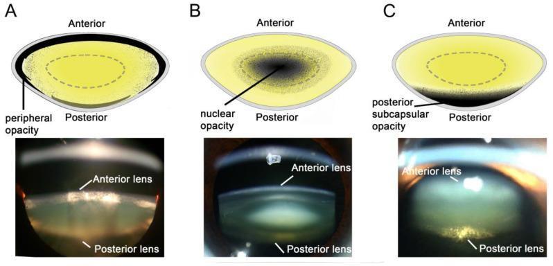
            "Cataract types: (A) Cortical  (B) Nuclear  (C) Posterior subcapsular",
            font_size=12, color=LIGHT_BLUE, italic=True)

add_bullet_textbox(slide, 0.3, 5.9, 12.7, 1.4,
    ["CCC (Continuous Curvilinear Capsulorrhexis) creates a circular opening in anterior capsule for safe nucleus delivery",
     "Stop-and-chop / divide-and-conquer techniques fragment the nucleus under phaco tip vacuum before aspiration",
     "Irrigation/Aspiration (I/A) removes cortical remnants before IOL implantation in the capsular bag"],
    font_size=13, bg_color=hex_to_rgb("0D1E35"), bullet_color=LIGHT_BLUE)

# ═══════════════════════════════════════════════════════════════════════════════
# SLIDE 10 — Summary & Key Points
# ═══════════════════════════════════════════════════════════════════════════════
slide = prs.slides.add_slide(blank)
add_rect(slide, 0, 0, 13.333, 7.5, GREY_BG)
add_rect(slide, 0, 0, 13.333, 1.1, DARK_BLUE)
add_rect(slide, 0, 1.1, 0.08, 6.4, GOLD)
add_rect(slide, 12.8, 1.1, 0.533, 6.4, GOLD)

add_textbox(slide, 0.3, 0.15, 12, 0.8,
            "SUMMARY — KEY POINTS FOR EXAMINATION",
            font_size=26, bold=True, color=WHITE)

# Three columns of key facts
col1 = [
    "Lens is biconvex, avascular, transparent",
    "Capsule thinnest posteriorly (4 µm) — risk zone",
    "Anterior capsule thickest at pre-equator (21 µm)",
    "Epithelium: only nucleated cell, absent posteriorly",
    "Germinative zone → new fibres throughout life",
    "Crystallins (α, β, γ) maintain transparency",
    "α-Crystallin acts as molecular chaperone",
]
col2 = [
    "Glucose → 85% via EM (glycolysis) → ATP",
    "HMS shunt → NADPH → reduced GSH (antioxidant)",
    "Sorbitol pathway: in DM → osmotic cataract",
    "GSH = most important lens antioxidant",
    "Na⁺/K⁺ ATPase pump on epithelium is critical",
    "Ca²⁺ elevated in cataract (activates proteases)",
    "Ascorbic acid in aqueous protects vs UV",
]
col3 = [
    "Accommodation: ciliary contraction → zonule slack → convex lens",
    "Presbyopia ~45 yrs due to nuclear hardening",
    "Age: water-insoluble proteins ↑ → sclerosis",
    "CCC is cornerstone of modern phaco surgery",
    "Posterior capsule: thinnest, most vulnerable",
    "In-the-bag IOL placement optimal for pseudophakia",
    "Brunescent nucleus: Grade IV–V, high phaco energy",
]

add_bullet_textbox(slide, 0.3, 1.25, 4.1, 5.9, col1, font_size=13,
                   title="Anatomy & Structure", title_color=DARK_BLUE, bg_color=WHITE)
add_bullet_textbox(slide, 4.6, 1.25, 4.1, 5.9, col2, font_size=13,
                   title="Metabolism", title_color=MID_BLUE, bg_color=WHITE)
add_bullet_textbox(slide, 9.1, 1.25, 4.1, 5.9, col3, font_size=13,
                   title="Surgery & Ageing", title_color=hex_to_rgb("1A6B3A"), bg_color=WHITE)

# ── Save ──────────────────────────────────────────────────────────────────────
out_path = "/tmp/workspace/ophthalmology-lens-ppt/Lens_Anatomy_Metabolism_AK_Khurana.pptx"
buf = BytesIO()
prs.save(buf)
with open(out_path, "wb") as f:
    f.write(buf.getvalue())

print(f"Saved to: {out_path}")
print(f"Slides: {len(prs.slides)}")
cd /tmp/workspace/ophthalmology-lens-ppt && python build_ppt.py 2>&1
ls /tmp/skills/shared/scripts/ 2>/dev/null || ls /tmp/skills/ 2>/dev/null
~/ophthalmology-lens-ppt/build_ppt.py
Successfully replaced 1 instance(s) of the string in '/tmp/workspace/ophthalmology-lens-ppt/build_ppt.py'
cd /tmp/workspace/ophthalmology-lens-ppt && python build_ppt.py 2>&1
# Download images with requests directly and embed them python3 -c " import requests, base64 urls = [ 'https://cdn.orris.care/cdss_images/pmc_clinical_VQA_626e046c0202ed53b1368186e2f9058a27ba8ad994f0443e5b3445835242c424.jpg', 'https://cdn.orris.care/cdss_images/pmc_clinical_VQA_2152c4d52781f088c6abbe8781d9cd575a4a57a88e332234d3b603e2cc202fd6.jpg', 'https://cdn.orris.care/cdss_images/pmc_clinical_VQA_2991c0e41c9773d4dcfddc6a01b2d15e72335fb327a91a1e3433b1ebe57dca9b.jpg', ] for i, url in enumerate(urls): r = requests.get(url, timeout=20) if r.status_code == 200: with open(f'/tmp/workspace/ophthalmology-lens-ppt/img{i}.jpg', 'wb') as f: f.write(r.content) print(f'Downloaded img{i}.jpg ({len(r.content)} bytes)') else: print(f'Failed: {url} -> {r.status_code}') "
~/ophthalmology-lens-ppt/build_ppt.py
Successfully replaced 1 instance(s) of the string in '/tmp/workspace/ophthalmology-lens-ppt/build_ppt.py'
cd /tmp/workspace/ophthalmology-lens-ppt && python build_ppt.py 2>&1
Give me in ppt form to present for my seminar
Lens Anatomy Metabolism AK Khurana
Presentation · PPTX
| Slide | Title |
|---|---|
| 1 | Title Slide - Surgical Anatomy & Metabolism of the Lens (AK Khurana) |
| 2 | Introduction - Key features, embryology & dimensions of the lens |
| 3 | Surgical Anatomy - Gross Structure - Capsule, epithelium, substance, zonules |
| 4 | Lens Capsule - Surgical Significance - Regional thickness table + surgical pearls |
| 5 | Lens Nucleus Zones & Hardness Grading - Embryonic→Adult nucleus + Emery-Little grading |
| 6 | Metabolism - Energy & Glucose Pathways - EM glycolysis, HMS shunt, Sorbitol pathway |
| 7 | Metabolism - Proteins, Transport & Antioxidants - Crystallins, Na⁺/K⁺ pump, GSH system |
| 8 | Accommodation & Age-Related Changes - Helmholtz theory, presbyopia, nuclear sclerosis |
| 9 | Clinical Images - Phacoemulsification stages + cataract types (real surgical photos) |
| 10 | Summary - Key Points for Examination - Three-column rapid revision layout |
- Professional navy-blue + gold color scheme suitable for a medical seminar
- Content structured exactly as per AK Khurana's Comprehensive Ophthalmology
- Real intraoperative surgical photos on Slide 9 (phacoemulsification steps + cataract type classification)
- Emery-Little nucleus hardness grading color-coded (Grade I white → Grade V black)
- Glucose metabolism pathways clearly separated (glycolysis vs HMS vs sorbitol - with DM implications)
- Examination-ready summary slide with 21 key facts across anatomy, metabolism, and surgery