
Anatomy Thoracic Cavity Skeleton and joints of the thoracic cage. Movements of the thorax. Muscles and fascia of the thoracic wall. Chest anatomy.
Reading File
Finding Sources
Finding Sources
Finding Sources
Finding Sources
Finding Sources
Finding Sources
Now let me read the key sections from Gray's Anatomy for Students and the THIEME Atlas in parallel:
Reading File
Reading File
Reading File
Reading File
Reading File
Reading File
Reading File
Reading File
Now let me fetch the key anatomical images to include in the response:
Loading Image
Loading Image
Loading Image
Now let me get the skeleton/joints of the thoracic cage and movements section from Gray's:
Reading File
Loading Image
Reading File
Reading File
I now have comprehensive information to write a thorough answer. Let me compile it:
Loading Image
Here is a complete, integrated account of thoracic cavity anatomy drawn from Gray's Anatomy for Students and the THIEME Atlas of Anatomy.
Thoracic Cavity - Complete Anatomy
1. Skeleton of the Thoracic Cage
The thoracic cage is an osseocartilaginous structure formed by 12 thoracic vertebrae posteriorly, 12 pairs of ribs laterally, and the sternum anteriorly.
Sternum
The sternum is a flat bone consisting of three parts:
- Manubrium - the widest, uppermost part; articulates with the clavicles and the cartilages of rib I
- Body - the longest part; articulates laterally with the cartilages of ribs II-VII
- Xiphoid process - a small, variable, cartilaginous/bony inferior tip
The sternal angle (angle of Louis) is the junction between the manubrium and body - a palpable ridge that lies at the level of the intervertebral disc between TIV and TV. It marks the articulation of the costal cartilage of rib II (used clinically to count ribs, since rib I is hidden under the clavicle). It also marks the superior boundary of the pericardium, the bifurcation of the trachea, and the beginning/end of the aortic arch. - Gray's Anatomy for Students, p. 168
Ribs
The 12 pairs of ribs are classified as:
- True ribs (1-7): costal cartilages articulate directly with the sternum
- False ribs (8-10): costal cartilages join the cartilage of the rib above; ribs 8-10 form the costal margin with their fused cartilages
- Floating ribs (11-12): short; no anterior articulation
A typical rib (3-9) has a head, neck, tubercle, and body (shaft). The costal groove runs along the inferior inner margin of the shaft and protects the intercostal neurovascular bundle (vein, artery, nerve - from superior to inferior, VAN).
Ribs 1, 2, 10, 11, and 12 are atypical:
- Rib 1 is the shortest, broadest, most curved; it bears grooves for the subclavian artery and vein (separated by the scalene tubercle for attachment of the anterior scalene muscle)
- Rib 2 is transitional; has a roughened area for serratus anterior
Thoracic Apertures
Superior thoracic aperture (thoracic inlet): bounded by the body of TI posteriorly, medial margins of rib I laterally, and the manubrium anteriorly. The plane slopes obliquely (faces anterosuperiorly). Structures traversing it include the trachea, esophagus, great vessels, brachial plexus, and thoracic duct. - Gray's Anatomy for Students, p. 162
Inferior thoracic aperture (thoracic outlet): bounded by the body of TXII posteriorly, ribs XI-XII posterolaterally, the costal margin (ribs VII-X) anterolaterally, and the xiphoid process anteriorly. Closed by the diaphragm. The posterior margin is lower than the anterior, so the aperture tilts superiorly when viewed from the front.
2. Joints of the Thoracic Cage
Costovertebral Joints (Posterior)
Each rib articulates with the thoracic vertebral column at two synovial joints:
-
Joint of the head of the rib - between the articular facets on the head of the rib and the costal facets on adjacent vertebral bodies (and intervening disc). Ribs 1, 10, 11, and 12 articulate with only one vertebral body (monoarticular). Ribs 2-9 articulate with two adjacent bodies (biarticular).
-
Costotransverse joint - between the articular facet of the tubercle of the rib and the transverse process of the corresponding vertebra. Absent for ribs 11 and 12 (no transverse facets at these levels).
Sternocostal (Sternocostal / Chondrosternal) Joints (Anterior)
- 1st rib forms a synchondrosis (primary cartilaginous joint) with the manubrium - no movement
- Ribs 2-7 form synovial plane joints with the sternum - allow gliding
Costochondral Joints
The junction between a rib and its costal cartilage is a synchondrosis - no movement possible.
Interchondral Joints
Between adjacent costal cartilages of ribs 6-10; small synovial joints that participate in the costal margin.
Manubriosternal Joint (Sternal Angle)
A secondary cartilaginous joint (symphysis) allowing slight angular movement during respiration.
3. Movements of the Thorax During Breathing
The thorax must increase in three dimensions to generate negative intrathoracic pressure for inspiration. - Gray's Anatomy for Students, p. 200
Vertical Dimension - Diaphragmatic Descent
The diaphragm is the primary muscle of inspiration. When its muscle fibers contract, the domes flatten (descend), increasing vertical thoracic volume by up to 1.5 cm in quiet breathing and up to 7-10 cm in forced inspiration. The right dome is higher than the left (reaches rib V), partly because the liver elevates it.
Anteroposterior and Lateral Dimensions - Rib Elevation
Because the anterior ends of ribs are lower than the posterior ends:
- Elevation of the ribs moves the sternum anteriorly ("pump-handle movement") - increases anteroposterior diameter
- Eversion of the rib shafts moves the lateral walls superolaterally ("bucket-handle movement") - increases transverse diameter
These movements occur together during normal inspiration.
Upper ribs (1-6) move predominantly with pump-handle motion.
Lower ribs (7-10) move predominantly with bucket-handle motion (their more horizontal orientation favors lateral expansion).
Quiet inspiration: diaphragm + scalenes + external intercostals
Forced inspiration: accessory muscles add scalenes (elevate rib 1), sternocleidomastoid (elevates sternum), serratus anterior, pectoralis minor
Quiet expiration: passive (elastic recoil of lung + chest wall)
Forced expiration: internal intercostals + abdominal muscles (obliques + transversus + rectus abdominis) depress ribs and raise intra-abdominal pressure
4. Muscles of the Thoracic Wall
Intercostal Muscles (Three Layers)
There are three flat muscles in each intercostal space. The intercostal nerve, artery, and vein (VAN, superior to inferior) run in the plane between the internal and innermost layers. - Gray's Anatomy for Students, p. 188
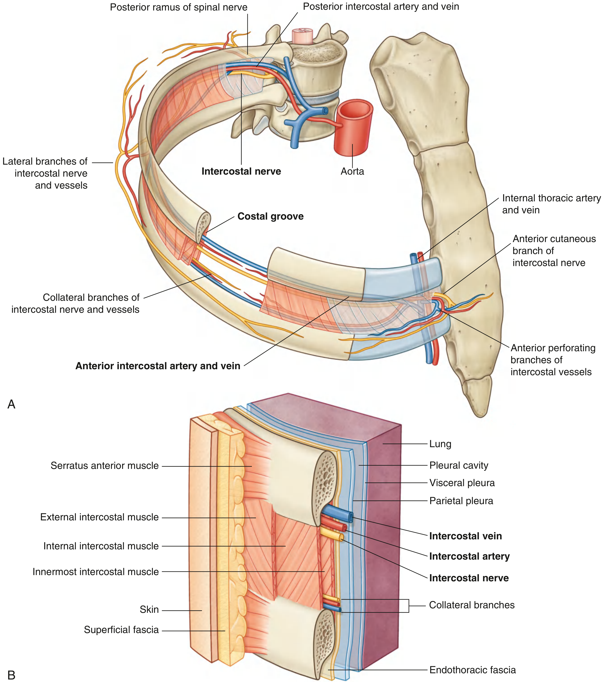
Fig. 3.27 - Intercostal Space: (A) Anterolateral view with neurovascular bundle, (B) Wall layers
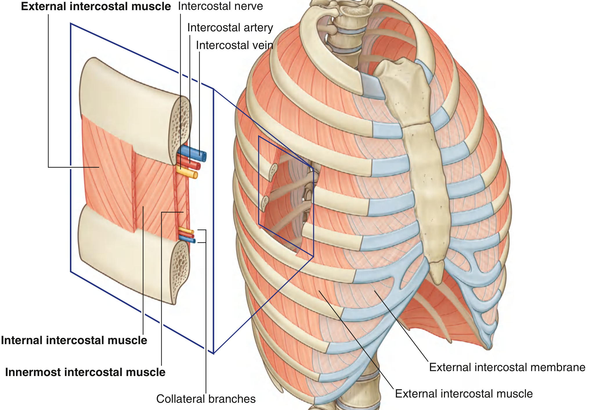
Fig. 3.28 - Intercostal Muscles showing three layers and the neurovascular plane
| Muscle | Attachments | Fiber Direction | Function | Innervation |
|---|---|---|---|---|
| External intercostal | Inferior margin of rib above → superior margin of rib below | Oblique anteroinferiorly (like hands in pockets) | Elevation of ribs; most active in inspiration | T1-T11 intercostal nn. |
| Internal intercostal | Lateral edge of costal groove above → superior margin of rib below | Oblique posteroinferiorly (perpendicular to external) | Depression of ribs; most active in expiration | T1-T11 intercostal nn. |
| Innermost intercostal | Medial edge of costal groove → internal superior margin of rib below | Same as internal intercostal | Acts with internal intercostals | T1-T11 intercostal nn. |
Key anatomical detail: The external intercostal muscle extends from the region of the rib tubercle anteriorly to the costal cartilage, where it continues as the external intercostal membrane. The internal intercostal extends from the sternum to the angle of the rib, where it continues as the internal intercostal membrane. - THIEME Atlas, p. 193
Subcostales (Subcostal Muscles)
- Located on the internal thoracic wall, near the vertebral column posteriorly
- Each spans two or three ribs (from the internal surface near the angle of a lower rib to the internal surface of the second or third rib above)
- Function: may depress ribs
- Innervation: related intercostal nerves
Transversus Thoracis (Sternocostalis)
- Origin: inferior posterior surface of the body of sternum, xiphoid process, and adjacent costal cartilages (IV-VII)
- Insertion: inferior margins and internal surfaces of costal cartilages of ribs II-VI
- Function: depresses costal cartilages; weakest of the respiratory muscles
- Innervation: related intercostal nerves
Scalene Muscles (Accessory Respiratory Muscles)
Though topographically in the neck, the scalenes are functionally key respiratory muscles. - THIEME Atlas, p. 193
| Muscle | Origin | Insertion | Action |
|---|---|---|---|
| Anterior scalene | Anterior tubercles C3-C6 | Scalene tubercle of rib 1 | Elevates rib 1 (inspiration); flexes/bends neck |
| Middle scalene | Posterior tubercles C3-C7 | First rib, posterior to subclavian groove | Elevates rib 1 |
| Posterior scalene | Posterior tubercles C5-C7 | Outer surface of rib 2 | Elevates rib 2 |
Serratus Posterior Muscles (Spinocostal Muscles)
- Serratus posterior superior: from nuchal ligament and spinous processes of CVII-TIII → ribs 2-5; elevates ribs (inspiration)
- Serratus posterior inferior: from spinous processes of TXI-LIII → ribs 9-12; depresses lower ribs (expiration); also prevents lower ribs being drawn up by the diaphragm
5. Fascia of the Thoracic Wall
Endothoracic Fascia
The thoracic cavity is lined internally by the endothoracic fascia - a layer of connective tissue between the deep thoracic wall muscles and the costal parietal pleura, to which it is firmly attached. It is analogous to the transversalis fascia of the abdomen. - THIEME Atlas, p. 193
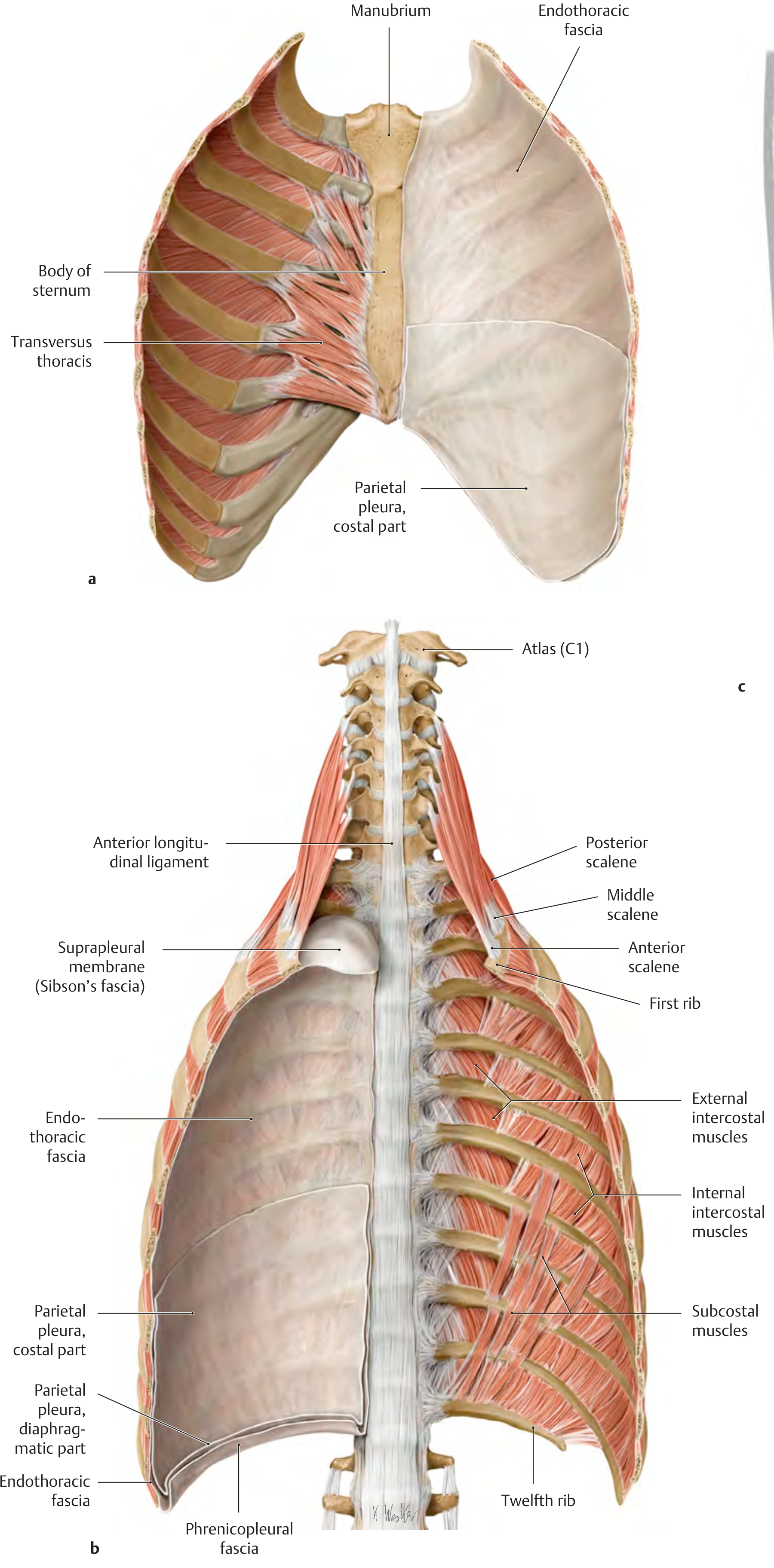
Thoracic wall and endothoracic fascia: (a) posterior view of anterior wall showing transversus thoracis; (b) posterior wall showing muscle layers and fascial relationships
Key regions:
- Suprapleural membrane (Sibson's fascia): a thickened condensation of endothoracic fascia that covers the cervical pleura (dome of pleura) at the thoracic inlet, protecting it and anchoring it to rib I and the transverse process of CVII
- Phrenicopleural fascia: the portion connecting the diaphragmatic parietal pleura to the upper surface of the diaphragm
- Costodiaphragmatic recess: a potential space between chest wall and diaphragm (between costal and diaphragmatic parietal pleura) that enlarges during inspiration as the diaphragm descends - the clinically important space where pleural fluid collects
6. Intercostal Neurovascular Bundle
Each intercostal space contains a neurovascular bundle running in the plane between the internal and innermost intercostal muscles, protected in the costal groove of the rib above.
Order from superior to inferior: Vein → Artery → Nerve (VAN mnemonic)
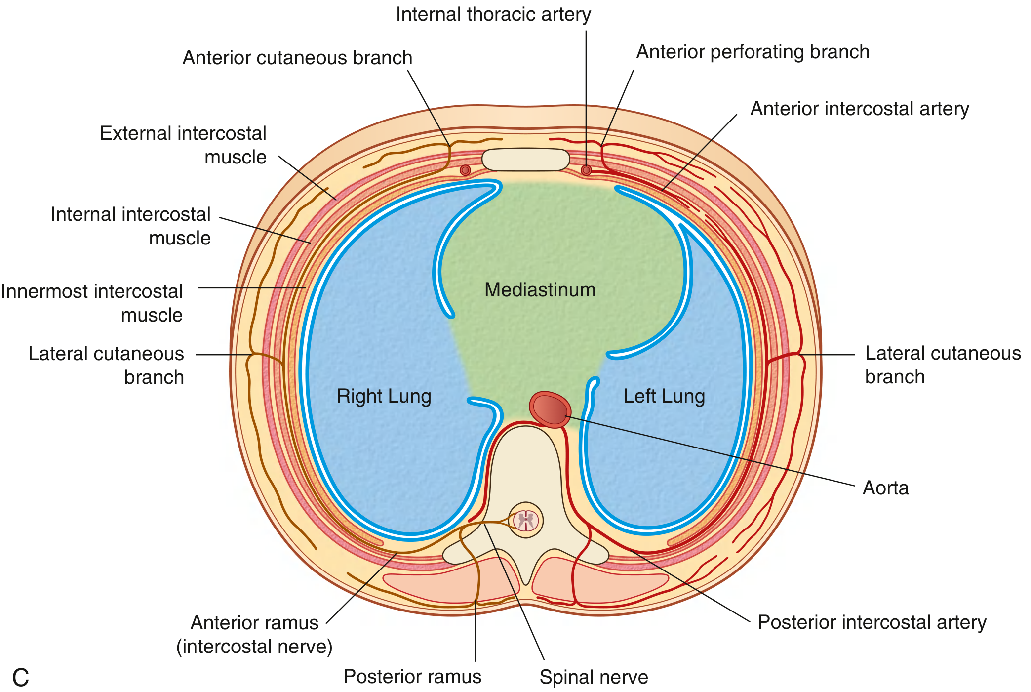
Fig. 3.27C - Transverse section showing three intercostal muscle layers, neurovascular bundle, mediastinum, and both lungs
Clinical point: When performing thoracentesis (needle aspiration of pleural fluid) or chest tube insertion, the needle should be directed over the superior margin of the lower rib (not under the upper rib) to avoid the neurovascular bundle.
Arterial supply:
- Posterior intercostal arteries (spaces 3-11): branches of the thoracic aorta
- Anterior intercostal arteries (spaces 1-6): branches of the internal thoracic artery; (spaces 7-9): branches of the musculophrenic artery
- The internal thoracic artery (internal mammary artery) descends 1 cm lateral to the sternum, giving anterior intercostal branches; it divides at rib VI into the superior epigastric and musculophrenic arteries
Venous drainage:
- Anterior intercostal veins → internal thoracic veins → brachiocephalic veins
- Posterior intercostal veins → azygos system (right side) or hemiazygos/accessory hemiazygos (left side) → SVC
Innervation (Intercostal Nerves):
The intercostal nerves are the anterior rami of T1-T11; T12 is the subcostal nerve.
Each typical intercostal nerve gives: lateral cutaneous branch (anterior + posterior divisions), and ends as the anterior cutaneous branch (pierces at the parasternal line). - Gray's Anatomy for Students, p. 193
7. Thoracic Cavity Subdivisions
The thoracic cavity contains three main compartments:
- Right pleural cavity - surrounds the right lung
- Left pleural cavity - surrounds the left lung
- Mediastinum - the central partition between them
The mediastinum extends from the sternum to the thoracic vertebrae and from the superior to inferior thoracic aperture. The sternal angle plane (TIV/V disc) divides it into:
- Superior mediastinum (above the plane): contains the great vessels, trachea, esophagus, thoracic duct, phrenic and vagus nerves, left recurrent laryngeal nerve
- Inferior mediastinum (below the plane):
- Anterior mediastinum - between sternum and pericardium (thymic remnants, fat, lymph nodes)
- Middle mediastinum - the pericardium and heart
- Posterior mediastinum - between pericardium and thoracic vertebrae (esophagus, descending aorta, azygos/hemiazygos veins, thoracic duct, sympathetic trunks)
8. Diaphragm - Floor of the Thoracic Cavity
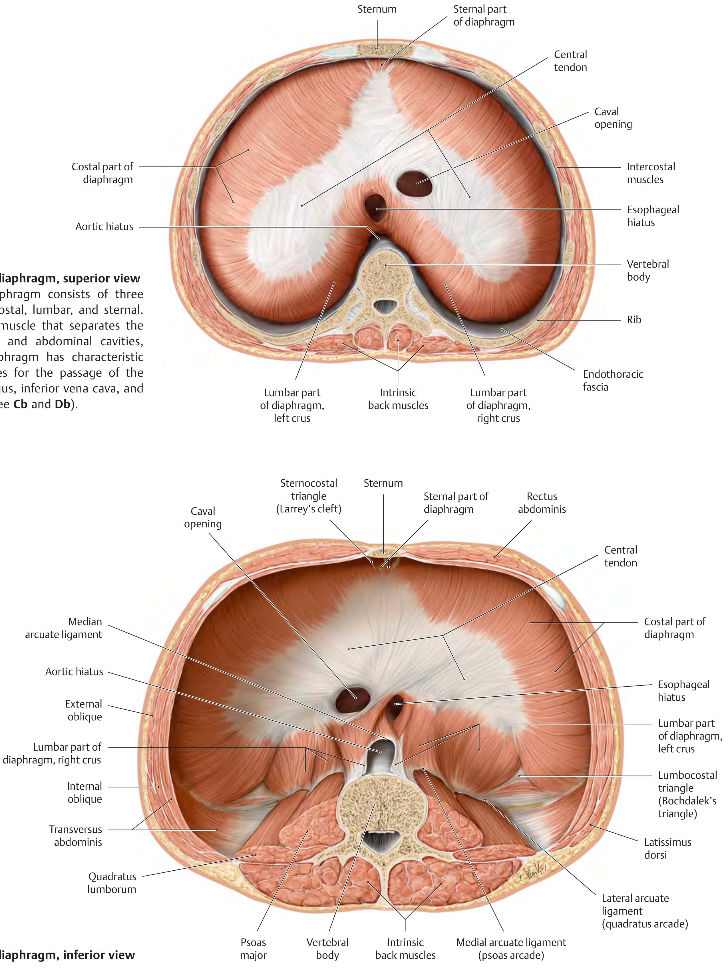
Diaphragm - superior view (top) and inferior view (bottom) showing openings, crura, and attachments
The diaphragm is a dome-shaped musculotendinous partition separating the thorax from the abdomen. It has three parts: costal, lumbar, and sternal. Muscle fibers converge into a central central tendon.
Three major openings:
| Opening | Level | Structures passing through |
|---|---|---|
| Caval opening | TVIII | Inferior vena cava, right phrenic nerve |
| Esophageal hiatus | TX | Esophagus, vagus nerves (anterior + posterior trunks) |
| Aortic hiatus | TXII | Aorta, thoracic duct, (sometimes azygos vein) |
Innervation: Phrenic nerves (C3, C4, C5 - "C3, 4, 5 keeps the diaphragm alive") innervate the diaphragm from its abdominal surface. The peripheral rim receives sensory supply from lower intercostal nerves (T5-T11). - Gray's Anatomy for Students, p. 200
Weak points (potential herniation sites):
- Sternocostal triangle (Larrey's cleft/Morgagni's hernia): between sternal and costal parts (anterior)
- Lumbocostal triangle (Bochdalek's triangle): between lumbar and costal parts (posterior) - most common site of congenital diaphragmatic hernia
Summary Table: Muscles of the Thoracic Wall
| Muscle | Location | Primary Action | Innervation |
|---|---|---|---|
| External intercostal | Intercostal spaces (superficial) | Raises ribs - inspiration | T1-T11 |
| Internal intercostal | Intercostal spaces (middle) | Depresses ribs - expiration | T1-T11 |
| Innermost intercostal | Intercostal spaces (deep) | With internal intercostals | T1-T11 |
| Subcostales | Posterior internal wall | Depress ribs | Intercostal nn. |
| Transversus thoracis | Anterior internal wall | Depresses costal cartilages | Intercostal nn. |
| Anterior scalene | Neck - rib 1 | Elevates rib 1 (inspiration) | C4-C6 |
| Middle scalene | Neck - rib 1 | Elevates rib 1 | C3-C8 |
| Posterior scalene | Neck - rib 2 | Elevates rib 2 | C6-C8 |
| Serratus posterior superior | Posterior wall - ribs 2-5 | Elevates ribs (inspiration) | T1-T4 |
| Serratus posterior inferior | Posterior wall - ribs 9-12 | Depresses lower ribs | T9-T12 |
| Diaphragm | Floor of thorax | Inspiration (primary) | Phrenic (C3-C5) |
Sources: Gray's Anatomy for Students (9780323934237); General Anatomy and Musculoskeletal System - THIEME Atlas of Anatomy (9781626237186)
This is a shared conversation. Sign in to Orris to start your own chat.