
Events of muscle contraction
Reading File
Finding Sources
Finding Sources
Searching PubMed
"muscle contraction"[MeSH Terms] AND skeletal
Searching the Web
skeletal muscle contraction sliding filament excitation contraction coupling physiology
Reading File
Reading File
Searching Images
diagram excitation-contraction coupling skeletal muscle T tubule sarcoplasmic reticulum calcium troponin cross bridge cycle
Events of skeletal muscle contraction
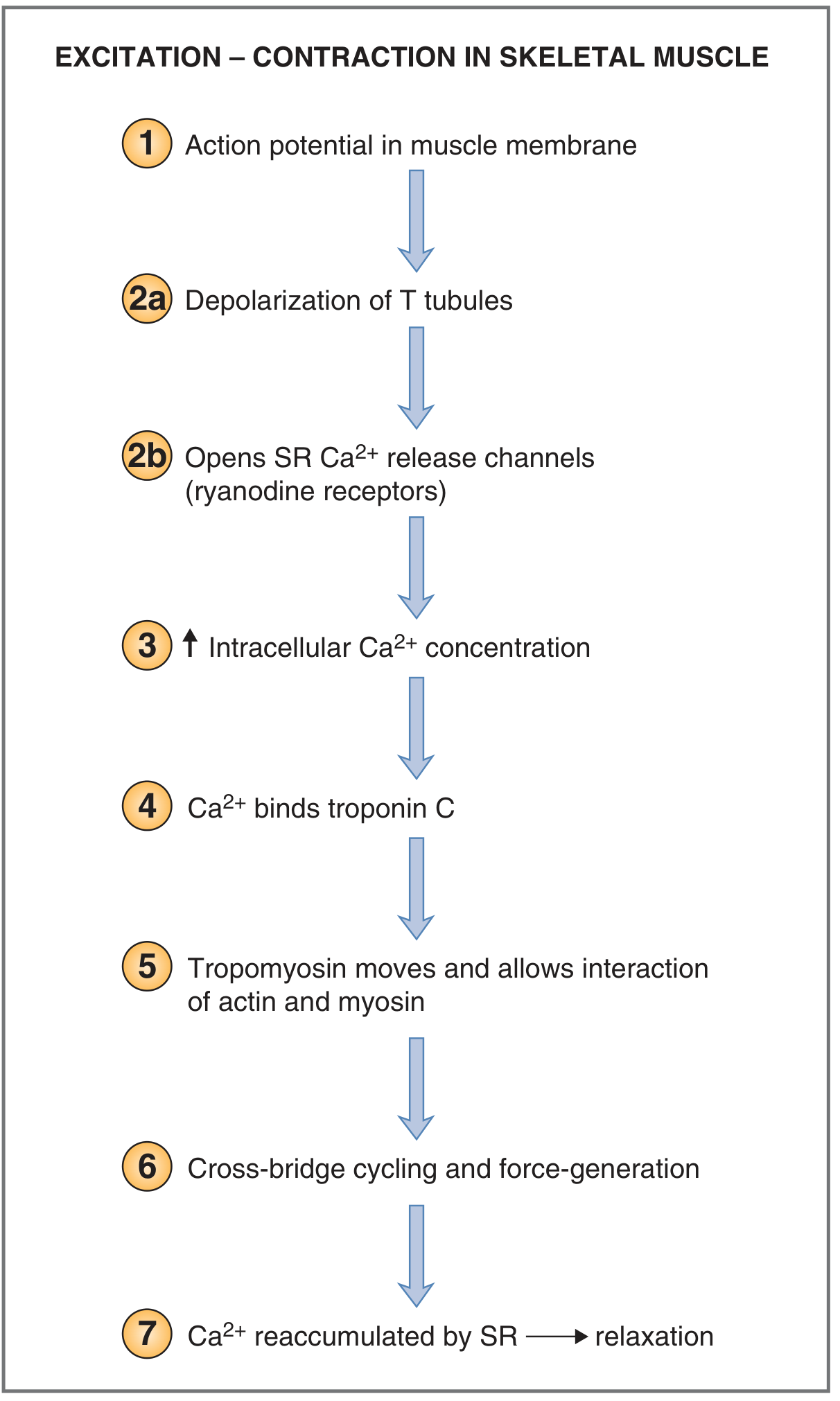
-
Nerve impulse arrives at the neuromuscular junction.
The motor neuron releases acetylcholine (ACh). -
End-plate potential and muscle action potential develop.
ACh binds nicotinic receptors on the motor end plate, causing Na⁺ influx. If threshold is reached, an action potential spreads across the sarcolemma. -
Action potential travels down T-tubules.
T-tubules conduct depolarization deep into the muscle fiber. -
Calcium is released from the sarcoplasmic reticulum (SR).
T-tubule depolarization activates dihydropyridine receptors, which open ryanodine receptors in the adjacent SR. This releases Ca²⁺ into the sarcoplasm. In skeletal muscle, extracellular Ca²⁺ entry is not required for this step.
Costanzo Physiology, 7th ed., p. 44. -
Ca²⁺ binds troponin C.
The troponin complex changes its shape. -
Tropomyosin moves away from actin’s myosin-binding sites.
This exposes the active sites on actin, permitting actin-myosin interaction. -
Cross-bridge cycle produces shortening.
- Energized myosin head, carrying ADP + Pi, binds actin.
- Pi, then ADP, are released.
- The myosin head pivots in the power stroke, pulling thin filaments toward the center of the sarcomere.
- A new ATP binds myosin, causing myosin to detach from actin.
- ATP is hydrolyzed to ADP + Pi, re-cocking the myosin head.
- The cycle repeats as long as Ca²⁺ and ATP are available.
-
Sarcomere shortens.
Thin filaments slide past thick filaments. The I band and H zone decrease, while the A band remains constant. The filaments themselves do not shorten. The actomyosin cycle converts ATP’s chemical energy into force.
Histology: A Text and Atlas with Correlated Cell and Molecular Biology, pp. 816-818. -
Relaxation occurs.
ACh is broken down by acetylcholinesterase, action potentials stop, and SERCA pumps Ca²⁺ back into the SR. Ca²⁺ dissociates from troponin, tropomyosin again blocks actin binding sites, cross-bridge cycling stops, and the muscle relaxes.
One-line sequence
Motor neuron AP → ACh release → muscle AP → T-tubules → SR Ca²⁺ release → troponin C → tropomyosin displacement → actin-myosin cross-bridges → power strokes → contraction → Ca²⁺ reuptake → relaxation.
This timing is consistent with the NCBI physiology overview: the muscle action potential occurs first, followed by intracellular Ca²⁺ rise, then mechanical contraction.
This is a shared conversation. Sign in to Orris to start your own chat.