CSF pathways for radiology complete

Reading File
Finding Sources
Finding Sources
Searching Images

CSF cerebrospinal fluid pathway ventricles subarachnoid space diagram
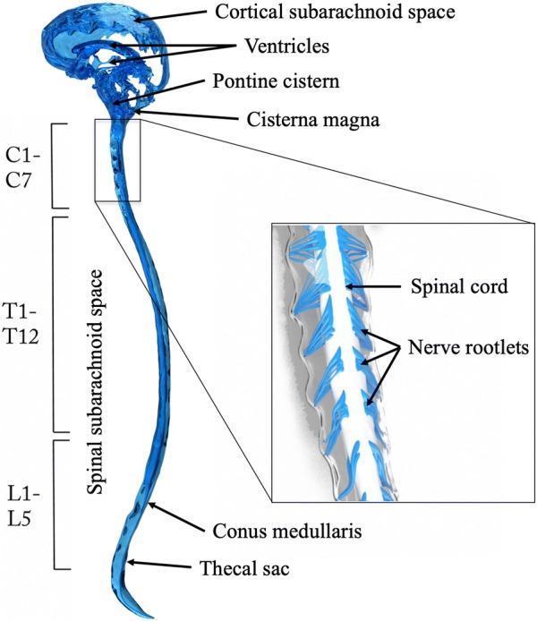
Anatomical diagram of the human cerebrospinal fluid (CSF) system, illustrating the continuous pathway between the intracranial and spinal compartments. The illustration highlights the brain's ventricles, the cortical subarachnoid space, and key basal cisterns including the pontine cistern and cisterna magna. The CSF pathway extends inferiorly into the spinal subarachnoid space (SSS), which is mapped along the vertebral levels: cervical (C1-C7), thoracic (T1-T12), and lumbar (L1-L5). Distal spinal structures identified include the conus medullaris and the thecal sac. A magnified inset provides a detailed view of the SSS, showing the central spinal cord and the lateral emergence of nerve rootlets within the fluid-filled space. This diagram serves as a physiological model for understanding CSF hydrodynamics and the anatomical relationship between the central nervous system and its protective fluid environment, suitable for neurology and neuroanatomy education.
Anatomical diagram of the human cerebrospinal fluid (CSF) system, illustrating the continuous pathway between the intracranial and spinal compartments. The illustration highlights the brain's ventricles, the cortical subarachnoid space, and key basal cisterns including the pontine cistern and cisterna magna. The CSF pathway extends inferiorly into the spinal subarachnoid space (SSS), which is mapped along the vertebral levels: cervical (C1-C7), thoracic (T1-T12), and lumbar (L1-L5). Distal spinal structures identified include the conus medullaris and the thecal sac. A magnified inset provides a detailed view of the SSS, showing the central spinal cord and the lateral emergence of nerve rootlets within the fluid-filled space. This diagram serves as a physiological model for understanding CSF hydrodynamics and the anatomical relationship between the central nervous system and its protective fluid environment, suitable for neurology and neuroanatomy education.

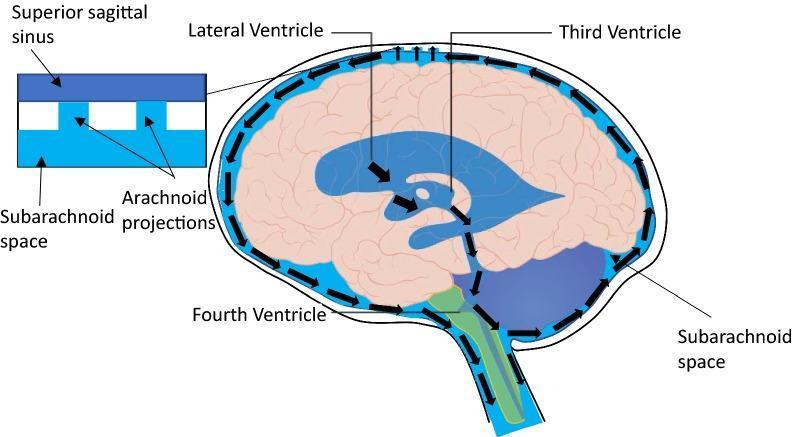
This medical anatomical diagram illustrates the conventional physiological pathway of cerebrospinal fluid (CSF) flow within the central nervous system. The illustration features a sagittal view of the human brain and spinal cord, with arrows indicating the unidirectional movement of CSF. The pathway begins with CSF production in the lateral ventricles, moving through the third and fourth ventricles. From the ventricular system, CSF enters the subarachnoid space, where it circulates around the external surfaces of the brain and extends down the spinal canal. A magnified inset detailed at the top left demonstrates the mechanism of CSF drainage, showing the relationship between the subarachnoid space and the superior sagittal sinus. This process is mediated by arachnoid projections (arachnoid villi/granulations), which allow CSF to be reabsorbed into the venous blood of the dural venous sinus. Key labeled structures include the lateral, third, and fourth ventricles, the subarachnoid space, and the superior sagittal sinus. This diagram serves as an educational resource for neuroanatomy and neurophysiology, specifically regarding intracranial pressure regulation and the glymphatic system context.
This medical anatomical diagram illustrates the conventional physiological pathway of cerebrospinal fluid (CSF) flow within the central nervous system. The illustration features a sagittal view of the human brain and spinal cord, with arrows indicating the unidirectional movement of CSF. The pathway begins with CSF production in the lateral ventricles, moving through the third and fourth ventricles. From the ventricular system, CSF enters the subarachnoid space, where it circulates around the external surfaces of the brain and extends down the spinal canal. A magnified inset detailed at the top left demonstrates the mechanism of CSF drainage, showing the relationship between the subarachnoid space and the superior sagittal sinus. This process is mediated by arachnoid projections (arachnoid villi/granulations), which allow CSF to be reabsorbed into the venous blood of the dural venous sinus. Key labeled structures include the lateral, third, and fourth ventricles, the subarachnoid space, and the superior sagittal sinus. This diagram serves as an educational resource for neuroanatomy and neurophysiology, specifically regarding intracranial pressure regulation and the glymphatic system context.

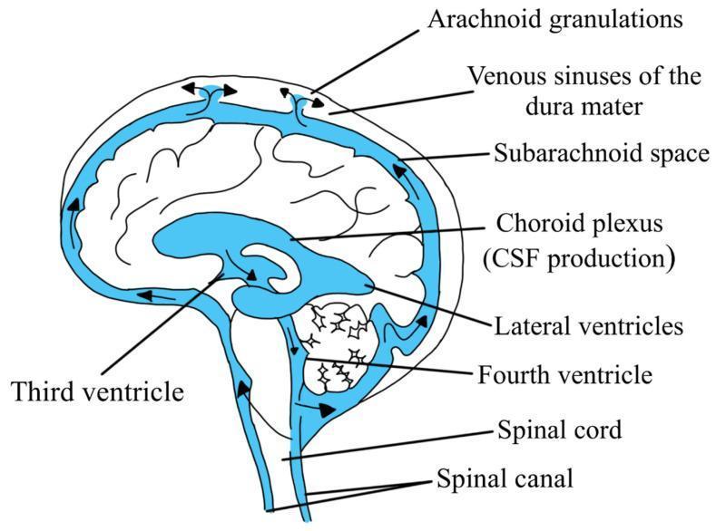
An anatomical diagram illustrating the production and circulation pathway of cerebrospinal fluid (CSF) in the human brain. The illustration depicts a sagittal view of the central nervous system with blue-shaded regions representing the CSF spaces. Production is identified at the choroid plexus within the lateral ventricles. Arrows demonstrate the flow through the ventricular system, including the third ventricle and fourth ventricle. The pathway continues as CSF exits into the subarachnoid space surrounding the brain and spinal cord, and into the spinal canal. Reabsorption is shown at the superior aspect via arachnoid granulations into the venous sinuses of the dura mater. Labeled structures include the lateral and third ventricles, fourth ventricle, subarachnoid space, spinal cord, spinal canal, and the dural venous sinuses. This educational material serves as a visual guide for neuroanatomy and physiological fluid dynamics.
An anatomical diagram illustrating the production and circulation pathway of cerebrospinal fluid (CSF) in the human brain. The illustration depicts a sagittal view of the central nervous system with blue-shaded regions representing the CSF spaces. Production is identified at the choroid plexus within the lateral ventricles. Arrows demonstrate the flow through the ventricular system, including the third ventricle and fourth ventricle. The pathway continues as CSF exits into the subarachnoid space surrounding the brain and spinal cord, and into the spinal canal. Reabsorption is shown at the superior aspect via arachnoid granulations into the venous sinuses of the dura mater. Labeled structures include the lateral and third ventricles, fourth ventricle, subarachnoid space, spinal cord, spinal canal, and the dural venous sinuses. This educational material serves as a visual guide for neuroanatomy and physiological fluid dynamics.

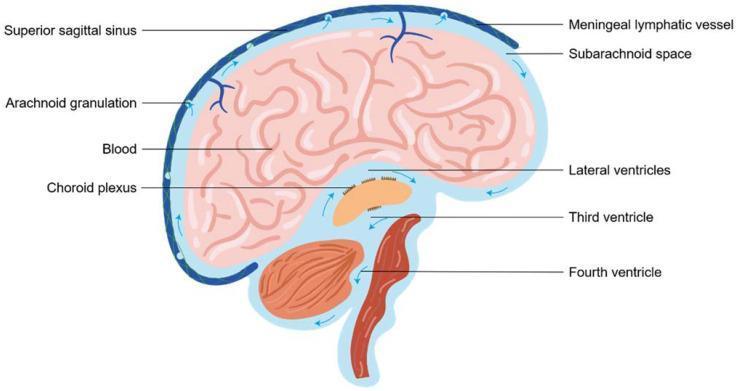
This medical anatomical diagram illustrates the production, circulation, and drainage pathways of cerebrospinal fluid (CSF) in the human brain via a sagittal section view. The illustration highlights the CSF flow starting from the choroid plexus within the lateral, third, and fourth ventricles. Blue arrows indicate the directional movement of fluid into the subarachnoid space (SAS) surrounding the cerebral cortex and cerebellum. Key drainage mechanisms are depicted: the traditional venous pathway where arachnoid granulations protrude into the superior sagittal sinus (containing blood), and the emerging lymphatic pathway via meningeal lymphatic vessels. The diagram provides a comprehensive overview of neuroanatomy and physiology related to intracranial pressure regulation and metabolite clearance, suitable for neuroscience and clinical neurology education.
This medical anatomical diagram illustrates the production, circulation, and drainage pathways of cerebrospinal fluid (CSF) in the human brain via a sagittal section view. The illustration highlights the CSF flow starting from the choroid plexus within the lateral, third, and fourth ventricles. Blue arrows indicate the directional movement of fluid into the subarachnoid space (SAS) surrounding the cerebral cortex and cerebellum. Key drainage mechanisms are depicted: the traditional venous pathway where arachnoid granulations protrude into the superior sagittal sinus (containing blood), and the emerging lymphatic pathway via meningeal lymphatic vessels. The diagram provides a comprehensive overview of neuroanatomy and physiology related to intracranial pressure regulation and metabolite clearance, suitable for neuroscience and clinical neurology education.

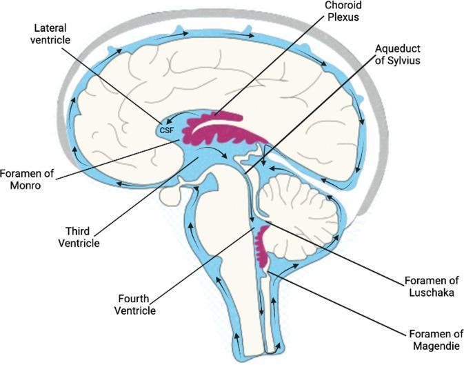
An anatomical diagram in sagittal view illustrating the human ventricular system and the physiological pathway of cerebrospinal fluid (CSF) flow. The diagram depicts the lateral ventricles, containing the choroid plexus (highlighted in pink), where CSF production occurs. Black arrows indicate the unidirectional flow of CSF from the lateral ventricles through the interventricular Foramen of Monro into the third ventricle. The pathway continues through the narrow Aqueduct of Sylvius into the fourth ventricle, located anterior to the cerebellum. From the fourth ventricle, CSF exits the ventricular system through the median Foramen of Magendie and the lateral Foramina of Luschka to enter the subarachnoid space. The diagram further illustrates the circulation of CSF around the external surface of the brain and spinal cord within the subarachnoid space (colored in light blue), showing its eventual movement toward the superior sagittal sinus for reabsorption. Key anatomical labels include the ventricles, foramina, and the brainstem/cerebellar silhouettes.
An anatomical diagram in sagittal view illustrating the human ventricular system and the physiological pathway of cerebrospinal fluid (CSF) flow. The diagram depicts the lateral ventricles, containing the choroid plexus (highlighted in pink), where CSF production occurs. Black arrows indicate the unidirectional flow of CSF from the lateral ventricles through the interventricular Foramen of Monro into the third ventricle. The pathway continues through the narrow Aqueduct of Sylvius into the fourth ventricle, located anterior to the cerebellum. From the fourth ventricle, CSF exits the ventricular system through the median Foramen of Magendie and the lateral Foramina of Luschka to enter the subarachnoid space. The diagram further illustrates the circulation of CSF around the external surface of the brain and spinal cord within the subarachnoid space (colored in light blue), showing its eventual movement toward the superior sagittal sinus for reabsorption. Key anatomical labels include the ventricles, foramina, and the brainstem/cerebellar silhouettes.

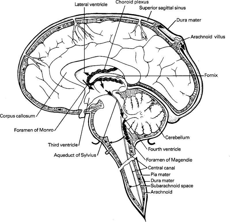
This medical anatomical diagram illustrates the production, circulation, and resorption of cerebrospinal fluid (CSF) within the human central nervous system. The illustration is a sagittal cross-section of the brain and upper spinal cord, highlighting the ventricular system and meningeal layers. Key anatomical structures labeled include the lateral ventricle, choroid plexus (the primary site of CSF production), the Foramen of Monro, third ventricle, Aqueduct of Sylvius, and fourth ventricle. The pathway of CSF flow is indicated by directional arrows, tracing the fluid's movement from the ventricles through the Foramen of Magendie into the subarachnoid space and the central canal. The diagram also depicts the three meningeal layers: the outermost dura mater, the arachnoid mater, and the innermost pia mater, with the subarachnoid space clearly delineated between the arachnoid and pia. Educational focus is placed on CSF resorption via the arachnoid villi (granulations) into the superior sagittal sinus. Supporting landmarks such as the corpus callosum, fornix, and cerebellum provide spatial context for neuroanatomical study.

This medical anatomical diagram illustrates the production, circulation, and resorption of cerebrospinal fluid (CSF) within the human central nervous system. The illustration is a sagittal cross-section of the brain and upper spinal cord, highlighting the ventricular system and meningeal layers. Key anatomical structures labeled include the lateral ventricle, choroid plexus (the primary site of CSF production), the Foramen of Monro, third ventricle, Aqueduct of Sylvius, and fourth ventricle. The pathway of CSF flow is indicated by directional arrows, tracing the fluid's movement from the ventricles through the Foramen of Magendie into the subarachnoid space and the central canal. The diagram also depicts the three meningeal layers: the outermost dura mater, the arachnoid mater, and the innermost pia mater, with the subarachnoid space clearly delineated between the arachnoid and pia. Educational focus is placed on CSF resorption via the arachnoid villi (granulations) into the superior sagittal sinus. Supporting landmarks such as the corpus callosum, fornix, and cerebellum provide spatial context for neuroanatomical study.

Finding Sources
Finding Sources
Searching Images

hydrocephalus obstructive communicating MRI brain ventricles
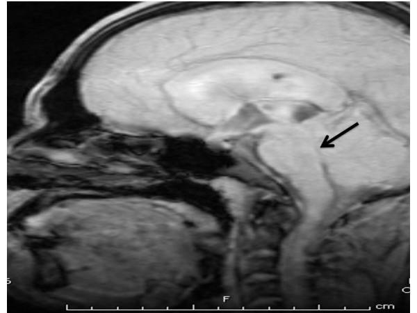
Midsagittal T1-weighted MRI of the brain demonstrating features of obstructive hydrocephalus. The image shows significant dilation of the lateral ventricles and the third ventricle. A black arrow points to the aqueduct of Sylvius (cerebral aqueduct) located between the third and fourth ventricles, indicating the site of aqueductal stenosis. The narrowing at this level leads to the proximal ventricular enlargement seen. The surrounding brainstem, including the pons and medulla, as well as the cerebellum, are visualized. This radiological finding is clinically significant for diagnosing non-communicating hydrocephalus, which in this context is associated with neurocysticercosis causing physical obstruction of the cerebrospinal fluid (CSF) flow path. The image provides a clear anatomical reference for identifying ventricular proportions and midbrain landmarks in neurosurgical and neuroradiological education.
Midsagittal T1-weighted MRI of the brain demonstrating features of obstructive hydrocephalus. The image shows significant dilation of the lateral ventricles and the third ventricle. A black arrow points to the aqueduct of Sylvius (cerebral aqueduct) located between the third and fourth ventricles, indicating the site of aqueductal stenosis. The narrowing at this level leads to the proximal ventricular enlargement seen. The surrounding brainstem, including the pons and medulla, as well as the cerebellum, are visualized. This radiological finding is clinically significant for diagnosing non-communicating hydrocephalus, which in this context is associated with neurocysticercosis causing physical obstruction of the cerebrospinal fluid (CSF) flow path. The image provides a clear anatomical reference for identifying ventricular proportions and midbrain landmarks in neurosurgical and neuroradiological education.

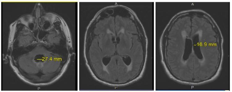
This diagnostic image set consists of three axial brain MRI slices (T2-weighted/FLAIR sequences) demonstrating features of obstructive or communicating hydrocephalus. The first slice (left) shows a transverse measurement of the fourth ventricle at 27.4 mm, indicating significant dilation within the posterior fossa. The middle and right slices display moderate ventriculomegaly of the lateral ventricles, with the right slice highlighting a transverse measurement of 16.9 mm. Notable periventricular T2/FLAIR hyperintensity is visible surrounding the margins of the lateral ventricles, particularly at the frontal and occipital horns. This visual finding is characteristic of transependymal CSF flow (interstitial edema), often seen in acute or subacute hydrocephalus where increased intraventricular pressure forces cerebrospinal fluid into the adjacent white matter. The images serve as an educational example of neuroradiological markers for hydrocephalus and the assessment of CSF dynamics.
This diagnostic image set consists of three axial brain MRI slices (T2-weighted/FLAIR sequences) demonstrating features of obstructive or communicating hydrocephalus. The first slice (left) shows a transverse measurement of the fourth ventricle at 27.4 mm, indicating significant dilation within the posterior fossa. The middle and right slices display moderate ventriculomegaly of the lateral ventricles, with the right slice highlighting a transverse measurement of 16.9 mm. Notable periventricular T2/FLAIR hyperintensity is visible surrounding the margins of the lateral ventricles, particularly at the frontal and occipital horns. This visual finding is characteristic of transependymal CSF flow (interstitial edema), often seen in acute or subacute hydrocephalus where increased intraventricular pressure forces cerebrospinal fluid into the adjacent white matter. The images serve as an educational example of neuroradiological markers for hydrocephalus and the assessment of CSF dynamics.

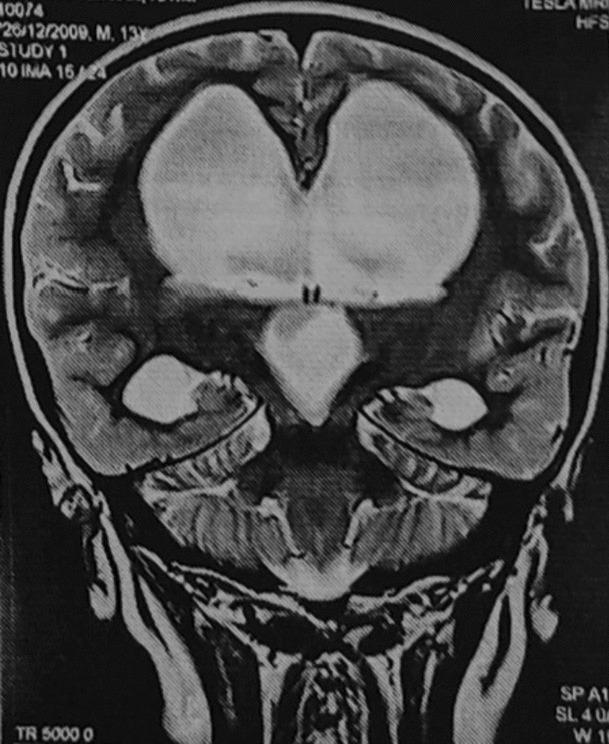
A T2-weighted coronal magnetic resonance imaging (MRI) scan of a human brain demonstrating significant obstructive hydrocephalus. The image clearly shows marked, symmetric dilation of both lateral ventricles and the third ventricle. These cerebrospinal fluid (CSF)-filled spaces exhibit characteristic high signal intensity (bright white) against the lower signal intensity (darker grey) of the surrounding brain parenchyma. The third ventricle appears widened and inferiorly displaced. The cortical mantle and white matter show signs of compression due to the massive ventricular enlargement. This diagnostic image illustrates the radiological features associated with CSF flow obstruction, such as from a third ventricular cyst or aqueductal stenosis, leading to non-communicating hydrocephalus. The visualization of the cerebellum and brainstem inferiorly provides anatomical context for the degree of supratentorial pressure.
A T2-weighted coronal magnetic resonance imaging (MRI) scan of a human brain demonstrating significant obstructive hydrocephalus. The image clearly shows marked, symmetric dilation of both lateral ventricles and the third ventricle. These cerebrospinal fluid (CSF)-filled spaces exhibit characteristic high signal intensity (bright white) against the lower signal intensity (darker grey) of the surrounding brain parenchyma. The third ventricle appears widened and inferiorly displaced. The cortical mantle and white matter show signs of compression due to the massive ventricular enlargement. This diagnostic image illustrates the radiological features associated with CSF flow obstruction, such as from a third ventricular cyst or aqueductal stenosis, leading to non-communicating hydrocephalus. The visualization of the cerebellum and brainstem inferiorly provides anatomical context for the degree of supratentorial pressure.

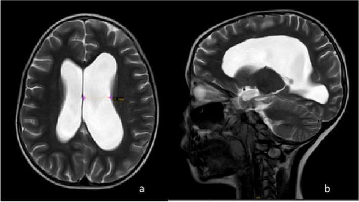
T2-weighted magnetic resonance imaging (MRI) of the brain in axial (a) and midsagittal (b) planes, demonstrating communicating hydrocephalus. The axial view displays a significant enlargement of the bilateral lateral ventricles, with cerebrospinal fluid (CSF) appearing as a high-intensity (bright white) signal relative to the darker brain parenchyma. Notably, the left lateral ventricle shows more pronounced dilatation compared to the right. The midsagittal view further highlights the global enlargement of the ventricular system, including prominent dilatation of the third and fourth ventricles, alongside widening of the suprasellar and prepontine cisterns. There is no evidence of an obstructive mass in the depicted sections. These neuroimaging findings are characteristic of altered CSF dynamics and increased intracranial pressure, clinically relevant in the context of neurological conditions like Guillain-Barré syndrome (GBS) which may present with elevated CSF protein levels leading to impaired resorption.

T2-weighted magnetic resonance imaging (MRI) of the brain in axial (a) and midsagittal (b) planes, demonstrating communicating hydrocephalus. The axial view displays a significant enlargement of the bilateral lateral ventricles, with cerebrospinal fluid (CSF) appearing as a high-intensity (bright white) signal relative to the darker brain parenchyma. Notably, the left lateral ventricle shows more pronounced dilatation compared to the right. The midsagittal view further highlights the global enlargement of the ventricular system, including prominent dilatation of the third and fourth ventricles, alongside widening of the suprasellar and prepontine cisterns. There is no evidence of an obstructive mass in the depicted sections. These neuroimaging findings are characteristic of altered CSF dynamics and increased intracranial pressure, clinically relevant in the context of neurological conditions like Guillain-Barré syndrome (GBS) which may present with elevated CSF protein levels leading to impaired resorption.

Reading File
Reading File
Finding Sources
Reading File
Reading File
Finding Sources
Your credits are exhausted. You can purchase a top-up or subscribe to a plan to continue using Orris. If you're already subscribed, your daily credits will reset tomorrow.

Complete cns pathway

Your credits are exhausted. You can purchase a top-up or subscribe to a plan to continue using Orris. If you're already subscribed, your daily credits will reset tomorrow.
This is a shared conversation. Sign in to Orris to start your own chat.