
teach me embryology development during 1st week
Reading File
Finding Sources
Finding Sources
Searching Images
first week embryo development fertilization cleavage blastocyst implantation diagram
Finding Sources
Finding Sources
Finding Sources
Reading File
Reading File
Reading File
Reading File
Reading File
Reading File
Reading File
Loading Image
Loading Image
Loading Image
I now have all the content I need for a thorough, well-illustrated answer. Let me compile it.
First Week of Human Embryonic Development
Sources: The Developing Human: Clinically Oriented Embryology (Moore et al.) and Langman's Medical Embryology
Overview: Stages 1-4 (Days 1-7)
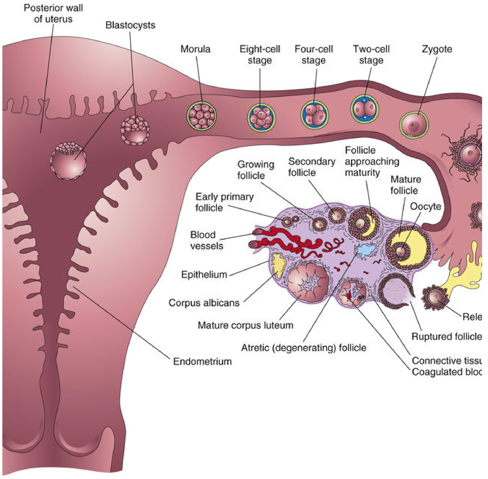
The first week covers everything from ovulation to the initial embedding of the blastocyst in the uterine wall. Four key events define it:
- Fertilization (Day 1) - zygote forms
- Cleavage (Days 2-3) - rapid mitotic divisions
- Morula (Day 3-4) - compact ball of 12-32 cells
- Blastocyst formation and implantation (Days 4-7)
Here is the classic summary diagram showing all stages in the uterine tube and uterus:
Step 1: Ovulation and Gamete Viability
- The secondary oocyte (arrested at metaphase II) is expelled from the Graafian follicle and swept by the fimbriae of the uterine tube into the ampulla.
- The oocyte can only be fertilized within 12-24 hours after ovulation; it degenerates shortly afterward.
- Sperm remain viable in the female genital tract for up to 48 hours (some sources say up to 5 days in cervical crypts).
- Of 200-300 million sperm deposited in the vagina, only 300-500 reach the ampulla. Only 1% enter the cervix.
Step 2: Capacitation and the Acrosome Reaction
Before a sperm can fertilize, it must undergo two preparation steps:
Capacitation
- Takes approximately 7 hours in the female reproductive tract (mainly the uterine tube).
- A glycoprotein coat and seminal plasma proteins are removed from the membrane overlying the acrosomal region.
- Sperm become hyperactivated - only capacitated sperm can penetrate the corona radiata and undergo the acrosome reaction.
Acrosome Reaction
- Triggered by binding to zona pellucida proteins (ZP3).
- The acrosomal membrane fuses with the sperm plasma membrane, releasing hydrolytic enzymes (acrosin, trypsin-like substances).
- These enzymes digest a path through the zona pellucida.
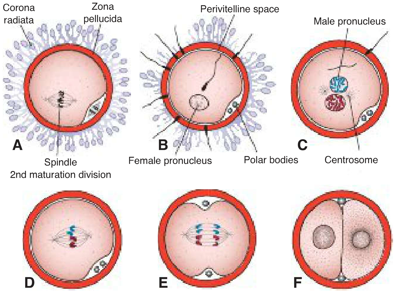
Step 3: Fertilization - The Three Phases
Site: Ampulla of the uterine tube
Phase 1: Penetration of the Corona Radiata
- Capacitated sperm pass freely through the corona cells aided by enzymes from the acrosome reaction and mechanical movement of the sperm tail.
Phase 2: Penetration of the Zona Pellucida
- The zona pellucida is a glycoprotein shell surrounding the oocyte.
- ZP3 acts as the sperm receptor and induces the acrosome reaction.
- ZP2 binds sperm pre-acrosin after the acrosome reaction.
- Acrosin and trypsin-like enzymes digest a path for the sperm head.
Phase 3: Fusion of Sperm and Oocyte Cell Membranes
- Sperm proteins (IZUMO on sperm; Juno on oocyte) are the key fusogenic pair.
- Fusion triggers the cortical reaction: cortical granules release enzymes that modify ZP2 and ZP3, hardening the zona pellucida - this is the block to polyspermy (zona reaction).
- A wave of Ca2+ sweeps through the oocyte cytoplasm simultaneously.
- Sperm penetration completes the secondary oocyte's meiosis II, expelling the second polar body.
- Male and female pronuclei form, each containing 23 chromosomes (haploid).
- The pronuclei approach each other, their membranes break down, and the chromosomes arrange on the first mitotic spindle - the zygote (46 chromosomes, diploid) is formed.
Results of Fertilization
| Result | Significance |
|---|---|
| Restores diploid number (46 chromosomes) | Genetic diversity |
| Determines chromosomal sex | XX (female) or XY (male) |
| Initiates cleavage | Starts embryonic development |
| Second meiotic division completed | Oocyte fully matures |
Step 4: Cleavage (Days 2-3)
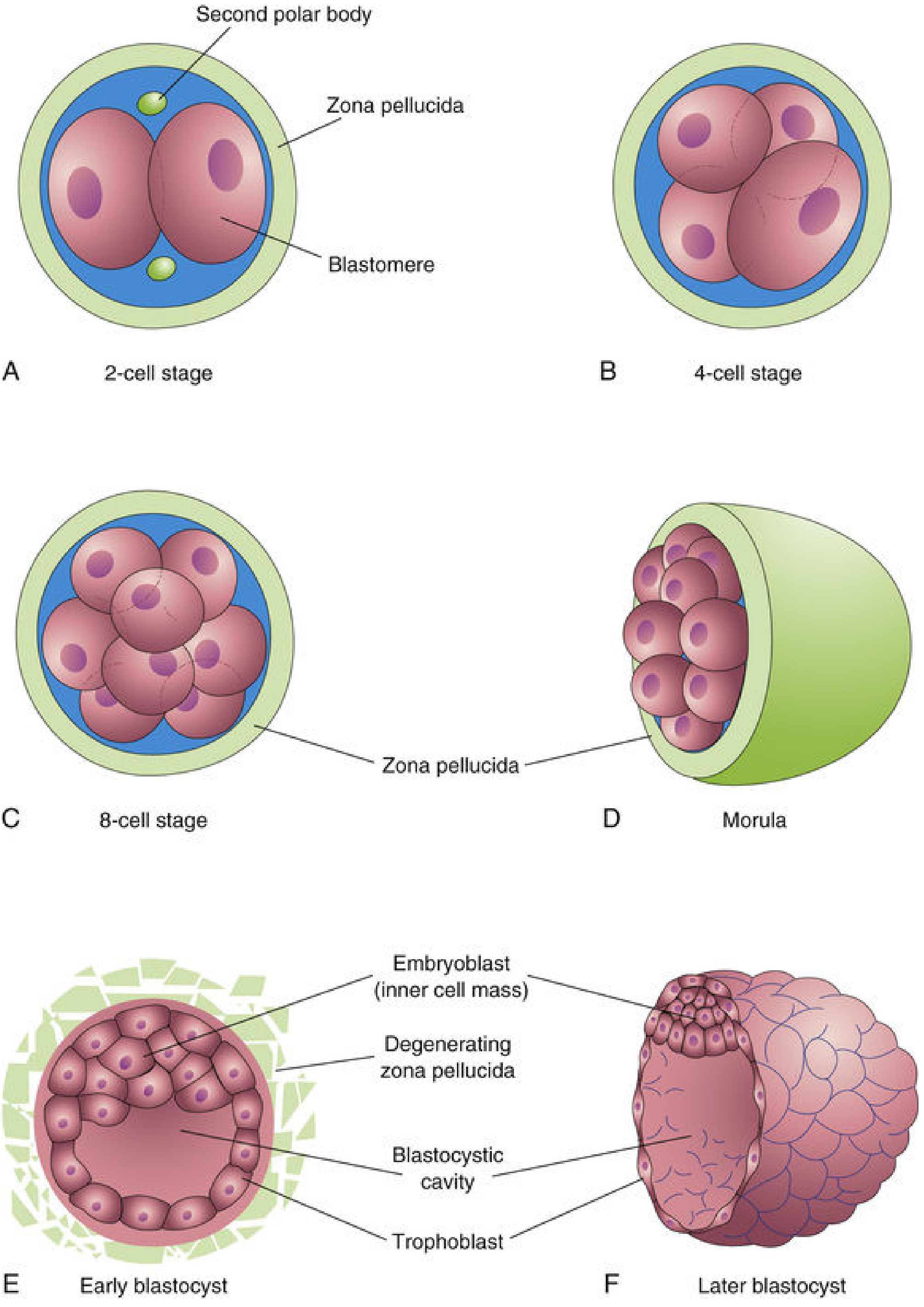
- Cleavage = rapid, repeated mitotic divisions of the zygote.
- Each daughter cell (blastomere) is smaller than the parent - no net growth yet (all divisions occur within the zona pellucida).
- Timeline:
- ~30 hours: 2-cell stage
- ~40 hours: 4-cell stage
- ~3 days: 12-16 cell morula
Compaction (9-cell stage onward)
- Blastomeres flatten and tightly align via E-cadherin-catenin (adherens junctions) and gap junctions.
- Cell polarity (apical vs. basolateral domains) develops.
- Hippo signaling drives segregation of the inner cells (future embryoblast) from outer cells (future trophoblast).
Step 5: Morula (Day 3-4)
- At 12-32 cells, the embryo is called the morula (Latin: mulberry).
- Outer cells form the outer cell mass (trophoblast) - will contribute to the placenta.
- Inner cells form the inner cell mass (embryoblast) - will form the embryo proper (all three germ layers).
- The morula enters the uterine cavity around day 3-4.
Step 6: Blastocyst Formation (Day 4-5)
-
Fluid from the uterine cavity seeps through the zona pellucida into the intercellular spaces of the inner cell mass.
-
The spaces coalesce into a single cavity: the blastocele (blastocystic cavity).
-
The embryo is now a blastocyst:
- Embryoblast (inner cell mass) clusters at one pole
- Trophoblast flattens to form the outer epithelial wall
- Blastocele fills with fluid
-
By day 4-5, the zona pellucida degenerates (hatching of the blastocyst), allowing the embryo to expand and begin implantation.
Step 7: Implantation (Days 6-7)
- The blastocyst adheres to the posterior wall of the body of the uterus in most cases.
- The endometrium must be in the secretory (luteal) phase to receive the embryo - this is the window of implantation.
Molecular Mechanism of Implantation
- Apposition - loose contact between trophoblast and uterine epithelium, mediated by L-selectin on trophoblast and carbohydrate receptors on the uterine epithelium ("blastocyst capture").
- Adhesion - firm attachment via integrins on trophoblast binding to extracellular matrix molecules laminin and fibronectin.
- Invasion - trophoblastic cells over the embryoblast pole penetrate between epithelial cells of the uterine mucosa on about day 6.
Trophoblast Differentiation
By the end of week 1, the trophoblast over the embryoblast pole begins to differentiate into:
- Cytotrophoblast (inner, mitotically active layer)
- Syncytiotrophoblast (outer, invasive layer - multinucleated, no cell boundaries; this develops more in week 2)
Timeline Summary
| Day | Event | Carnegie Stage |
|---|---|---|
| 0 | Ovulation; fertilization in ampulla | Stage 1 |
| 1-2 | 2-cell → 4-cell cleavage | Stage 2 |
| 3 | 8-16 cell morula; enters uterine cavity | Stage 3 |
| 4 | Late morula; zona pellucida hatching | Stage 3 |
| 4-5 | Blastocyst forms; zona degenerates | Stage 3 |
| 6-7 | Implantation begins | Stage 4 |
Key Clinical Points
- Ectopic pregnancy: If fertilization occurs but the embryo cannot travel to the uterus (e.g., tubal damage), it implants in the tube → potentially life-threatening rupture. The most common site is the ampulla.
- Spontaneous abortion rate: 50-70% of all fertilized ova abort spontaneously, most before clinical recognition. Over 50% are due to chromosomal abnormalities - this is natural selection of defective embryos.
- IVF: Embryos (4-8 cell or blastocyst stage) are transferred into the uterus. The success of implantation depends on endometrial receptivity.
- Identical (monozygotic) twins: If the inner cell mass splits at the blastocyst stage, both twins share one chorion (monochorionic). Splitting at the 2-cell stage gives dichorionic identical twins.
- Sex determination: Determined at fertilization by the sperm - X-bearing sperm → female (XX); Y-bearing sperm → male (XY). The oocyte always carries an X chromosome.
The Developing Human: Clinically Oriented Embryology, pp. 116-141 | Langman's Medical Embryology, pp. 58-65
testme giving mcqs like those of inicet and neet pg
Here are 10 INICET/NEET PG-style MCQs on First Week Embryology. Try answering before reading the explanations!
MCQ 1
The usual site of fertilization in humans is:
A. Isthmus of uterine tube
B. Ampulla of uterine tube
C. Fimbriated end of uterine tube
D. Body of uterus
🔍 Click for Answer
Answer: B - Ampulla of uterine tube
The ampulla is the widest part of the uterine tube, closest to the ovary. Chemical signals (chemoattractants) from the oocyte and cumulus cells guide capacitated sperm here. Fertilization does NOT occur in the uterine body.
Tip: If the oocyte is not fertilized in the ampulla, it degenerates as it passes toward the uterus.
MCQ 2
During capacitation of spermatozoa, which of the following occurs?
A. Acrosome reaction is triggered
B. Glycoprotein coat is removed from the acrosomal region
C. Second polar body is expelled
D. ZP3 binding initiates calcium release
🔍 Click for Answer
Answer: B - Glycoprotein coat is removed from the acrosomal region
Capacitation takes ~7 hours in the female reproductive tract. It involves removal of a glycoprotein coat and seminal plasma proteins from the plasma membrane overlying the acrosome. Only after this can sperm undergo the acrosome reaction.
- Option A (acrosome reaction) is triggered by ZP3 binding - this happens AFTER capacitation
- Option C (second polar body) is expelled after sperm entry into the oocyte
- Option D describes the cortical reaction, not capacitation
MCQ 3
Block to polyspermy is achieved by:
A. Zona reaction - modification of ZP2 and ZP3 by cortical granule enzymes
B. Acrosome reaction depleting acrosin
C. Hyperpolarization of the oocyte membrane
D. Destruction of sperm flagella by uterine secretions
🔍 Click for Answer
Answer: A - Zona reaction - modification of ZP2 and ZP3 by cortical granule enzymes
Upon sperm-oocyte fusion, a Ca2+ wave triggers cortical granule exocytosis. The released enzymes digest the sperm receptors ZP2 and ZP3, hardening the zona pellucida and preventing further sperm from penetrating. This is called the zona reaction (permanent block to polyspermy).
Note: There is also a fast, transient membrane depolarization block, but the zona reaction is the definitive block.
MCQ 4
At what cell stage does compaction occur in the human embryo?
A. 2-cell stage
B. 4-cell stage
C. 9-cell stage onward
D. 32-cell stage (morula)
🔍 Click for Answer
Answer: C - 9-cell stage onward
After the 9-cell stage, blastomeres tightly align themselves against each other via E-cadherin-catenin (adherens junctions) and gap junctions. This maximizes cell-to-cell contact and is a prerequisite for inner cell mass (embryoblast) segregation. Hippo signaling drives the separation of inner (embryoblast) vs. outer (trophoblast) cells.
MCQ 5
The inner cell mass (embryoblast) of the blastocyst gives rise to:
A. Trophoblast and placenta
B. Amnion only
C. All tissues of the embryo proper
D. Chorion
🔍 Click for Answer
Answer: C - All tissues of the embryo proper
- Inner cell mass (embryoblast) → embryo proper (all three germ layers + amniotic membrane)
- Outer cell mass (trophoblast) → placenta (chorion, cytotrophoblast, syncytiotrophoblast)
This distinction is HIGH YIELD. The trophoblast never contributes to the embryo itself.
MCQ 6
A woman's oocyte remains fertilizable for how long after ovulation?
A. 6 hours
B. 12-24 hours
C. 48-72 hours
D. 5-7 days
🔍 Click for Answer
Answer: B - 12-24 hours
- Oocyte is usually fertilized within 12 hours after ovulation
- Cannot be fertilized after 24 hours; degenerates shortly thereafter
- Sperm survive up to 48 hours in the female tract (some up to 5 days in cervical crypts)
Clinical relevance: The fertile window is approximately 5 days before ovulation + the day of ovulation itself, owing to sperm longevity.
MCQ 7
On which day does the blastocyst begin implanting into the uterine endometrium?
A. Day 3
B. Day 4-5
C. Day 6
D. Day 10
🔍 Click for Answer
Answer: C - Day 6
- Day 4-5: Blastocyst forms; zona pellucida degenerates (hatching)
- Day 6: Trophoblastic cells over the embryoblast pole begin penetrating between uterine epithelial cells
- Day 6-7: Initial implantation establishes by end of Week 1
The endometrium must be in the secretory (luteal) phase for implantation to succeed. Inadequate progesterone from the corpus luteum is a common cause of implantation failure.
MCQ 8
The sperm protein that pairs with Juno on the oocyte surface to enable gamete fusion is:
A. Acrosin
B. IZUMO
C. SED1
D. SPAM1
🔍 Click for Answer
Answer: B - IZUMO
The IZUMO-Juno pair is the key fusogenic receptor pair for sperm-egg membrane fusion:
- IZUMO (sperm) + Juno (oocyte) = fusion
- SED1 binds ZP3 during the acrosome reaction
- Acrosin/trypsin digest the zona pellucida
- SPAM1 (sperm adhesion molecule) is involved in zona penetration
This is a frequently asked molecular embryology question in recent INICET papers.
MCQ 9
Which of the following correctly describes the morula?
A. A fluid-filled cavity appears within it
B. It consists of 12-32 blastomeres surrounded by zona pellucida
C. It implants in the uterus
D. The zona pellucida has already degenerated
🔍 Click for Answer
Answer: B - It consists of 12-32 blastomeres surrounded by zona pellucida
- Morula = 12-32 cells, still within the zona pellucida
- Fluid cavity (blastocele) appears AFTER morula → making it a blastocyst
- Zona pellucida degenerates at the blastocyst stage (~day 4-5)
- The blastocyst (not morula) implants
MCQ 10
A couple conceives via IVF. The embryo is transferred at the blastocyst stage. Which cell lineage of the blastocyst will form the fetal contribution to the placenta?
A. Embryoblast
B. Trophoblast
C. Epiblast
D. Hypoblast
🔍 Click for Answer
Answer: B - Trophoblast
The trophoblast (outer cell mass) differentiates into:
- Cytotrophoblast - inner, mitotically active
- Syncytiotrophoblast - outer invasive layer, secretes hCG
These form the chorion and placenta. The embryoblast forms the embryo proper; epiblast and hypoblast are second-week derivatives of the embryoblast (bilaminar disc).
hCG secreted by the syncytiotrophoblast is what pregnancy tests detect - it maintains the corpus luteum and progesterone production.
Score Yourself
| Score | Performance |
|---|---|
| 9-10 | Excellent - NEET PG ready |
| 7-8 | Good - review weak areas |
| 5-6 | Average - re-read the topic |
| <5 | Go back to basics first |
Want more MCQs on a specific subtopic (e.g., implantation, fertilization mechanisms, trophoblast differentiation), or shall I move to Second Week Embryology?
This is a shared conversation. Sign in to Orris to start your own chat.