
Axis and planes biomechanics
Axes and Planes in Biomechanics
The Cardinal Body Planes
| Plane | Also Called | Orientation | What it Divides |
|---|---|---|---|
| Sagittal | Median plane (when central) | Vertical, front-to-back | Left half / Right half |
| Frontal | Coronal plane | Vertical, side-to-side | Anterior / Posterior |
| Transverse | Axial / Horizontal plane | Horizontal | Superior / Inferior |
- The midsagittal (median) plane is the single sagittal plane that divides the body into exactly equal left and right halves.
- Any plane parallel to the forehead or coronal suture of the skull is a frontal (coronal) plane.
- The transverse plane is perpendicular to the longitudinal body axis and produces cross-sectional "slices."
The Cardinal Body Axes
| Axis | Also Called | Orientation | Formed by Intersection of |
|---|---|---|---|
| Vertical (Longitudinal) | Craniocaudal axis | Top to bottom | Sagittal + Frontal planes |
| Sagittal (Anteroposterior) | AP axis | Front to back | Sagittal + Transverse planes |
| Transverse (Horizontal) | Mediolateral axis | Side to side | Frontal + Transverse planes |
Movements in Each Plane (Around Each Axis)
| Movement | Plane it occurs IN | Axis it rotates AROUND |
|---|---|---|
| Flexion / Extension | Sagittal plane | Transverse (mediolateral) axis |
| Abduction / Adduction | Frontal plane | Sagittal (AP) axis |
| Internal / External rotation; Pronation / Supination | Transverse plane | Vertical (longitudinal) axis |
| Circumduction | All three planes | All three axes (combined) |
Degrees of Freedom
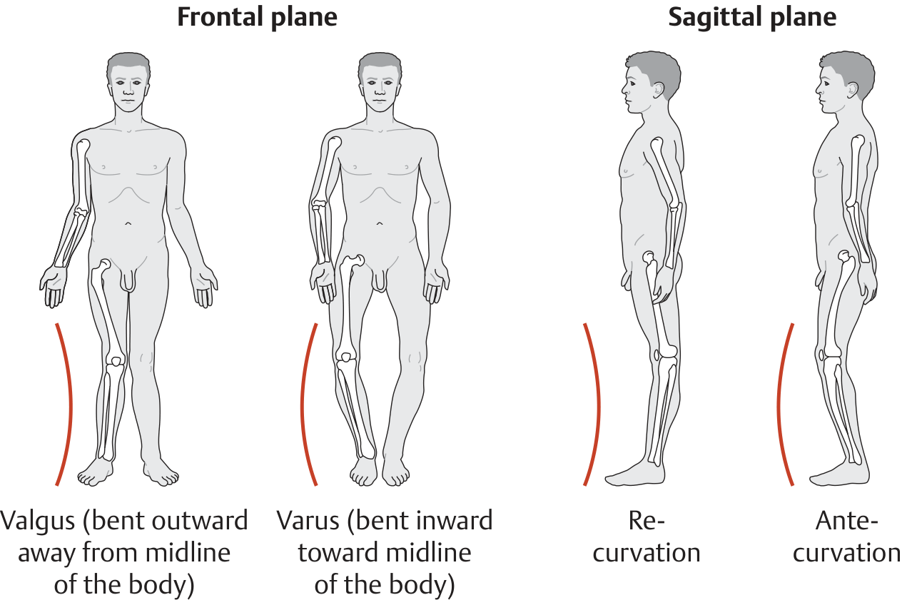
Axis Deviation and Clinical Significance
-
Frontal plane deviations = Varus or Valgus
- Varus: the distal bone points toward the body's midline (e.g., genu varum = bow-legs). The axis deviation is concave to the vertical body axis.
- Valgus: the distal bone points away from the midline (e.g., genu valgum = knock-knee). The axis deviation is convex to the vertical body axis.
-
Sagittal plane deviations = Recurvation or Antecurvation (hyperextension vs. forward bend deformity).
Center of Gravity and Line of Gravity
- The whole-body center of gravity lies just below the sacral promontory at the level of S2.
- The line of gravity runs vertically along the midsagittal plane.
- Anterior view: passes through the midsagittal plane.
- Lateral view: passes through the external auditory canal → dens of the axis (C2) → anatomic junctions of the spine → whole-body center of gravity → hip joint → anterior to the knee joint → anterior to the ankle joint.
- Any displacement of the line of gravity changes the demand on muscles and ligaments to maintain balance.
Instant Center of Rotation
- In a geometrically simple joint, it is fixed.
- In the knee, the ICR changes throughout the arc of motion, tracing a curved path - this is why the knee both rolls and slides to maintain congruence.
- The ICR normally lies on a line perpendicular to the tangent of the joint surface at the point of contact.
- Rolling: the ICR is at the rolling surface; no slipping occurs.
- Sliding: the wheel (or joint surface) rotates about a stationary axis - pure translation with no rolling.
- Most joints use a combination of rolling and sliding.
Coupled Motions
- Classic example: lateral bending of the spine is automatically accompanied by axial rotation in the same direction (and vice versa).
- The shoulder complex: during elevation of the arm, the clavicle at the sternoclavicular joint undergoes coupled posterior rotation together with scapular motion via the coracoclavicular ligaments.
Quick Reference Summary
Sagittal plane   → Flexion/Extension  (around transverse axis)
Frontal plane    → Abd/Adduction      (around sagittal/AP axis)
Transverse plane → Rotation           (around vertical/long axis)

Vertical axis    = intersection of Sagittal + Frontal planes
Sagittal axis    = intersection of Sagittal + Transverse planes
Transverse axis  = intersection of Frontal + Transverse planes
Axis and planes biomechanics images assignment
What format should the assignment be in? · What should the assignment cover? · What is the academic level?
mkdir -p /home/daytona/workspace/axes-planes-assignment/images && cd /home/daytona/workspace/axes-planes-assignment/images && curl -s -o axis_deviation.png "https://cdn.orris.care/cdss_images/2e8211de7642c967cadc186a6f354d03d46990420817127516787e954f3a1dc5.png" && curl -s -o line_of_gravity.png "https://cdn.orris.care/cdss_images/1da414f838ce2b3968ac825f4e5cbc5a584c18f11c16118e43ab8768df16807a.png" && curl -s -o hip_movements.png "https://cdn.orris.care/cdss_images/a02e871caf93b8f339745e36f17a4c31ce6b431264b94f56da12815ecc8c1f8a.png" && curl -s -o knee_ankle.png "https://cdn.orris.care/cdss_images/00f60eee7d2773ecaa52055002a6ca2f6f27237f63fff654bb40a7b98cbcb99e.png" && curl -s -o skull_planes.png "https://cdn.orris.care/cdss_images/dd39500c7f02c59135daf5eef91b6de57576a87bd4b190e9e7c6eeba625918a0.png" && ls -la
~/axes-planes-assignment/create_assignment.js
const {
  Document, Packer, Paragraph, TextRun, Table, TableRow, TableCell,
  ImageRun, HeadingLevel, AlignmentType, BorderStyle, WidthType,
  VerticalAlign, PageBreak, ShadingType
} = require('docx');
const fs = require('fs');
const path = require('path');

const imgDir = '/home/daytona/workspace/axes-planes-assignment/images';

function loadImg(filename) {
  return fs.readFileSync(path.join(imgDir, filename));
}

function heading1(text) {
  return new Paragraph({
    text,
    heading: HeadingLevel.HEADING_1,
    spacing: { before: 400, after: 200 },
  });
}

function heading2(text) {
  return new Paragraph({
    text,
    heading: HeadingLevel.HEADING_2,
    spacing: { before: 300, after: 150 },
  });
}

function body(text) {
  return new Paragraph({
    children: [new TextRun({ text, size: 24, font: 'Calibri' })],
    spacing: { before: 100, after: 100 },
  });
}

function bullet(text) {
  return new Paragraph({
    children: [new TextRun({ text, size: 24, font: 'Calibri' })],
    bullet: { level: 0 },
    spacing: { before: 60, after: 60 },
  });
}

function subbullet(text) {
  return new Paragraph({
    children: [new TextRun({ text, size: 22, font: 'Calibri' })],
    bullet: { level: 1 },
    spacing: { before: 40, after: 40 },
  });
}

function caption(text) {
  return new Paragraph({
    children: [new TextRun({ text, size: 20, italics: true, color: '444444', font: 'Calibri' })],
    alignment: AlignmentType.CENTER,
    spacing: { before: 60, after: 200 },
  });
}

function image(data, w, h, desc) {
  return new Paragraph({
    children: [
      new ImageRun({
        data,
        transformation: { width: w, height: h },
        altText: { title: desc, description: desc, name: desc },
      })
    ],
    alignment: AlignmentType.CENTER,
    spacing: { before: 200, after: 100 },
  });
}

function makeTable(headers, rows) {
  const headerCells = headers.map(h =>
    new TableCell({
      children: [new Paragraph({
        children: [new TextRun({ text: h, bold: true, size: 22, font: 'Calibri', color: 'FFFFFF' })],
        alignment: AlignmentType.CENTER,
      })],
      shading: { type: ShadingType.SOLID, color: '1F5C99', fill: '1F5C99' },
      verticalAlign: VerticalAlign.CENTER,
    })
  );

  const dataRows = rows.map((row, ri) => {
    const cells = row.map((cell) =>
      new TableCell({
        children: [new Paragraph({
          children: [new TextRun({ text: cell, size: 22, font: 'Calibri' })],
          alignment: AlignmentType.LEFT,
        })],
        shading: ri % 2 === 0
          ? { type: ShadingType.SOLID, color: 'EAF2FB', fill: 'EAF2FB' }
          : { type: ShadingType.SOLID, color: 'FFFFFF', fill: 'FFFFFF' },
        verticalAlign: VerticalAlign.CENTER,
      })
    );
    return new TableRow({ children: cells });
  });

  return new Table({
    rows: [new TableRow({ children: headerCells, tableHeader: true }), ...dataRows],
    width: { size: 100, type: WidthType.PERCENTAGE },
  });
}

function spacer() {
  return new Paragraph({ text: '', spacing: { before: 100, after: 100 } });
}

function pageBreak() {
  return new Paragraph({ children: [new PageBreak()] });
}

// Load images
const skullPlanes = loadImg('skull_planes.png');
const axisDeviation = loadImg('axis_deviation.png');
const lineOfGravity = loadImg('line_of_gravity.png');
const hipMovements = loadImg('hip_movements.png');
const kneeAnkle = loadImg('knee_ankle.png');

const doc = new Document({
  styles: {
    paragraphStyles: [
      {
        id: 'Heading1',
        name: 'Heading 1',
        basedOn: 'Normal',
        next: 'Normal',
        run: { bold: true, size: 32, color: '1F5C99', font: 'Calibri' },
        paragraph: { spacing: { before: 400, after: 200 } },
      },
      {
        id: 'Heading2',
        name: 'Heading 2',
        basedOn: 'Normal',
        next: 'Normal',
        run: { bold: true, size: 26, color: '2E74B5', font: 'Calibri' },
        paragraph: { spacing: { before: 280, after: 140 } },
      },
    ],
  },
  sections: [{
    properties: {
      page: {
        margin: { top: 1080, bottom: 1080, left: 1080, right: 1080 },
      },
    },
    children: [
      // ─── TITLE PAGE ───────────────────────────────────────────────────
      spacer(), spacer(), spacer(),
      new Paragraph({
        children: [new TextRun({ text: 'AXES AND PLANES IN BIOMECHANICS', bold: true, size: 48, color: '1F5C99', font: 'Calibri' })],
        alignment: AlignmentType.CENTER,
        spacing: { before: 200, after: 400 },
      }),
      new Paragraph({
        children: [new TextRun({ text: 'A Comprehensive Illustrated Assignment', size: 28, italics: true, color: '444444', font: 'Calibri' })],
        alignment: AlignmentType.CENTER,
        spacing: { before: 0, after: 200 },
      }),
      new Paragraph({
        children: [new TextRun({ text: 'Subject: Biomechanics & Kinesiology', size: 24, font: 'Calibri', color: '666666' })],
        alignment: AlignmentType.CENTER,
        spacing: { before: 0, after: 100 },
      }),
      new Paragraph({
        children: [new TextRun({ text: 'Date: July 2026', size: 24, font: 'Calibri', color: '666666' })],
        alignment: AlignmentType.CENTER,
        spacing: { before: 0, after: 600 },
      }),
      pageBreak(),

      // ─── SECTION 1: INTRODUCTION ──────────────────────────────────────
      heading1('1. Introduction'),
      body('Biomechanics is the science of movement of living bodies. To describe and analyse human movement precisely, a standardised three-dimensional reference framework is used — three cardinal planes and three cardinal axes, all mutually perpendicular to one another and based on the three spatial coordinates (x, y, z). Understanding this framework is fundamental to clinical assessment, rehabilitation, and sports science.'),
      spacer(),
      body('All descriptions assume the anatomical position: the body is erect, feet together, upper limbs at the sides with the palms facing forward.'),

      // ─── SECTION 2: CARDINAL PLANES ──────────────────────────────────
      pageBreak(),
      heading1('2. The Three Cardinal Planes'),
      body('Although any number of planes can be drawn through the human body, only three are designated as cardinal planes. Each plane divides the body into two portions and defines a direction of movement.'),
      spacer(),

      heading2('2.1 Sagittal Plane'),
      bullet('Orientation: Vertical plane passing through the body from front to back (anteroposteriorly).'),
      bullet('Named after the sagittal suture of the skull.'),
      bullet('The mid-sagittal plane (= median plane) divides the body into equal left and right halves.'),
      bullet('Any parallel plane is also a sagittal plane (parasagittal plane).'),
      bullet('Movements occurring in this plane: Flexion and Extension.'),
      spacer(),

      heading2('2.2 Frontal (Coronal) Plane'),
      bullet('Orientation: Vertical plane passing side to side through the body.'),
      bullet('Named after the coronal suture of the skull / the forehead (Latin: frons).'),
      bullet('Divides the body into anterior (front) and posterior (back) portions.'),
      bullet('Movements occurring in this plane: Abduction and Adduction; Lateral Flexion (spine).'),
      spacer(),

      heading2('2.3 Transverse Plane'),
      bullet('Also called: Axial plane or Horizontal plane.'),
      bullet('Orientation: Horizontal plane perpendicular to the long axis of the body.'),
      bullet('Divides the body into superior (upper) and inferior (lower) portions.'),
      bullet('Movements occurring in this plane: Internal and External Rotation; Pronation and Supination.'),
      spacer(),

      // Skull planes diagram
      image(skullPlanes, 380, 340, 'Sagittal and coronal planes illustrated on the skull'),
      caption('Fig. 1 — Sagittal and coronal planes shown on the skull, named after their respective sutures. Source: THIEME Atlas of General Anatomy and Musculoskeletal System.'),

      spacer(),
      makeTable(
        ['Plane', 'Orientation', 'Divides Body Into', 'Movements'],
        [
          ['Sagittal', 'Vertical, front-to-back', 'Left / Right halves', 'Flexion, Extension'],
          ['Frontal (Coronal)', 'Vertical, side-to-side', 'Anterior / Posterior', 'Abduction, Adduction, Lateral Flexion'],
          ['Transverse (Axial)', 'Horizontal', 'Superior / Inferior', 'Rotation, Pronation, Supination'],
        ]
      ),
      caption('Table 1 — Summary of the three cardinal planes.'),

      // ─── SECTION 3: CARDINAL AXES ────────────────────────────────────
      pageBreak(),
      heading1('3. The Three Cardinal Axes'),
      body('Each axis is a straight line about which rotation occurs. Every axis lies at the intersection of two planes and is therefore perpendicular to the plane in which the associated movement takes place.'),
      spacer(),

      heading2('3.1 Vertical (Longitudinal) Axis'),
      bullet('Runs craniocaudally (top to bottom) through the body.'),
      bullet('In the standing position it is perpendicular to the ground.'),
      bullet('Lies at the intersection of the sagittal and frontal (coronal) planes.'),
      bullet('Movements around this axis: Internal/External rotation; Supination/Pronation. These occur in the transverse plane.'),
      spacer(),

      heading2('3.2 Sagittal (Anteroposterior) Axis'),
      bullet('Runs from front to back (or back to front) through the body.'),
      bullet('Lies at the intersection of the sagittal and transverse planes.'),
      bullet('Also called the AP axis or dorsoventral axis.'),
      bullet('Movements around this axis: Abduction and Adduction; Lateral Flexion. These occur in the frontal plane.'),
      spacer(),

      heading2('3.3 Transverse (Mediolateral) Axis'),
      bullet('Runs from side to side through the body.'),
      bullet('Lies at the intersection of the frontal and transverse planes.'),
      bullet('Also called the horizontal axis or coronal axis.'),
      bullet('Movements around this axis: Flexion and Extension. These occur in the sagittal plane.'),
      spacer(),

      makeTable(
        ['Axis', 'Also Called', 'Direction', 'Formed by Intersection of', 'Movements'],
        [
          ['Vertical', 'Longitudinal / Craniocaudal', 'Top to bottom', 'Sagittal + Frontal planes', 'Rotation (transverse plane)'],
          ['Sagittal', 'Anteroposterior (AP)', 'Front to back', 'Sagittal + Transverse planes', 'Abd/Adduction (frontal plane)'],
          ['Transverse', 'Mediolateral / Horizontal', 'Side to side', 'Frontal + Transverse planes', 'Flex/Extension (sagittal plane)'],
        ]
      ),
      caption('Table 2 — Summary of the three cardinal axes.'),

      // ─── SECTION 4: PLANES-AXES-MOVEMENTS ────────────────────────────
      pageBreak(),
      heading1('4. Relationship Between Planes, Axes, and Movements'),
      body('A key rule: movement occurs IN a plane but AROUND the axis perpendicular to that plane. The axis and the plane are always perpendicular to each other.'),
      spacer(),

      makeTable(
        ['Movement', 'Plane of Occurrence', 'Axis of Rotation', 'Examples'],
        [
          ['Flexion', 'Sagittal plane', 'Transverse (mediolateral) axis', 'Elbow bend, knee bend, spinal forward bend'],
          ['Extension', 'Sagittal plane', 'Transverse (mediolateral) axis', 'Straightening knee, spinal extension'],
          ['Abduction', 'Frontal plane', 'Sagittal (AP) axis', 'Raising arm sideways, hip abduction'],
          ['Adduction', 'Frontal plane', 'Sagittal (AP) axis', 'Bringing arm back to side, hip adduction'],
          ['Internal Rotation', 'Transverse plane', 'Vertical (longitudinal) axis', 'Inward rotation of the femur, shoulder'],
          ['External Rotation', 'Transverse plane', 'Vertical (longitudinal) axis', 'Outward rotation of the femur, shoulder'],
          ['Pronation', 'Transverse plane', 'Vertical (longitudinal) axis', 'Rotating forearm so palm faces down'],
          ['Supination', 'Transverse plane', 'Vertical (longitudinal) axis', 'Rotating forearm so palm faces up'],
          ['Circumduction', 'All three planes', 'All three axes (combined)', 'Shoulder / hip circular movement'],
          ['Lateral Flexion', 'Frontal plane', 'Sagittal (AP) axis', 'Side-bending of the spine'],
          ['Dorsiflexion', 'Sagittal plane', 'Transverse (mediolateral) axis', 'Pulling toes up toward shin'],
          ['Plantarflexion', 'Sagittal plane', 'Transverse (mediolateral) axis', 'Pointing the foot downward'],
        ]
      ),
      caption('Table 3 — Comprehensive movements with their planes and axes.'),

      // ─── SECTION 5: HIP MOVEMENTS IMAGE ─────────────────────────────
      pageBreak(),
      heading1('5. Illustrated Joint Movements'),
      heading2('5.1 Hip Joint Movements'),
      body('The hip is a ball-and-socket joint with movement possible in all three planes around all three axes, making it capable of circumduction. Key movements include:'),
      bullet('Flexion and Extension — in the sagittal plane, around the transverse axis (Fig. 2A).'),
      bullet('Abduction and Adduction — in the frontal plane, around the sagittal axis (Fig. 2B).'),
      bullet('Internal and External Rotation — in the transverse plane, around the vertical axis (Fig. 2C).'),
      bullet('Circumduction — combination of all planes and axes (Fig. 2D).'),
      spacer(),
      image(hipMovements, 480, 360, 'Hip joint movements - flexion, extension, abduction, adduction, rotation, circumduction'),
      caption('Fig. 2 — Hip joint movements. (A) Flexion/Extension in the sagittal plane. (B) Abduction/Adduction in the frontal plane. (C) Internal/External rotation in the transverse plane. (D) Circumduction. Source: Gray\'s Anatomy for Students.'),

      // ─── SECTION 5.2: KNEE AND ANKLE ────────────────────────────────
      heading2('5.2 Knee and Ankle Joint Movements'),
      body('The knee is a bicondylar hinge joint. Its primary movements are in the sagittal plane, though limited rotation is possible. The ankle also primarily moves in the sagittal plane.'),
      bullet('Knee flexion and extension: sagittal plane, transverse axis.'),
      bullet('Ankle dorsiflexion and plantarflexion: sagittal plane, transverse axis.'),
      spacer(),
      image(kneeAnkle, 420, 280, 'Knee flexion extension and ankle dorsiflexion plantarflexion'),
      caption('Fig. 3 — (A) Knee flexion and extension. (B) Ankle dorsiflexion and plantarflexion. Both movements occur in the sagittal plane around the transverse axis. Source: Gray\'s Anatomy for Students.'),

      // ─── SECTION 6: AXIS DEVIATION ────────────────────────────────────
      pageBreak(),
      heading1('6. Axis Deviation and Clinical Significance'),
      body('Joint deformities cause axis deviation — a malalignment between the two articulating bones — visible in both the frontal and sagittal planes. Understanding these deviations is clinically important for diagnosis and surgical planning.'),
      spacer(),

      heading2('6.1 Frontal Plane Deviations'),
      bullet('Valgus deformity: axis deviation is convex to the vertical body axis. The distal bone points away from the midline.'),
      subbullet('Example: Genu valgum (knock-knee) — tibia points away from midline.'),
      subbullet('Clinical significance: increased medial compartment loading at the knee.'),
      bullet('Varus deformity: axis deviation is concave to the vertical body axis. The distal bone points toward the midline.'),
      subbullet('Example: Genu varum (bow-leggedness) — tibia points toward midline.'),
      subbullet('Clinical significance: increased lateral compartment loading at the knee.'),
      spacer(),

      heading2('6.2 Sagittal Plane Deviations'),
      bullet('Recurvation (hyperextension deformity): the joint bends backward beyond the neutral position.'),
      bullet('Antecurvation (flexion deformity): the joint is held in a forward-bent position.'),
      spacer(),

      image(axisDeviation, 480, 260, 'Valgus, varus, recurvation and antecurvation axis deviations'),
      caption('Fig. 4 — Axis deviations of the lower limb. Valgus and varus are frontal plane deformities; recurvation and antecurvation are sagittal plane deformities. Source: THIEME Atlas of General Anatomy and Musculoskeletal System.'),

      // ─── SECTION 7: CENTER AND LINE OF GRAVITY ─────────────────────
      pageBreak(),
      heading1('7. Center of Gravity and Line of Gravity'),
      body('The line of gravity is the vertical line passing through the whole-body center of gravity. It relates directly to the cardinal planes and axes:'),
      spacer(),

      heading2('7.1 Whole-Body Center of Gravity'),
      bullet('Located just below the sacral promontory at the level of the second sacral vertebra (S2).'),
      bullet('Shifts with body position and load.'),
      spacer(),

      heading2('7.2 Line of Gravity — Anterior View'),
      bullet('Runs vertically along the midsagittal plane.'),
      bullet('Passes through the midline from head to feet.'),
      spacer(),

      heading2('7.3 Line of Gravity — Lateral View'),
      bullet('Passes through the external auditory canal.'),
      bullet('Passes through the dens of the axis (C2).'),
      bullet('Passes through the cervicothoracic and thoracolumbar junctions.'),
      bullet('Passes through the whole-body center of gravity.'),
      bullet('Passes through the hip joint.'),
      bullet('Passes anterior to the knee joint (maintaining extension during standing).'),
      bullet('Passes anterior to the ankle joint.'),
      spacer(),

      image(lineOfGravity, 420, 520, 'Whole-body center of gravity and line of gravity anterior and lateral views'),
      caption('Fig. 5 — Whole-body center of gravity and line of gravity. (a) Anterior view. (b) Lateral view showing landmarks the line passes through. Source: THIEME Atlas of General Anatomy and Musculoskeletal System.'),

      // ─── SECTION 8: DEGREES OF FREEDOM ─────────────────────────────
      pageBreak(),
      heading1('8. Degrees of Freedom'),
      body('In biomechanics, degrees of freedom (DOF) describe the number of independent ways a rigid body can move in space. For a body segment moving in three-dimensional space, there are six degrees of freedom:'),
      spacer(),
      bullet('3 Rotational DOF: rotation about the x, y, and z axes (corresponding to the three cardinal axes).'),
      bullet('3 Translational DOF: translation along the x, y, and z axes (linear sliding in each direction).'),
      spacer(),
      body('In practice for most joint analyses:'),
      bullet('Translational movements are often relatively small and can be ignored in simpler analyses.'),
      bullet('The 3 rotational DOF are the primary descriptors of joint motion.'),
      bullet('A hinge joint (e.g. elbow humero-ulnar joint) has 1 DOF — only flexion/extension.'),
      bullet('A condylar joint (e.g. wrist) has 2 DOF — flexion/extension and abduction/adduction.'),
      bullet('A ball-and-socket joint (e.g. hip, shoulder) has 3 DOF — full triplanar rotation.'),
      spacer(),

      makeTable(
        ['Joint Type', 'Example', 'Degrees of Freedom', 'Permitted Movements'],
        [
          ['Hinge joint', 'Elbow (humero-ulnar)', '1', 'Flexion, Extension'],
          ['Pivot joint', 'Atlanto-axial joint', '1', 'Rotation (axial)'],
          ['Condylar (ellipsoid)', 'Wrist joint', '2', 'Flex/Ext, Abd/Add, Circumduction (limited)'],
          ['Saddle joint', 'CMC joint of thumb', '2', 'Flex/Ext, Abd/Add, Circumduction'],
          ['Ball-and-socket', 'Hip, Glenohumeral', '3', 'Flex/Ext, Abd/Add, Rotation, Circumduction'],
          ['Plane joint', 'Acromioclavicular', '2-3', 'Gliding / sliding in multiple directions'],
        ]
      ),
      caption('Table 4 — Degrees of freedom by joint type.'),

      // ─── SECTION 9: INSTANT CENTER OF ROTATION ──────────────────────
      pageBreak(),
      heading1('9. Instant Center of Rotation'),
      body('The instant center of rotation (ICR) is the point about which a joint rotates at any given instant in time. Understanding the ICR has important implications for joint mechanics and arthroplasty design.'),
      spacer(),
      bullet('For geometrically simple joints (e.g., a perfect hinge), the ICR is fixed throughout motion.'),
      bullet('For the knee joint, the ICR migrates posteriorly as the knee flexes, following a curved path — a consequence of its complex geometry and the interplay between the cruciate ligaments.'),
      bullet('The ICR normally lies on a line perpendicular to the tangent of the joint surface at the point of contact.'),
      spacer(),

      heading2('9.1 Rolling and Sliding'),
      body('Almost all joints use a combination of rolling and sliding rather than pure rolling or pure sliding:'),
      bullet('Pure rolling: the instant center is at the rolling surfaces; contacting points have zero relative velocity; no slipping occurs between the two surfaces.'),
      bullet('Pure sliding: the joint surface rotates about a stationary axis; there is no translatory motion.'),
      bullet('Combined roll-slide: most joints (e.g., the knee) roll and slide simultaneously. At the knee, rolling predominates early in flexion; sliding predominates as flexion increases.'),

      // ─── SECTION 10: COUPLED MOTIONS ────────────────────────────────
      pageBreak(),
      heading1('10. Coupled Motions'),
      body('Coupled motions occur when rotation about one axis obligatorily produces rotation about a second axis. The two associated movements and their forces are said to be "coupled."'),
      spacer(),
      bullet('Spinal coupling: lateral bending of the spine (frontal plane, sagittal axis) is automatically accompanied by axial rotation (transverse plane, vertical axis). This is the classic example of coupled motion.'),
      bullet('Shoulder girdle coupling: during arm elevation, posterior rotation of the clavicle at the sternoclavicular joint is coupled with scapular upward rotation via the coracoclavicular ligaments.'),
      bullet('Subtalar joint: inversion and eversion of the subtalar joint are coupled to plantarflexion/dorsiflexion through the oblique axis of the joint.'),
      spacer(),
      body('Coupled motions are clinically significant because correcting a deformity in one plane may inadvertently affect alignment in another plane.'),

      // ─── SECTION 11: JOINT REACTION FORCE ───────────────────────────
      pageBreak(),
      heading1('11. Joint Reaction Forces and Biomechanical Principles'),
      body('Joint reaction force (R) is the resultant force within a joint produced in response to all external and internal forces acting on it. Muscle contraction is the single largest contributing factor. Key principles include:'),
      spacer(),
      bullet('R is correlated with predisposition to degenerative joint disease.'),
      bullet('Joint contact stress = Force / Contact Area. Stress can be minimised by decreasing R or increasing the contact area.'),
      bullet('High joint congruency (good fit of articular surfaces) increases contact area and distributes force more evenly.'),
      bullet('Low congruency reduces contact area, increasing stress and predisposing the joint to cartilage degeneration.'),
      spacer(),

      heading2('11.1 Practical Examples'),
      makeTable(
        ['Joint', 'Activity', 'Approximate Joint Reaction Force'],
        [
          ['Hip', 'Level walking', '3x body weight'],
          ['Hip', 'Stair climbing', '5-6x body weight'],
          ['Knee (tibiofemoral)', 'Level walking', '3x body weight'],
          ['Knee (tibiofemoral)', 'Stair walking', '4x body weight'],
          ['Patellofemoral', 'Normal walking', '0.5x body weight'],
          ['Patellofemoral', 'Squatting / jogging', '7x body weight'],
        ]
      ),
      caption('Table 5 — Joint reaction forces during various activities. Source: Miller\'s Review of Orthopaedics 9th Edition.'),

      // ─── SECTION 12: SUMMARY ────────────────────────────────────────
      pageBreak(),
      heading1('12. Summary'),
      body('The axes and planes framework provides a universal language for describing and analysing human movement:'),
      spacer(),
      makeTable(
        ['Concept', 'Key Point'],
        [
          ['Sagittal plane', 'Vertical, front-to-back; site of flexion and extension.'],
          ['Frontal plane', 'Vertical, side-to-side; site of abduction and adduction.'],
          ['Transverse plane', 'Horizontal; site of rotation, pronation, and supination.'],
          ['Vertical axis', 'Top-to-bottom; rotation in the transverse plane occurs around it.'],
          ['Sagittal (AP) axis', 'Front-to-back; abduction/adduction in the frontal plane occurs around it.'],
          ['Transverse axis', 'Side-to-side; flexion/extension in the sagittal plane occurs around it.'],
          ['Degrees of freedom', 'Up to 6 DOF; 3 rotational + 3 translational. Most joint analyses use 3 rotational DOF.'],
          ['Instant center of rotation', 'Migrates in complex joints (e.g., knee); key to understanding roll-slide mechanics.'],
          ['Coupled motion', 'One plane movement obligatorily accompanied by movement in another plane (e.g., spinal lateral bend + axial rotation).'],
          ['Valgus/Varus', 'Frontal plane axis deviations. Valgus = distal bone away from midline; Varus = toward midline.'],
          ['Recurvation/Antecurvation', 'Sagittal plane axis deviations. Recurvation = hyperextension; Antecurvation = flexion deformity.'],
        ]
      ),
      caption('Table 6 — Summary of key concepts.'),

      spacer(),
      new Paragraph({
        children: [new TextRun({ text: 'References', bold: true, size: 24, font: 'Calibri' })],
        spacing: { before: 400, after: 100 },
      }),
      body('1. THIEME Atlas of General Anatomy and Musculoskeletal System — Cardinal body planes and axes.'),
      body('2. Miller\'s Review of Orthopaedics, 9th Edition — Biomechanics, degrees of freedom, joint reaction forces.'),
      body('3. Gray\'s Anatomy for Students — Joint types, movements, locomotion.'),
      body('4. Firestein & Kelley\'s Textbook of Rheumatology — Kinematics.'),
      body('5. Rockwood and Green\'s Fractures in Adults, 10th Edition — Long bone anatomic axes.'),
    ],
  }],
});

Packer.toBuffer(doc).then(buffer => {
  fs.writeFileSync('/home/daytona/workspace/axes-planes-assignment/Axes_and_Planes_Biomechanics_Assignment.docx', buffer);
  console.log('Document created successfully.');
});
cd /home/daytona/workspace/axes-planes-assignment && node create_assignment.js 2>&1
python /home/daytona/skills/docx/scripts/office/validate.py /home/daytona/workspace/axes-planes-assignment/Axes_and_Planes_Biomechanics_Assignment.docx 2>&1 && ls -lh /home/daytona/workspace/axes-planes-assignment/Axes_and_Planes_Biomechanics_Assignment.docx
ls -lh /home/daytona/workspace/axes-planes-assignment/Axes_and_Planes_Biomechanics_Assignment.docx && python3 -c "import zipfile; z = zipfile.ZipFile('/home/daytona/workspace/axes-planes-assignment/Axes_and_Planes_Biomechanics_Assignment.docx'); print('Valid ZIP. Files:', z.namelist()[:8])"
Axes and Planes Biomechanics Assignment
Word Document · DOCX
| Section | Content |
|---|---|
| 1. Introduction | Overview of biomechanical reference framework |
| 2. The Three Cardinal Planes | Sagittal, Frontal, Transverse — with skull planes diagram |
| 3. The Three Cardinal Axes | Vertical, Sagittal, Transverse axes — with summary table |
| 4. Planes, Axes & Movements | Full 12-movement table linking plane → axis → example |
| 5. Illustrated Joint Movements | Hip movements diagram (flex/ext/abd/add/rotation/circumduction) + Knee & ankle diagram |
| 6. Axis Deviation | Valgus/Varus/Recurvation/Antecurvation illustrated with clinical significance |
| 7. Center & Line of Gravity | Anterior + lateral line of gravity diagram with landmark labels |
| 8. Degrees of Freedom | 1-3 DOF by joint type, with table |
| 9. Instant Center of Rotation | Rolling vs. sliding, knee ICR migration |
| 10. Coupled Motions | Spine, shoulder, subtalar examples |
| 11. Joint Reaction Forces | Forces at hip, knee, patellofemoral with activity table |
| 12. Summary | Condensed recap table + 5 textbook references |