
Montgomery T tube
Reading File
The Montgomery T-tube is a silicone stent used to maintain subglottic and tracheal airway patency after laryngotracheal reconstruction or in cases of subglottic/tracheal stenosis. It has a characteristic T-shape with one limb exiting through a tracheostomy and two limbs sitting in the tracheal lumen above and below the stenosis.
Searching sources...
Finding Sources
Finding Sources
Searching the Web
Montgomery T tube tracheal stent indications placement complications
Reading File
Reading File
Loading Image
Reading File
Finding Sources
Reading File
Here is a comprehensive overview of the Montgomery T-Tube:
Montgomery T-Tube
What It Is
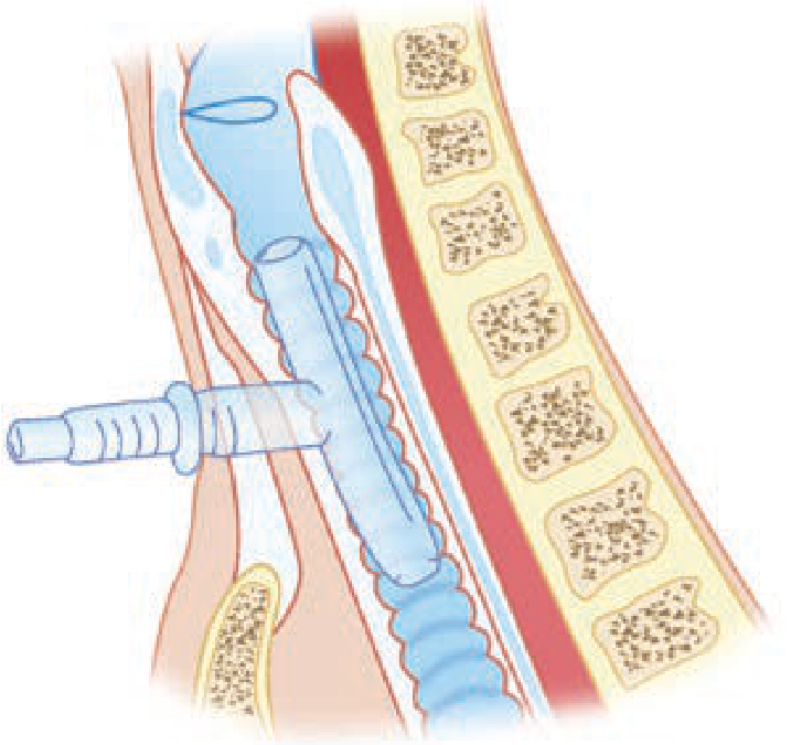
The Montgomery T-tube (also called the "Safe-T-Tube") is a silicone, T-shaped airway stent that functions simultaneously as a tracheal stent and a tracheostomy tube. It is a modification of a standard tracheostomy tube but has no inner cannula. The "T" shape consists of:
- Upper (vertical) limb - extends superiorly through the subglottis/stenotic segment toward the glottis
- Lower (vertical) limb - extends inferiorly in the trachea
- Horizontal (external) limb - exits through the tracheostomy stoma for access, suctioning, and ventilation
The diagram below illustrates the T-tube positioned in the trachea with the horizontal limb exiting through the tracheostomy:
Material & Design
- Made of implant-grade silicone - soft, pliable, and non-adherent smooth surface
- Available in clear or radiopaque material
- MRI-safe
- Comes in multiple series: Pediatric, Standard, Thoracic, Extra-Long, and Tapered
- A safety ring-and-groove system secures a plug to cap the external limb, allowing the patient to breathe and speak normally via the upper airway when plugged
Indications
The T-tube is used in benign and malignant tracheal/laryngotracheal disease:
| Category | Examples |
|---|---|
| Acute airway trauma | Blunt laryngotracheal injury |
| Tracheal/subglottic stenosis | Post-intubation, post-tracheostomy stenosis |
| Reconstructed/reconstituted trachea | After segmental resection and anastomosis |
| Stenosis not amenable to surgery | Long-segment stenosis, poor surgical candidates |
| Combined laryngotracheal stenosis | After simultaneous glottis + subglottis repair |
| Cervical trachea defects | When the cervical trachea cannot be repaired or reconstructed |
It is most commonly used for subglottic and upper tracheal stenosis and serves as an alternative to a solid stent when phonation is desired - the hollow lumen allows air passage to the vocal cords when the external limb is plugged. - Cummings Otolaryngology, p. 1217
As the Tintinalli's Emergency Medicine textbook notes: "The mainstay of treatment for symptomatic tracheal stenosis is surgical resection and anastomosis. When surgical treatment is not possible, the option exists for insertion of airway stents as a bridge to surgery, as a treatment for patients who are not surgical candidates, and in cases where there is a long segment of stenosis."
Contraindications
- Aspiration by the patient (risk of material entering the lung through the stent)
- Patients requiring positive pressure-assisted respiration (the T-tube is not compatible with standard mechanical ventilation via the tracheal route without special adaptors)
Advantages Over Solid Stents
| Feature | Solid Stent | Montgomery T-tube |
|---|---|---|
| Phonation | No | Yes (when external limb plugged) |
| Endotracheal intubation from above | Possible (through lumen) | Possible (through lumen) |
| Tracheostomy dependency | Yes (stent blocks airway above stoma) | No - upper limb patent |
| Suctioning access | Limited | Both limbs accessible |
Placement
- Inserted by an ENT or thoracic surgeon experienced with the device
- Placed under general anesthesia, typically with rigid bronchoscopy or direct laryngoscopy
- The external limb is plugged with the provided ring/plug system when the patient breathes spontaneously through the upper airway
- For anesthesia: a balloon catheter is used to occlude the upper end of the T-tube to create a closed system for ventilation
Nursing & Post-Operative Care
- Humidification and regular suctioning of both upper and lower limbs are essential - mucous plugging and crusting are the most common complications - Tintinalli's, Ch. 247
- Daily saline nebulizers are prescribed
- Carbocysteine may be added if mucus plugging is problematic
- The patient must be educated about the single-lumen design and its inherent higher risk of obstruction compared to double-cannula tubes
Complications
Early (perioperative)
- Postoperative fever
- Bleeding
- Excessive mucus secretion
- Irritating cough
- Short-term dyspnea
Late (long-term)
- Granulation tissue formation - most common at the upper edge, often due to friction from tube displacement during swallowing or speaking
- Mucous plugging / obstruction - from impaired expectoration
- Secondary infection / microbial colonization
- Tube dislodgement - migration from coughing or swallowing
- Ischemic mucosal injury - if pressure on mucosa exceeds 20-40 mmHg capillary pressure
Emergency Management of T-Tube Obstruction
- Suction both the upper and lower vertical limbs
- If obstruction persists after suctioning - remove the entire T-tube
- Re-establish airway via tracheostomy with an appropriately sized tracheostomy tube or endotracheal tube
- Do NOT attempt to pass an endotracheal tube blindly if the T-tube is still in situ
- Consult specialist (ENT/thoracic surgery) for re-insertion - Tintinalli's Emergency Medicine, Ch. 247
Relationship to Other Tracheal Stents
The Dumon tube is the other most commonly used silicone stent. Unlike the Montgomery T-tube, the Dumon is secured endoluminally via external studs and also has bronchial versions. The T-tube's unique advantage is its external access limb and the ability to support phonation.
A stent can also be fashioned from just the vertical limb of a T-tube (oversewn at the top, secured with a suture through the anterior commissure), used specifically as an endolaryngeal stent for laryngeal injuries - Cummings Otolaryngology, p. 1201; Scott-Brown's Otorhinolaryngology, Ch. 76.
Tracheal reconstruction
Tracheal reconstruction covers surgical restoration of tracheal continuity and lumen after resection, trauma, or stenosis. Key techniques include segmental resection with primary anastomosis, slide tracheoplasty, and staged reconstruction with stenting.
Searching sources...
Finding Sources
Finding Sources
Searching the Web
tracheal reconstruction surgery techniques segmental resection anastomosis 2024
Reading File
Finding Sources
Reading File
Reading File
Reading File
Finding Sources
Finding Sources
Reading File
Reading File
Reading File
Finding Sources
Reading File
Finding Sources
Reading File
Loading Image
Here is a comprehensive overview of Tracheal Reconstruction:
Tracheal Reconstruction
Anatomy Relevant to Surgery
The adult trachea is approximately 11 cm long (range 10-13 cm) and contains 14-20 tracheal rings. With neck hyperextension, about 50% of the trachea is cervical. The blood supply enters laterally in a segmental pattern - this is critical to preserve during dissection. The recurrent laryngeal nerves lie in the tracheoesophageal groove bilaterally and enter the larynx just medial to the inferior cornua of the thyroid cartilages.
Importantly, approximately 50% of the tracheal length (5-7 cm) can be safely resected and anastomosed primarily with appropriate mobilization - Cummings Otolaryngology, Ch. 112.
Surgical Access by Level
| Tracheal Location | Approach |
|---|---|
| Cervical & upper 2/3 intrathoracic | Low collar (transverse cervical) incision ± partial sternotomy |
| Lower 1/3 intrathoracic | Right posterolateral thoracotomy (4th ICS) |
| Carina | Median sternotomy (preferred for exposure) |
| Extended access (mid-trachea) | T-shaped incision with vertical arm to angle of Louis + partial manubrium division |
The partial upper sternotomy (not full sternotomy) is preferred because the carina lies at the level of the angle of Louis - full sternotomy adds no useful exposure while the great vessels obstruct the view. - Current Surgical Therapy 14e
Tracheal Resection and Primary Anastomosis
This is the gold standard for both benign stenosis and resectable tracheal tumors.
Patient Positioning
- Supine with inflatable pad beneath shoulders to extend the neck maximally
- The pad is deflated after resection to allow neck flexion for a tension-free anastomosis
Key Intraoperative Steps
- Subplatysmal flaps developed superiorly to thyroid cartilage, inferiorly to clavicular heads
- Strap muscles separated in midline; thyroid isthmus divided and retracted
- Tracheal mobilization - dissection kept immediately adjacent to the tracheal wall (preserves recurrent laryngeal nerves and lateral blood supply)
- Lesion localization - intraoperative bronchoscopy + 25-gauge needle through the tracheal wall at the upper and lower extents of the stenosis
- Stay sutures placed in proximal and distal ends before division to assist alignment and gauge tension
- Distal airway management - proximal ETT withdrawn; sterile armored ETT placed in distal trachea across the surgical field for ventilation during resection
- Frozen section of resection margins (especially for malignant tumors)
Anastomosis Technique
- Posterior membranous wall: continuous running absorbable suture
- Anterior cartilaginous wall: interrupted simple absorbable sutures (e.g., 4-0 Vicryl) placed circumferentially, every 5 mm, knots tied outside the lumen
- Sutures are placed through the cartilage ~4 mm from the cut edge and 4 mm apart
- Anastomosis tested by insufflation to 20-30 cm H₂O while submerged in saline - must be completely airtight
Wound Closure
- Sternohyoid muscle or thyroid isthmus used to cover and buttress all suture lines
- If innominate artery was dissected, a pedicled strap muscle flap is interposed between the artery and trachea
- Flat suction drains placed in pretracheal and substernal spaces
Tension Reduction: Mobilization Maneuvers
Anastomotic tension is the single most important technical factor determining outcome. Several progressive maneuvers are used:
| Maneuver | Additional Length Gained |
|---|---|
| Neck flexion (deflate shoulder pad) | Significant - initial step |
| Cervical approach + tracheal mobilization | 4-5 cm total |
| Montgomery suprahyoid release | ~1-2 cm |
| Right hilar release (U-incision around inferior pulmonary vein + pericardial incision) | ~1 cm additional |
Montgomery Suprahyoid Release
The muscles inserting on the superior aspect of the hyoid (between the lesser cornua) are divided. The hyoid bone is then divided just lateral to the lesser cornu on both sides. This detaches the hyoid from the skull base musculature and allows the larynx to descend, gaining 1-2 cm of extra tracheal mobility. - Current Surgical Therapy 14e
Guardian (Chin-to-Chest) Stitch
After closure, a heavy suture is placed from the submental skin crease to the presternal skin, tying with the neck in moderate flexion. This prevents sudden neck hyperextension in the first postoperative week, which would place stress on the fresh anastomosis. Hyperflexion must be avoided as it can cause spinal cord ischemia and quadriplegia. - Current Surgical Therapy 14e
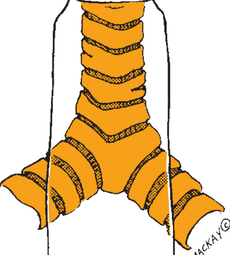
Illustration of the trachea and carina
Subglottic / Cricoid Involvement
When stenosis involves the cricoid:
- Anterior-only involvement: anterior cricoid arch resected, distal trachea beveled to match the defect - recurrent laryngeal nerves are spared
- Circumferential involvement: may require laryngectomy, or the oblique cricoid resection (Pearson technique): resection line begins anteriorly at the inferior thyroid cartilage border and passes posteriorly below the cricoid plate, below the recurrent nerve entry point; the distal trachea is "telescoped" in front of the residual cricoid shell (thyrotracheal anastomosis)
- In pediatric CTR (cricotracheal resection): laryngeal release is usually not required - Scott-Brown's Otorhinolaryngology
Carinal Resection and Reconstruction
Carinal resection is technically the most demanding form of tracheal surgery. Tumors involving more than 4 cm of tracheal length generally preclude resection.
Options for reconstruction (described by Grillo, 1982):
- Carinal restitution (for small/central tumors only): medial walls of right and left mainstem bronchi are approximated to create a neo-carina, then anastomosed to the distal trachea
- Standard carinal reconstruction (most common): trachea anastomosed end-to-end to one mainstem bronchus (usually left); the other bronchus anastomosed to the lateral wall of the trachea above the first anastomosis
- The left mainstem bronchus is relatively fixed by the aorta, limiting approximation - this is why most carinal reconstructions are right-sided approaches
Postoperative Management
- Extubation in the OR is preferred after tracheal resection
- If airway is unsatisfactory: small ETT with cuff deflated left for 24-48 hours, with IV dexamethasone (4 mg q6h), diuretics, fluid restriction, head elevation to reduce laryngeal edema
- Re-examine under anesthesia at 48 hours; if still problematic, a small tracheostomy tube placed 2 rings below the anastomosis (not through it)
- Contraindications to repair (Sabiston): (1) inadequately treated laryngeal problem; (2) need for permanent ventilatory support (ALS, myasthenia gravis, quadriplegia); (3) high-dose steroids; (4) inflamed/recent tracheostomy
Tracheal Substitutes (When Primary Anastomosis Is Not Possible)
Four approaches exist, all facing the challenge of restoring blood supply and mucociliary barrier function - Cummings Otolaryngology, Ch. 112:
| Approach | Key Features | Limitations |
|---|---|---|
| Allotransplantation | Donor trachea revascularized via forearm flap, then orthotopically transplanted | Prolonged 2-stage process; long-term immunosuppression needed; not suitable for malignancy |
| Autologous tissue | Tubularized vascularized pedicle flap from another site | No immunosuppression needed; built-in blood supply; but cartilaginous rigidity difficult to replicate |
| Bioprosthesis | Marlex mesh, Dacron, silicone | Historical; non-porous materials doomed to failure due to infection and poor integration |
| Tissue engineering | Stem cell-seeded scaffolds (cadaveric or synthetic nanocomposite) | Promising early cases reported; revascularization within 1 week; still experimental |
The published literature shows that nonbiologic prostheses uniformly fail due to the fragile segmental blood supply and high infection risk. Allotransplantation is considered most promising for true replacement but is limited by immunosuppression requirements. - Fischer's Mastery of Surgery, Ch. on Carinal Resection
Contraindications to Surgical Resection (Tracheal Tumors)
- Invasion of critical mediastinal structures (aorta, esophagus, superior vena cava)
- Tumor involving an extensive tracheal length precluding tension-free anastomosis
- Bilateral recurrent laryngeal nerve sacrifice (requires permanent tracheostomy)
- Poor functional status / inability to undergo major surgery
Palliative / Endoscopic Options for Unresectable Disease
When surgical resection is not feasible, the following bronchoscopic/endoscopic techniques offer palliation:
- Dilation (bougie or balloon) - temporizing only
- Rigid bronchoscopic debridement / coring out
- Nd:YAG laser vaporization - best for pedunculated, vascular tumors; can be done through flexible scope under topical anesthesia
- Photodynamic therapy (PDT) - useful for circumferential or submucosal tumors
- Cryotherapy
- Brachytherapy
- Tracheal stenting (Dumon or Montgomery T-tube) - maintains lumen, especially in long-segment or subglottic stenosis
Outcomes
- For benign stenosis (post-intubation): success rates >90% with segmental resection and primary anastomosis - Cummings Otolaryngology
- Primary tracheal malignancies: surgical resection with primary anastomosis is the therapy of choice when feasible; adenoid cystic carcinoma and squamous cell carcinoma are the two most common types
- Subglottic involvement and impaired vocal cord movement are associated with poorer outcomes
- Anastomotic complications (dehiscence, restenosis) are the most feared postoperative problems and are directly related to anastomotic tension and devascularization
Slide tracheoplasty
Slide tracheoplasty is a surgical technique for long-segment congenital tracheal stenosis where the trachea is transected at its midpoint, the upper segment incised anteriorly and lower segment incised posteriorly, then the two segments slid together and anastomosed - doubling the cross-sectional area while halving the length.
Searching sources...
Finding Sources
Finding Sources
Searching the Web
slide tracheoplasty congenital tracheal stenosis technique outcomes
Reading File
Reading File
Reading File
Reading File
Reading File
Loading Image
Loading Image
Finding Sources
Reading File
Reading File
Excellent - I now have comprehensive information from multiple authoritative sources. Here is the complete overview:
Slide Tracheoplasty
Background and Indication
Slide tracheoplasty is the operation of choice for long-segment congenital tracheal stenosis (LSCTS) caused by complete tracheal rings. It was first described by Tsang and Goldstraw in 1989 and has become the dominant surgical approach for this condition, replacing patch tracheoplasty and other earlier techniques.
Complete tracheal rings are circumferential cartilaginous rings lacking the normal posterior membranous wall. They occur in approximately 1 in 64,500 live births and are strongly associated with:
| Associated Condition | Frequency |
|---|---|
| Left pulmonary artery (LPA) sling | ~48% |
| Intracardiac anomalies (e.g. VSD, tetralogy of Fallot, ASD) | ~47% |
| Tracheal bronchus | 13% |
| Lung agenesis | 9% |
| Down syndrome | 8% |
| Anorectal/duodenal malformation | 7% |
| VACTERL | 6% |
- K.J. Lee's Essential Otolaryngology, p. 1035
Concept and Geometric Principle
The elegance of slide tracheoplasty is its geometry:
- The stenotic segment is divided at its midpoint
- The proximal half is incised anteriorly in the midline
- The distal half is incised posteriorly in the midline
- The two segments are slid together (overlapped), matching the open anterior of one to the open posterior of the other
This produces three simultaneous beneficial effects:
- Length reduced by half (minimizing anastomotic tension compared to full resection)
- Circumference nearly doubled
- Cross-sectional luminal area quadrupled
The entire lumen remains native mucosa-lined - no foreign patch material is needed. - Cummings Otolaryngology, Ch. 210
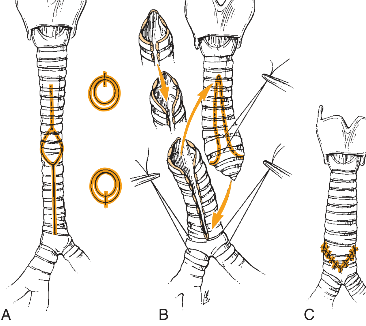
Slide tracheoplasty: (A) area of stenosis with planned incisions; (B) trachea divided and segments slid together; (C) completed reconstruction
Advantages Over Other Techniques
| Feature | Slide Tracheoplasty | Resection & Anastomosis | Patch Tracheoplasty |
|---|---|---|---|
| Preserves native mucosa | Yes | Yes | Partial (patch is foreign) |
| Usable for entire-trachea stenosis | Yes | No (tension too great) | Yes |
| Mobilization required | Less (segments travel only half the stenosis length) | Extensive | Moderate |
| Cross-sectional area gain | 4x | None | Variable |
| Risk of restenosis at anastomosis | Lower (oblique anastomosis) | Higher (end-to-end) | Variable |
| Foreign material | None | None | Yes |
The oblique nature of the anastomotic line is thought to be less liable to post-operative stenosis than a straight end-to-end anastomosis. - Scott-Brown's Otorhinolaryngology Vol 2, Ch. 31
Preoperative Assessment
- Flexible or rigid bronchoscopy: delineates the proximal and distal extents of stenosis, the degree of narrowing, and identifies associated bronchomalacia/bronchial stenosis
- Intraoperative localization trick: place the scope at the proximal end of stenosis, turn off the room lights - if the light is visible superior to the clavicles, a cervical approach is feasible; if inferior, a thoracic approach is needed
- CT with contrast + 3D reconstruction: maps the full extent of stenosis and identifies associated vascular anomalies (especially LPA sling)
- Cardiac echocardiogram: for concurrent cardiac defects requiring simultaneous repair
Surgical Technique (Detailed)
Setup and Access
- Performed on cardiopulmonary bypass (CPB) or ECMO - this is standard for LSCTS
- CPB improves tracheal exposure by allowing retraction of the innominate artery away from the underlying trachea
- Allows simultaneous repair of concurrent cardiovascular anomalies
- Cervical approach (low collar incision): accesses upper two-thirds of the trachea
- Intrathoracic approach: original description; used for more distal stenosis
Localization of the Stenosis
- An assistant performs bronchoscopy (rigid or flexible) while the surgeon places a 30-gauge needle into the airway at the proximal and distal extents
- The length of stenosis is measured and the midpoint is marked on the anterior airway surface
- Alternatively, transillumination can be used
Tracheal Division
- Circumferential dissection of the trachea is performed only at the midpoint of the stenosis (lateral blood supply preserved above and below)
- Retention sutures placed in the distal segment before division
- Bevelled transection commencing on the anterior trachea proximal to the midpoint, extending over two rings distally, with the posterior transection point at or just distal to the midpoint
- Result: the transection itself is oblique, not flat
Incisions on Each Segment
- Distal trachea: split in the midline posteriorly to just beyond the distal extent of the stenosis
- Proximal trachea: split anteriorly to just beyond the proximal extent - typically up to the thyroid cartilage
- The most damaged/stenotic tissue at the midpoint may be resected if desired
- The right-angled edges are trimmed to allow better approximation
Anastomosis
- Suture: double-armed PDS (polydioxanone), typically 4-0 with RB-1 needles in older children/adults
- Running suture technique (not interrupted) - no attempt is made to keep sutures extraluminal, but the suture is carefully tightened as the anastomosis proceeds
- Completed with a single proximal anterior knot
- Anastomosis is leak-tested then sealed with fibrin glue
- Patient is typically extubated at the end of the procedure
Optional Adjunct Maneuvers for Tension
- Suprahyoid release for upper segment mobilization
- Right intrapericardial hilar pulmonary release for lower segment
- Pedicled thymic lobe interposed between the reconstructed trachea and the innominate artery (protection)
Special Cases
- Pre-existing tracheostomy: can be incorporated into the anterior proximal incision
- Combined subglottic stenosis (SGS): if SGS co-exists and a graft to the anterior cricoid is feasible, the lower trachea can be slid into a split anterior cricoid instead of using a costal cartilage graft
- Slide into posterior cricoid: technically possible but challenging with unpredictable results - not recommended - Scott-Brown's Otorhinolaryngology Vol 2
When to Operate
- Severely symptomatic patients in the first few months of life
- Patients with poor exercise tolerance around age 5 years
- Short-segment stenosis (1-2 rings): resection and anastomosis is still preferred over slide tracheoplasty
- Long-segment stenosis (extending the entire trachea in extreme cases): slide tracheoplasty is the only option since resection would be impossible
Postoperative Care
- Multiple flexible bronchoscopies for bronchoalveolar lavage required following surgery (secretion management)
- Often requires several balloon dilations until the trachea fully relaxes and remodels
- Absorbable PDS stents (preferred over metallic stents) used for post-operative malacia, particularly bronchomalacia
- Granulation tissue at the anastomosis: treated with balloon dilation ± endoscopic debridement
- Over 50% of patients can be extubated within 48 hours in experienced centers
Outcomes
| Outcome Measure | Slide Tracheoplasty | Other Repairs (e.g. Patch) |
|---|---|---|
| Survival rate (modern era) | ~90% overall; >70% even with bronchomalacia/bronchial stenosis | ~70% |
| Success rate (published series) | 81% | 44% |
| Mortality rate (published series) | 14% | 30% |
| Need for tracheostomy salvage | 0.67% | 5% |
- Cummings Otolaryngology, Ch. 210 (Ywakim & El-Hakim series: slide n=149 vs other repairs n=122)
Poor prognostic indicators:
- Pre-operative bronchomalacia
- Bronchial stenosis (though survival still >70% in these groups)
- Scott-Brown's Otorhinolaryngology Vol 2, Ch. 31
Concerns about restricting post-operative tracheal growth have been resolved - the trachea grows normally after slide tracheoplasty.
Comparison with Patch Tracheoplasty
Patch tracheoplasty was historically used before slide tracheoplasty became the standard. It involves opening the anterior trachea longitudinally and widening it with an interpositional patch (pericardium, costal cartilage, etc.). An innovative variation uses the resected native stenotic trachea as an autograft - the excised stenotic segment is reintroduced anteriorly as a respiratory-epithelium-lined autograft (Backer technique), preserving cartilage contour and growth potential. Slide tracheoplasty has largely replaced patch techniques due to superior outcomes.
This is a shared conversation. Sign in to Orris to start your own chat.