Now I have comprehensive content from multiple authoritative sources. Let me compile the full detailed answer.
Oral Submucous Fibrosis (OSMF)
1. Definition
Oral submucous fibrosis (OSMF) is a chronic, insidious, progressive, premalignant condition of the oral mucosa and oropharynx characterized by a progressive fibroelastic change and inflammation of the lamina propria and the juxtaepithelial connective tissue, leading to formation of collagenous bands in the submucosa, resulting in marked rigidity of the oral and pharyngeal mucosa. It is a potentially malignant disorder (PMD).
"Submucous fibrosis represents a multifactorial disorder with the considered chief etiologic factor being the consistent and habitual use of areca (betel) nut... Submucous fibrosis is considered a premalignant condition, and transformation rates as high as 7.6% were reported from India over a 17-year period."
- Cummings Otolaryngology Head & Neck Surgery
Malignant transformation risk: 7-30% (varying across studies).
2. Applied Anatomy
The disease primarily affects structures lined by non-keratinized or partially keratinized squamous epithelium. The sites involved, in order of frequency:
| Site | Relevance |
|---|
| Buccal mucosa (bilateral) | Most common; fibrous bands form vertically, limiting mouth opening |
| Soft palate & uvula | Fibrosis leads to stenosis; uvula may become fibrosed and shrunken |
| Tonsillar pillars & fauces | "Webbing" of pillars; narrowing of fauces |
| Labial mucosa & commissures | Perioral fibrosis; microstomia |
| Tongue (ventral & lateral) | Reduced tongue mobility (limited protrusion, lateral movement) |
| Floor of mouth | Blanched, rigid mucosa |
| Hard palate | Less common; leathery texture |
| Pharynx & esophagus | Rare; dysphagia if involved |
Anatomical consequence: The submucosa, particularly the lamina propria and deeper connective tissue, is replaced by dense, avascular collagen. The overlying epithelium becomes atrophic with loss of rete ridges. Fibrosis of the pterygomandibular raphe and medial pterygoid muscle involvement leads to trismus.
Muscles involved in trismus:
- Medial pterygoid (most affected - fibrous change leads to reduced mandibular mobility)
- Buccinator (fibrosis limits buccal distension)
- Temporalis, masseter (late stages)
3. Etiology
Primary - Areca nut (betel nut):
- Areca nut alkaloids (arecoline, arecaidine, guvacoline, guvacine) stimulate fibroblasts, upregulate collagen synthesis, and inhibit collagenase
- Forms: betel quid (paan), paan masala, gutka, mawa
Other contributing factors:
- Chili (capsaicin): Mucosal irritation, especially in western Pacific populations
- Tobacco: Synergistic carcinogen
- Nutritional deficiencies: Iron, B vitamins (B12, folate), zinc - impair mucosal integrity
- Genetic susceptibility: HLA-A10, B7, DR3; mutations in TGF-β pathway
- Immunological: Increased IgG, IgA; altered T-cell responses (Th1/Th2 imbalance)
- Autoimmune component: Autoantibodies against oral mucosal antigens
4. Pathophysiology
The fundamental defect is failure of collagen remodeling - excessive collagen deposition with reduced breakdown.
Molecular cascade:
Areca nut alkaloids (especially arecoline)
        ↓
Mucosal irritation → Inflammatory response
        ↓
Upregulation of TGF-β1, bFGF, PDGF
        ↓
Fibroblast activation and proliferation
        ↓
↑ Collagen synthesis (Types I and III)
↓ Collagenase activity (MMP inhibition)
        ↓
Accumulation of abnormal cross-linked collagen
        ↓
Hyalinization of connective tissue
        ↓
Obliteration of blood vessels → Ischemia
        ↓
Atrophy of overlying epithelium
        ↓
Loss of rete ridges, epithelial dysplasia → SCC
Key molecular players:
- Inducible nitric oxide synthetase (iNOS): Upregulated in early OSMF - mediates the initial erythematous phase
- TGF-β1: Master fibrogenic cytokine - key driver
- Lysyl oxidase: Cross-links collagen, making it resistant to enzymatic degradation
- Reduced MMP-1 (collagenase-1): Less degradation of collagen
- Copper: Elevated salivary and tissue copper (from areca nut) activates lysyl oxidase and stimulates collagen synthesis
Histopathological progression:
- Early: Juxtaepithelial inflammatory infiltrate, dilated vessels, edema, thin delicate collagen fibers
- Intermediate: Moderate fibrosis, moderate inflammation, beginning hyalinization
- Late: Dense hyalinized collagen, atrophic epithelium with loss of rete ridges, reduced vascularity, muscle atrophy, variable dysplasia
"Dominating the histopathology is a juxtaposition of atrophic epithelium surfacing a subjacent fibrosis... Early connective tissue alterations are characterized by delicate and loosely arranged collagen fibers with progressive degrees of hyalinization until in the late stages, complete hyalinization of the supportive connective tissue is apparent. Variable degrees of chronic inflammation occur in the form of lymphocytes and plasma cells... mild dysplasia was present in 46% of cases, moderate dysplasia in 52%, and severe dysplasia in 2%."
5. Clinical Presentation
Symptoms (in order of progression):
Early stage:
- Burning sensation and intolerance to spicy food
- Vesiculation and ulceration of the oral mucosa
- Erythema of the mucosa (related to upregulated iNOS)
- Hypersalivation or xerostomia
Intermediate stage:
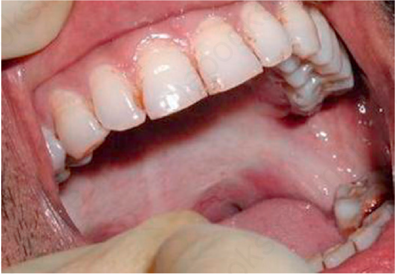
- Progressive pallor of the mucosa (blanching - pale, marble-like appearance)
- Palpable fibrous bands in the buccal mucosa, soft palate, fauces
- Reduced mouth opening (trismus) - measured by inter-incisal distance (IID)
- Reduced tongue mobility
- Hearing loss (if eustachian tube fibrosis involved)
Late stage:
- Severe trismus - IID < 20 mm
- Microstomia
- Difficulty in eating, swallowing (dysphagia), and speech
- Leukoplakic patches
- Development of SCC (thickening of epithelium, ulceration, induration)
Signs:
- Blanching: Diffuse pale/white mucosa, often described as "marble-like"
- Fibrous bands: Vertical, palpable, cord-like, most often in buccal mucosa and fauces
- Restricted mouth opening: Measured with IID (inter-incisal distance)
- Shrunken uvula: "Bud-like" uvula
- Sunken cheeks: Fibrosis of buccinator
- Petechiae and melanosis: Can coexist
- Loss of stippling of the gingiva
"The irritation produced first causes a thickening of the palate, tonsillar pillars, and fauces secondary to dermal and muscular fibrosis. As the disease progresses, opening of the mouth and protrusion of the tongue develop, such that eating, swallowing, and speech are impaired. Later, ulceration and leukoplakic areas occur, and finally, in approximately 7% of patients, malignant transformation to squamous cell carcinoma (SCC) develops."
- Andrews' Diseases of the Skin
6. Classification
Pindborg & Sirsat (1966) - Histological:
| Stage | Features |
|---|
| Stage I (Early) | Very early fibrosis, inflammatory infiltrate, dilated vessels |
| Stage II (Moderately Advanced) | Slight fibrosis with early hyalinization |
| Stage III (Advanced) | Dense fibrosis and hyalinization; atrophic epithelium |
| Stage IV (End stage) | Bone involvement |
Khanna & Andrade (1995) - Clinical (Mouth Opening):
| Group | Interincisal distance | Features |
|---|
| Group I | > 35 mm | No functional limitation (very early) |
| Group II | 26-35 mm | Slight difficulty, burning sensation |
| Group III | 15-25 mm | Restricted mouth opening with palpable fibrous bands |
| Group IV | < 15 mm | Severe trismus, leathery mucosa, total inability to eat |
Passi et al. (2020) / WHO clinical staging (functional):
- Stage I: Stomatitis; inter-incisal opening > 35 mm
- Stage II: Blanching ± fibrous bands; IID 26-35 mm
- Stage III: Blanching + fibrous bands + IID < 25 mm
Ranganathan & Mishra (2006) - Combined clinical-pathological grading (widely used):
| Grade | IID | Mucosal findings | Histopathology |
|---|
| Grade 1 | > 40 mm | Erythema, burning | Mild fibrosis |
| Grade 2 | 20-40 mm | Blanching, fibrous bands | Moderate fibrosis |
| Grade 3 | < 20 mm | Leathery, whitish mucosa | Dense hyaline fibrosis |
| Grade 4 | Severe | Malignant changes | Dysplasia/SCC |
7. Investigations
Clinical assessment:
- Interincisal distance (IID): Gold standard clinical measurement - normal >40 mm
- Tongue protrusion measurement
- Cheek flexibility assessment
Biopsy and histopathology (mandatory):
- Incisional biopsy from the most representative area (blanched, fibrotic zone)
- Stains:
- H&E: Shows hyalinized collagen, atrophic epithelium, inflammatory infiltrate
- Masson's trichrome: Highlights collagen - stains green/blue
- Van Gieson: Collagen appears red
- Assess: Degree of fibrosis, hyalinization, epithelial atrophy, dysplasia
Laboratory investigations:
- CBC: Rule out anemia (iron deficiency common)
- Serum iron, ferritin, TIBC
- Serum B12 and folate
- Serum copper: Often elevated
- Immunological: Serum IgG, IgA; T-cell subsets (CD4/CD8 ratio)
- Blood glucose: Rule out diabetes (impairs healing if surgery planned)
Imaging:
- OPG (orthopantomogram): Baseline dental assessment, rule out bony involvement
- MRI: Best modality to assess soft tissue fibrosis extent, pterygomandibular raphe, muscle involvement
- CT scan: If malignant transformation suspected, lymph node assessment
- Ultrasound elastography: Research tool - assesses tissue stiffness
Special tests:
- Exfoliative cytology: Screening tool - not diagnostic
- Toluidine blue staining: Identifies dysplastic areas before biopsy
- Autofluorescence: Identifies areas of dysplasia/malignancy
- PCR/molecular testing: TGF-β, p53, K-ras mutations if malignant transformation suspected
8. Differential Diagnosis
| Condition | Key Differentiating Features |
|---|
| Oral lichen planus | Wickham's striae (white reticulate pattern), bilateral buccal mucosa, autoimmune (T-cell mediated), no trismus, skin lesions possible |
| Leukoplakia | White patch, non-palpable fibrous bands, no trismus, single site common, smoking-related |
| Oral cancer (SCC) | Induration, ulceration, cervical lymphadenopathy, rapid progression, may complicate OSMF |
| Scleroderma (progressive systemic sclerosis) | Systemic disease - skin tightening, Raynaud's, telangiectasia, anti-Scl-70 or anti-centromere antibodies; microstomia but different pathology |
| Pemphigus vulgaris | Nikolsky's sign positive, flaccid bullae, acantholysis on histology, no fibrosis |
| Cicatricial pemphigoid | Subepidermal bulla, scarring, conjunctival involvement, DIF positive (linear IgG, C3) |
| Epidermolysis bullosa | Genetic, blistering on trauma, starts in childhood |
| Temporal mandibular joint (TMJ) ankylosis | Bony/fibrous ankylosis on imaging, no mucosal changes, history of trauma/infection |
| Tetanus (trismus) | Acute, history of wound, generalised muscle spasm, no mucosal changes |
| Submandibular space infection | Acute, swelling, pain, fever, no pallor or bands |
9. Treatment
Management is difficult, especially in advanced stages. The gold standard first step is cessation of areca/tobacco habit.
A. Non-Surgical (Medical) Management
1. Habit cessation (essential)
- Cessation of areca nut, gutka, paan masala
- Tobacco cessation counseling
2. Physiotherapy
- Forcible mouth opening exercises (jaw stretching)
- Balloon/tongue depressor exercises
- Dynasplint Trismus System
3. Intralesional injections (most widely used medical treatment)
| Agent | Mechanism | Notes |
|---|
| Triamcinolone acetonide (10 mg/mL) | Anti-inflammatory, inhibits fibroblast activity | 1-2 mL per site, weekly × 8-10 sessions |
| Dexamethasone (4 mg/mL) | Anti-inflammatory | Less used; may be combined with hyaluronidase |
| Hyaluronidase (1500 IU) | Depolymerizes hyaluronic acid, softens tissue | Combined with steroids for synergistic effect |
| Collagenase (enzyme) | Direct collagen degradation | Promising results; limited availability |
| Placental extract (Placentrex) | Anti-fibrotic, improves vascularity | 2 mL intralesional, alternate days |
| Pentoxifylline | Reduces TGF-β, vasodilator, anti-fibrotic | 400 mg TDS orally; improves microcirculation |
"More recently collagenase and pentoxifylline administration in separate studies has been proposed."
4. Nutritional supplements
- Lycopene 16 mg/day: Antioxidant, shown to improve symptoms - supported by 2024 systematic review (PMID 38155549)
- Vitamin A, C, E: Antioxidants
- Iron, B12, zinc supplementation if deficient
- Spirulina: Antioxidant; intralesional use studied
5. Topical agents
- Betamethasone mouth rinse
- Topical retinoids
6. Other systemic agents
- IFN-γ: Downregulates collagen synthesis (investigational)
- Turmeric / curcumin: Anti-inflammatory
- Traditional Chinese Medicine (Dan shen/Salvia miltiorrhiza): 2024 network meta-analysis (PMID 37551736) showed benefit
A 2024 Cochrane systematic review (
PMID 38415846) examined interventions for OSMF - pentoxifylline and dexamethasone had the most evidence, but overall evidence quality remains low. No single intervention is definitively superior.
B. Surgical Management
Indicated when IID < 25-30 mm or when medical treatment fails.
Principle: Release fibrous bands + interposition of tissue to prevent re-fibrosis
Techniques:
| Procedure | Description | Use |
|---|
| Simple fibrotomy | Excision of fibrous bands under LA | Early-moderate disease, Grade 2 |
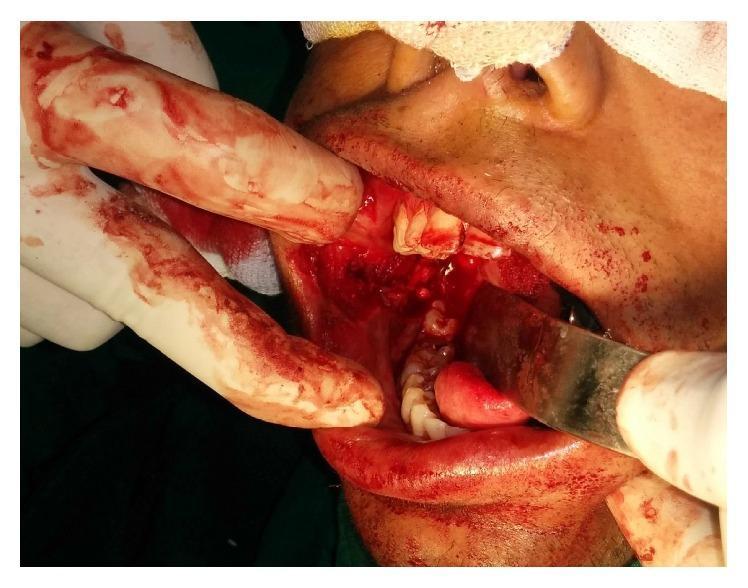
| Fibrotomy + split-thickness skin graft (STSG) | Defect covered with STSG from thigh | Moderate-severe disease |
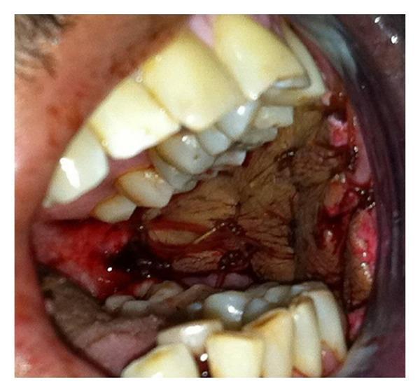
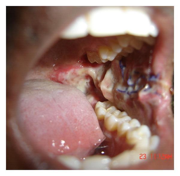
| Fibrotomy + pedicled buccal fat pad (BFP) graft | BFP interpositioning | Good vascularity; Grade 3 |
| Fibrotomy + nasolabial flap | Inferiorly based nasolabial flap | Suitable for anterior buccal defects |
| Fibrotomy + tongue flap | Used for posterior defects | Grade 3-4 |
| Temporalis myotomy/coronoidectomy | For severe trismus with coronoid involvement | Grade 4 |
| Laser surgery (CO₂ / Nd:YAG) | Band ablation, reduced bleeding, good healing | Minimally invasive option |
Key principle: Simple excision without interposition leads to re-fibrosis in 3-6 months.
Post-surgical physiotherapy with jaw opening exercises is mandatory to maintain results.
C. Follow-up and Malignant Surveillance
- All OSMF patients are at risk of SCC
- Transformation rates: 7.6% over 17 years (Indian cohort data)
- KJ Lee's: "Risk of malignancy: ranges from 7% to 30%"
- 6-monthly review with biopsy of any suspicious area (ulceration, induration, rapid change)
- Toluidine blue staining at each visit
Summary Table
| Parameter | Key Points |
|---|
| Definition | Chronic premalignant fibrotic disorder of oral mucosa due to areca nut |
| Anatomy | Buccal mucosa, soft palate, fauces, lips, tongue, floor of mouth |
| Pathophysiology | Arecoline → TGF-β → ↑collagen synthesis, ↓collagenase → hyaline fibrosis |
| Histology | Atrophic epithelium + hyalinized subepithelial collagen |
| Classification | Khanna & Andrade (clinical); Pindborg & Sirsat (histological) |
| Key presentation | Burning, blanching, fibrous bands, trismus, restricted tongue |
| Investigations | IID measurement, biopsy (H&E, Masson's trichrome), CBC, iron studies |
| DD | Lichen planus, leukoplakia, scleroderma, SCC, cicatricial pemphigoid |
| Treatment | Habit cessation + intralesional steroids/hyaluronidase + physiotherapy; surgery for Grade 3-4 |
| Malignant risk | 7-30%; 6-monthly surveillance mandatory |
Clinical photographs:
Fig: OSMF showing pallor/blanching of the gingival and buccal mucosa with restricted mouth opening (Andrews' Diseases of the Skin, courtesy Dr. Shyam Verma)
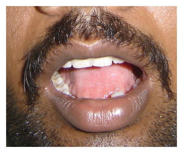
Fig: Severely restricted interincisal distance (trismus) in a patient with OSMF, before surgical intervention
Fig: Pedicled buccal fat pad graft used to cover the defect after fibrous band release in OSMF
Sources: Cummings Otolaryngology Head & Neck Surgery | KJ Lee's Essential Otolaryngology | Andrews' Diseases of the Skin | Scott-Brown's Otorhinolaryngology |
Cochrane Review - Interventions for OSMF, 2024 (PMID 38415846) |
Antioxidant treatments in OSMF, 2024 (PMID 38155549) |
TCM for OSMF meta-analysis, 2024 (PMID 37551736)