
Hypersensitivity reaction treatment protocol
"hypersensitivity reaction" AND treatment
hypersensitivity reaction types classification diagram
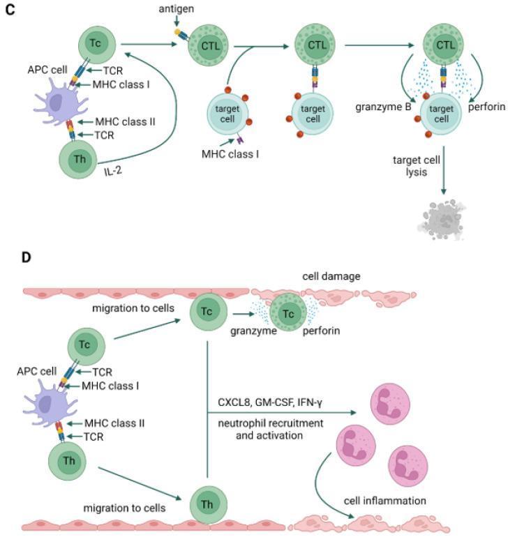
This pathophysiology diagram illustrates the cellular mechanisms of Type IVc and Type IVd hypersensitivity reactions. Panel C (Type IVc) depicts a cytotoxic T-cell-mediated pathway. It shows an antigen-presenting cell (APC) interacting with a cytotoxic T cell (Tc) via MHC class I and a helper T cell (Th) via MHC class II. Stimulated by IL-2, the Tc cell differentiates into a cytotoxic T lymphocyte (CTL). The CTL recognizes antigens on a target cell's MHC class I molecule, triggering the release of granzyme B and perforin, which culminates in target cell lysis. Panel D (Type IVd) illustrates a neutrophil-mediated delayed-type hypersensitivity reaction. Similar to Type IVc, an APC activates Tc and Th cells. These cells migrate to the site of cell damage. The Tc cells release granzyme and perforin, while the T lymphocytes (Tc and Th) secrete chemokines and cytokines including CXCL8 (IL-8), GM-CSF, and IFN-gamma. This signaling promotes neutrophil recruitment and activation, leading to tissue inflammation. These diagrams serve as educational resources for understanding the molecular immunology and T-cell-mediated mechanisms of delayed hypersensitivity disorders.
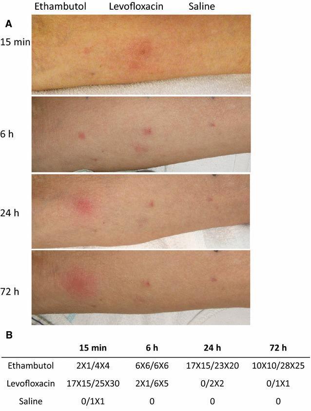
Educational clinical image and data table illustrating types of drug hypersensitivity reactions. Part A displays four sequential clinical photographs of a patient's forearm following intradermal skin testing with Ethambutol, Levofloxacin, and Saline (negative control) over a 72-hour period. At 15 minutes, Levofloxacin shows a significant erythematous wheal and flare reaction, characteristic of Type I IgE-mediated immediate hypersensitivity, which diminishes by 6 hours. In contrast, Ethambutol demonstrates a late-phase reaction (LPR), with minimal initial changes progressing to significant indurative erythema and edema that peaks at 24 hours and persists through 72 hours. Part B provides a quantitative data table measuring the diameters (in mm) of the wheal and induration/erythema at each time point (15 min, 6 h, 24 h, and 72 h), correlating clinical visuals with objective diagnostic measurements. This material is used to teach the temporal differences between immediate hypersensitivity and late-phase or delayed-type hypersensitivity reactions in clinical allergy and immunology.
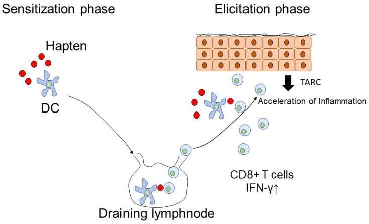
This pathophysiology diagram illustrates the two phases of a Type IV delayed hypersensitivity reaction in the skin: sensitization and elicitation. On the left, the 'Sensitization phase' shows haptens (small red circles) being captured by a cutaneous dendritic cell (DC), represented as a blue star-shaped cell. An arrow indicates the migration of this DC into a draining lymph node, where it presents the antigen to induce specific effector cells. On the right, the 'Elicitation phase' depicts a subsequent exposure under the epidermis (shown as cuboidal cells). Re-exposure triggers dendritic cell activation and the recruitment of CD8+ T cells and lymphocytes. Key molecular markers involved in the acceleration of inflammation are highlighted, including TARC (thymus and activation-regulated chemokine) and elevated IFN-γ (interferon-gamma). The diagram serves as an educational model for understanding the immune mechanisms behind allergic drug eruptions and contact dermatitis, emphasizing the progression from initial antigen recognition in lymph nodes to the localized cytotoxic inflammatory response in peripheral tissues.
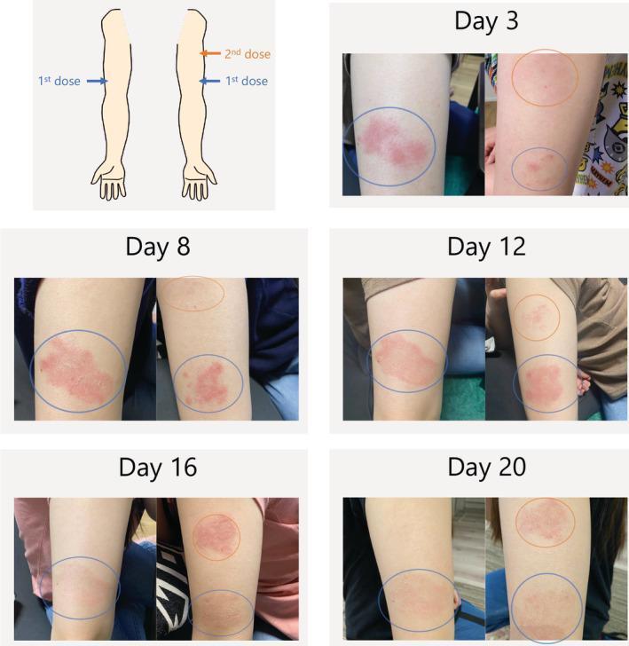
This clinical photographic sequence illustrates a timeline of a delayed cutaneous hypersensitivity reaction at injection sites following the administration of the biologic agent dupilumab. The material consists of an anatomical diagram indicating injection sites and a series of photographs from Day 3 to Day 20. The first dose site (blue circle) shows an erythematous, well-demarcated plaque that peaks in intensity and texture at Day 8 before gradually fading by Day 20. The second dose site (orange circle) exhibits a similar localized inflammatory response that develops later, peaking around Day 16. This progression demonstrates a classic delayed-type hypersensitivity reaction, potentially related to excipients like polysorbate 80. The image provides a clinical reference for monitoring drug-induced skin eruptions and distinguishes between immediate and delayed cutaneous adverse events in immunology and dermatology. It is suitable for medical education regarding the side effect profile of monoclonal antibodies.
anaphylaxis treatment algorithm epinephrine antihistamine
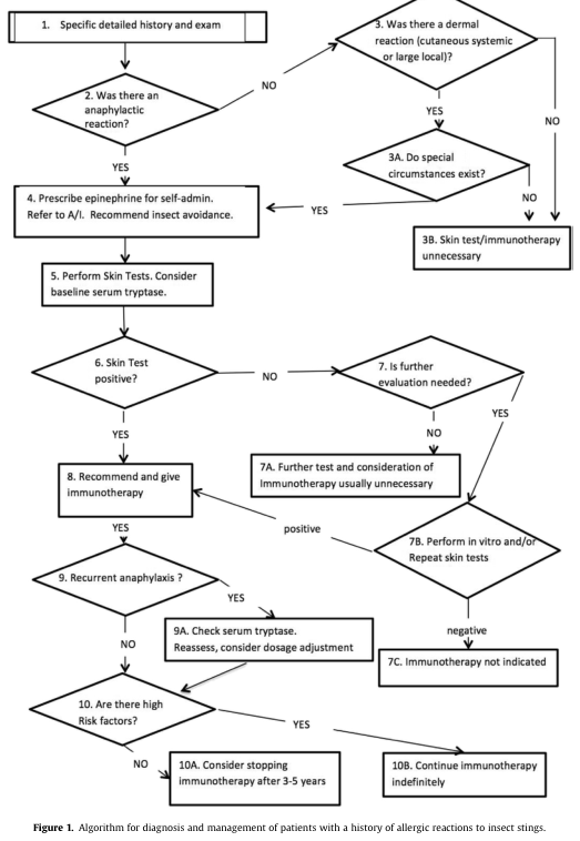
Summary : This figure presents a flowchart algorithm for the diagnosis and management of patients with a history of allergic reactions to insect stings, guiding clinicians through decision points based on patient history, reaction type, and test results. flowchart: # Nodes : • 1. Specific detailed history and exam (rectangle) • 2. Was there an anaphylactic reaction? (diamond) • 3. Was there a dermal reaction (cutaneous systemic or large local)? (diamond) • 3A. Do special circumstances exist? (diamond) • 3B. Skin test/immunotherapy unnecessary (rectangle) • 4. Prescribe epinephrine for self-admin. Refer to A/I. Recommend insect avoidance. (rectangle) • 5. Perform Skin Tests. Consider baseline serum tryptase. (rectangle) • 6. Skin Test positive? (diamond) • 7. Is further evaluation needed? (diamond) • 7A. Further test and consideration of Immunotherapy usually unnecessary (rectangle) • 7B. Perform in vitro and/or Repeat skin tests (rectangle) • 7C. Immunotherapy not indicated (rectangle) • 8. Recommend and give immunotherapy (rectangle) • 9. Recurrent anaphylaxis? (diamond) • 9A. Check serum tryptase. Reassess, consider dosage adjustment (rectangle) • 10. Are there high Risk factors? (diamond) • 10A. Consider stopping immunotherapy after 3-5 years (rectangle) • 10B. Continue immunotherapy indefinitely (rectangle) # Connectors : • Arrows indicate flow from one node to the next, with YES/NO branches at decision diamonds. • From node 2, YES leads to node 4, NO leads to node 3. • From node 3, YES leads to node 3A, NO leads to node 3B. • From node 3A, YES leads to node 4, NO leads to node 3B. • From node 4, arrow leads to node 5. • From node 5, arrow leads to node 6. • From node 6, YES leads to node 8, NO leads to node 7. • From node 7, YES leads to node 7B, NO leads to node 7A. • From node 7B, positive leads to node 8, negative leads to node 7C. • From node 8, arrow leads to node 9. • From node 9, YES leads to node 9A, NO leads to node 10. • From node 9A, arrow leads to node 10. • From node 10, YES leads to node 10B, NO leads to node 10A. # Layout : • The flowchart is arranged vertically, with decision diamonds branching to the right or left for YES/NO answers. • Merges and loops occur at nodes 8, 9, and 10, guiding further management based on test results and risk factors. # Analysis : • The algorithm prioritizes a detailed history and exam, then stratifies patients based on the severity and type of reaction (anaphylactic vs. dermal). • Skin testing and serum tryptase are central to further evaluation. • Immunotherapy is recommended for those with positive skin tests and significant reactions, with ongoing assessment for recurrent anaphylaxis and risk factors. • The flowchart provides clear guidance for when to stop or continue immunotherapy, emphasizing individualized patient management.
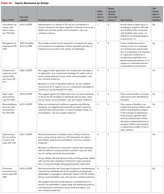
Table 36. Topics Reviewed by EvUps <table><thead><tr><th>Topic/PICO</th><th>Year last updated</th><th>Existing treatment recommendation</th><th>RCTs since last review, n</th><th>Observational studies since last review, n</th><th>Key findings</th><th>Sufficient data to warrant SysRev?</th></tr></thead><tbody><tr><td>Oral dilution for caustic substance ingestion (FA 202)</td><td>2010 CoSTR</td><td>Administration of a diluent in FA may be considered if a caustic substance has been ingested, if advised to do so by a health care provider (weak recommendation, very low-certainty evidence</td><td>1</td><td>0</td><td>Animal study of alkali injury of esophagus; irrigation with kefir and distilled water compared with distilled water alone; no differences in histopathological outcomes at 7 d</td><td>No</td></tr><tr><td>Recognition of anaphylaxis (FA 503)</td><td>2020 ScopRev; 2010 CoSTR</td><td>FA providers should not be expected to recognize the signs and symptoms of anaphylaxis without repeated episodes of training and encounters with victims of anaphylaxis.</td><td>0</td><td>8</td><td>Survey studies focused on training in the use of epinephrine autoinjectors and recognition of anaphylaxis and reported on improved confidence in recognizing anaphylaxis and administering epinephrine or on reasons for hesitation/nonuse of epinephrine autoinjectors.</td><td>No</td></tr><tr><td>Compression wraps for acute closed ankle joint injury (FA 511)</td><td>2020 CoSTR</td><td>We suggest either application of a compression bandage or no application of a compression bandage for adults with an acute closed ankle joint injury (weak recommendation, very low-certainty evidence). Because of a lack of identified evidence, we are unable to recommend for or against use of a compression bandage for closed joint injuries besides the ankle.</td><td>0</td><td>0</td><td></td><td>No</td></tr><tr><td>Open chest wound dressings (FA 525)</td><td>2015 CoSTR</td><td>We suggest against the application of an occlusive dressing or device by FA providers in individuals with an open chest wound (weak recommendation, very low-quality evidence).</td><td>0</td><td>0</td><td>Three animal studies of vented chest seals were identified but excluded.</td><td>No</td></tr><tr><td>Bronchodilators for acute asthma exacerbation (FA 534)</td><td>2015 CoSTR</td><td>When an individual with asthma is experiencing difficulty breathing, we suggest that trained FA providers assist the individual with administration of a bronchodilator (weak recommendation, very low-quality evidence).</td><td>0</td><td>0</td><td>One review of SysRevs concluded that among children with asthma exacerbations treated in the emergency department, albuterol/salbutamol delivered by metered-dose inhaler decreases hospital admission in younger children and emergency department length of stay in older children.</td><td>No</td></tr><tr><td>Optimal duration of cooling of burns with water (FA 770)</td><td>2021 CoSTR</td><td>We recommend the immediate active cooling of thermal burns using running water as a FA intervention for adults and children (strong recommendation, very low-certainty evidence). Because no difference in outcomes could be demonstrated with the different cooling durations studied, a specific duration of cooling cannot be recommended. Young children with thermal burns that are being actively cooled with running water should be monitored for signs and symptoms of excessive body cooling (good practice statement).</td><td>0</td><td>0</td><td></td><td>No</td></tr><tr><td>Preventive interventions for presyncope (FA 798)</td><td>2019 CoSTR</td><td>We recommend the use of any type of physical counterpressure maneuver by individuals with acute symptoms of presyncope attributable to vasovagal or orthostatic causes in the FA setting (strong recommendation, low- and very low-certainty evidence). We suggest that lower body physical counterpressure maneuvers are preferable to upper body and abdominal physical counterpressure maneuvers (weak recommendation, very low-certainty evidence).</td><td>0</td><td>0</td><td></td><td>No</td></tr></tbody></table>
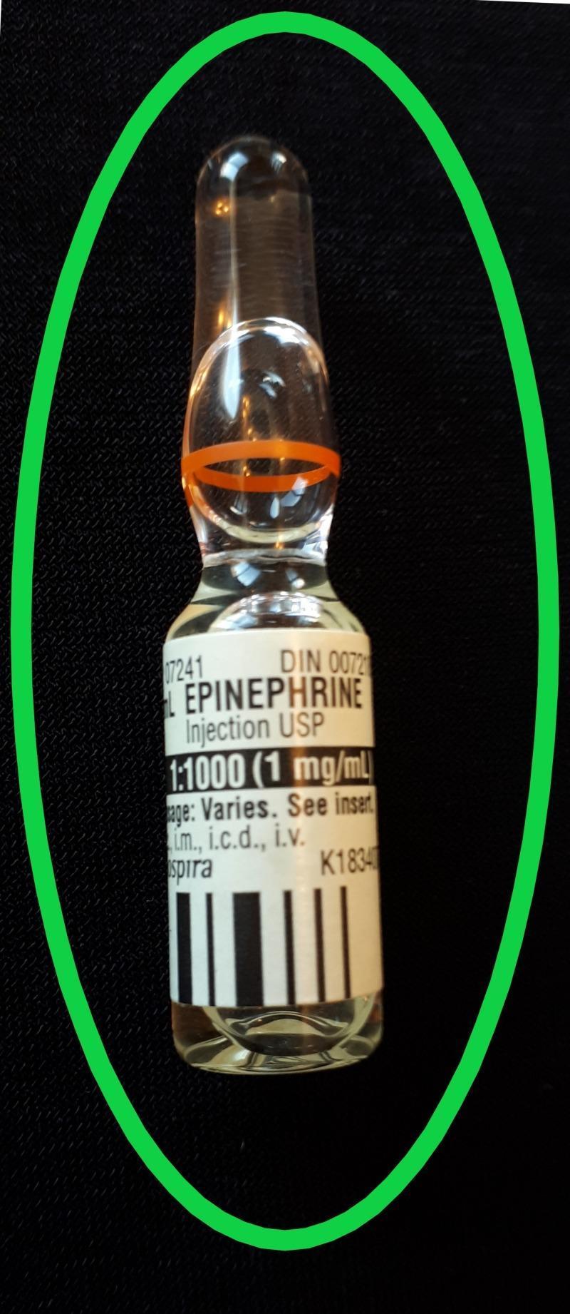
A clinical photograph of a single-use glass medication ampoule containing a clear solution of Epinephrine. The ampoule features a cylindrical body, a tapered neck with an orange identification ring indicating a break point, and a rounded, sealed top. The white adhesive label prominently identifies the contents as 'EPINEPHRINE Injection USP' with a concentration of '1:1000 (1 mg/mL)'. Regulatory and administrative information is visible, including 'DIN 0072' and 'Dosage: Varies. See insert', alongside abbreviations for administration routes: 'i.m., i.c.d., i.v.' (intramuscular, intracardiac, and intravenous). This specific formulation and volume (1 mg in 1 mL) is used as a critical teaching example for anaphylaxis management to differentiate it from larger-volume cardiac arrest syringes (1 mg in 10 mL), highlighting the importance of medication safety and concentration awareness in emergency medicine and pharmacology education.
Hypersensitivity Reaction Treatment Protocol
Gell-Coombs Classification - Quick Reference
| Type | Mechanism | Onset | Examples |
|---|---|---|---|
| Type I | IgE-mediated mast cell degranulation | Minutes | Anaphylaxis, allergic asthma, urticaria |
| Type II | Antibody-dependent cytotoxicity (IgG/IgM vs. cell surface) | Hours | Hemolytic anemia, Goodpasture's, transfusion reactions |
| Type III | Immune complex deposition | 6-21 days | Serum sickness, SLE, post-streptococcal GN |
| Type IV | T cell-mediated (delayed) | 48-72 h | Contact dermatitis, tuberculin reaction, DRESS |
Type I - IgE-Mediated (Immediate) Hypersensitivity
Mild-Moderate (Urticaria, Rhinitis, Localized Angioedema)
- Remove/stop the trigger (food, drug, insect venom)
- H1 antihistamine - diphenhydramine 25-50 mg IV/IM/PO q6h (adults); 1 mg/kg q6h (children)
- H2 blocker - ranitidine 50 mg IV over 5 min (adults); 0.5 mg/kg in children - adds synergistic blockade
- Oral corticosteroid for moderate reactions - prednisone 40-60 mg PO daily for 3-5 days
Severe - Anaphylaxis Protocol
Step 1 - Immediate Resuscitation (First-Line)
-
Epinephrine IM - thigh (vastus lateralis) - this is ALWAYS the first drug:
- Adults: 0.3-0.5 mg (0.3-0.5 mL of 1:1000 solution) IM
- Children: 0.01 mg/kg (0.01 mL/kg of 1:1000 solution) IM, max 0.5 mg
- EpiPen: 0.3 mg adult; EpiPen Jr: 0.15 mg for children <30 kg
- May repeat every 5-10 minutes if no response
- Thigh injections achieve higher and faster peak levels than deltoid
-
Airway management - secure early if angioedema is present; stridor warrants immediate intubation before complete obstruction
-
Oxygen - titrate to SpO2 ≥ 90%
-
IV access + cardiac monitoring + pulse oximetry
-
IV fluids - NS or Lactated Ringer's:
- Adults: 1-2 L IV bolus
- Children: 10-20 mL/kg bolus
-
Position - supine with legs elevated (unless dyspneic - then semi-recumbent)
-
Allergen decontamination - remove stinger, stop drug infusion
Step 2 - Second-Line Therapy (After epinephrine)
| Drug | Adult Dose | Pediatric Dose |
|---|---|---|
| Diphenhydramine (H1) | 25-50 mg IV/IM q6h | 1 mg/kg IV/IM q6h |
| Ranitidine (H2) | 50 mg IV over 5 min | 0.5 mg/kg IV over 5 min |
| Cimetidine (H2) | 300 mg IV | 4-8 mg/kg IV |
| Hydrocortisone | 250-500 mg IV | 5-10 mg/kg IV (max 500 mg) |
| Methylprednisolone | 80-125 mg IV | 1-2 mg/kg IV (max 125 mg) |
| Prednisone (PO) | 40-60 mg PO daily | 1-2 mg/kg/d divided BID |
Step 3 - Refractory Anaphylaxis
- IV Epinephrine bolus: 100 mcg over 5-10 min (mix 0.1 mg in 10 mL NS)
- IV Epinephrine infusion: start at 1 mcg/min; mix 1 mg in 50 mL NS at 0.5 mL/min; titrate
- Pediatric: 0.1-0.3 mcg/kg/min, max 1.5 mcg/kg/min
- Glucagon (if patient is on beta-blockers causing refractory hypotension): 1-2 mg IV/IM q5min, then infusion 5-15 mcg/min
- Vasopressors (norepinephrine, dopamine, vasopressin) for shock resistant to epinephrine
Additional for Bronchospasm
- Salbutamol (albuterol) nebulized 2.5-5 mg q20 min PRN
- Add ipratropium bromide 0.5 mg nebulized for severe bronchospasm
Biphasic Anaphylaxis Risk
- Monitor for 4-6 hours minimum after resolution
- Prolonged observation or admission if: protracted reaction, hypotension, airway involvement, unknown trigger, >1 dose of epinephrine used, beta-blocker use, lives alone, elderly, asthma history
- Risk factors for biphasic reaction: severe anaphylaxis or repeated epinephrine doses
Discharge Protocol
- Epinephrine autoinjector x2 - with training on use (carry at all times, at home and work)
- Antihistamine: diphenhydramine 25-50 mg PO q6-8h for 3-5 days
- Corticosteroid: prednisone 40-60 mg PO daily for 3-5 days
- MedicAlert identification bracelet
- Referral to allergist/immunologist
- Patients on beta-blockers should be switched to an alternative drug class
Type II - Antibody-Mediated Cytotoxic Hypersensitivity
General Treatment Principles
- Stop the offending agent (drug, transfusion)
- Corticosteroids - primary immunosuppressive therapy (e.g., prednisone 1 mg/kg/day for autoimmune cytopenias)
- IV Immunoglobulin (IVIG) - for ITP, myasthenia gravis crisis; blocks Fc-mediated phagocytosis
- Plasmapheresis - for Goodpasture syndrome, anti-GBM disease, myasthenic crisis; removes circulating antibodies
- Rituximab (anti-CD20) - for refractory autoimmune hemolytic anemia, ITP
- Splenectomy - second-line for refractory ITP or AIHA
- Supportive: transfusion support (crossmatch carefully), manage organ-specific complications
Type III - Immune Complex-Mediated Hypersensitivity
General Treatment
- Remove/treat the antigen source (stop the drug, treat the infection)
- NSAIDs - for mild serum sickness (fever, arthralgias, urticaria)
- Antihistamines - for urticaria/pruritus component
- Corticosteroids - for moderate-severe disease (prednisone 1-2 mg/kg/day; taper over weeks)
- Essential for lupus nephritis, RPGN, vasculitis
- Immunosuppressants (for chronic/severe conditions):
- Cyclophosphamide - for ANCA vasculitis, severe lupus nephritis
- Azathioprine / mycophenolate mofetil - maintenance therapy
- Rituximab - for ANCA vasculitis, refractory SLE
- Plasmapheresis - for cryoglobulinemia, severe lupus nephritis with anti-GBM overlap
- Avoid antigen re-exposure after serum sickness
Type IV - T Cell-Mediated (Delayed) Hypersensitivity
Contact Dermatitis
- Remove the allergen/irritant immediately
- Topical corticosteroids - mainstay of treatment
- Low-potency (hydrocortisone 1%) for face/intertriginous areas
- Mid/high-potency (triamcinolone, clobetasol) for body
- Oral antihistamines - for pruritus (sedating H1 blockers)
- Oral corticosteroids - for severe/widespread reactions: prednisone 40-60 mg daily, tapered over 2 weeks
Drug Hypersensitivity (DRESS/SJS/TEN)
- Immediately stop the causative drug - most important step
- Systemic corticosteroids (controversial in SJS/TEN but used in DRESS)
- IVIG - high-dose for SJS/TEN
- Cyclosporine - second-line for TEN
- Wound care - like burns in SJS/TEN; ICU management for extensive disease
Transplant Rejection
- Calcineurin inhibitors - tacrolimus, cyclosporine
- High-dose IV corticosteroids (methylprednisolone 500-1000 mg pulse) for acute rejection
- Anti-thymocyte globulin (ATG) for steroid-resistant acute rejection
- Biologics - basiliximab (anti-IL-2R), belatacept
Special Considerations
Angioedema (Non-histaminergic / Hereditary)
- Icatibant (bradykinin B2 receptor antagonist) - for hereditary angioedema (HAE) Type I/II
- Ecallantide (kallikrein inhibitor) - for HAE
- C1 esterase inhibitor (human or recombinant) - for HAE attacks and prophylaxis
- Fresh frozen plasma (FFP) - used with variable success in HAE, acquired C1 esterase inhibitor deficiency (ACID), and ACE inhibitor-induced angioedema
- Stop ACE inhibitor permanently in ACE inhibitor-induced angioedema
Summary Table: Treatment by Mechanism
| Type | First-Line | Second-Line | Adjuncts |
|---|---|---|---|
| I (Anaphylaxis) | Epinephrine IM 0.3-0.5 mg | H1/H2 blockers, corticosteroids | IV epi infusion, glucagon (beta-blockers), bronchodilators |
| I (Mild allergic) | Antihistamine, allergen removal | Oral corticosteroids | H2 blockers |
| II (Cytotoxic) | Stop agent, corticosteroids | IVIG, plasmapheresis | Rituximab, splenectomy |
| III (Immune complex) | Remove antigen, NSAIDs/steroids | Immunosuppressants | Plasmapheresis, rituximab |
| IV (Delayed) | Remove allergen, topical/systemic steroids | IVIG (SJS/TEN), cyclosporine | Calcineurin inhibitors (transplant) |
- Epinephrine first - always, in anaphylaxis. Antihistamines and steroids are never first-line.
- Corticosteroids are slow-acting (hours) - they prevent biphasic reactions but do not treat the acute phase.
- Allergen removal is universal across all four types.
- Non-histaminergic (bradykinin) angioedema requires specific agents (icatibant, C1-INH).
- Type II-IV reactions often require prolonged immunosuppression; taper carefully to avoid adrenal insufficiency.