
pilons fracture
pilon fracture distal tibia CT scan X-ray
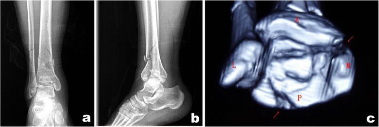
This composite figure displays diagnostic imaging of a pilon fracture (distal tibial fracture) in a 37-year-old male. Panels (a) and (b) are preoperative X-ray radiographs in anterior-posterior and lateral views, respectively. They reveal a complex, comminuted fracture of the right distal tibia and fibula with significant intra-articular involvement and visible displacement of bone fragments. Panel (c) is a 3D-reconstructed axial CT scan identifying key fracture components: a posterior malleolar fragment (P), a medial die-punch fragment (M), and an anterior fragment (A), alongside a lateral fragment (L). The imaging demonstrates a collapse of the articular surface and multi-planar disruption of the distal tibial plafond. Red arrows in the CT scan highlight areas of fragment displacement. These images illustrate the preoperative classification and mapping used to determine surgical approaches, such as posterolateral or anteromedial access, based on the spatial distribution of fragments relative to anatomical landmarks like the tarsal tunnel.
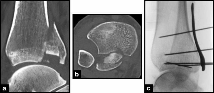
This composite figure illustrates a complex distal tibia fracture involving a dislocated intercalary fragment (ICF), presented through diagnostic imaging and intraoperative fluoroscopy. Panel (a) is a sagittal CT scan showing a large, displaced ICF at the distal tibia with a prominent fracture gap relative to the main tibial shaft and articular surface. Panel (b) provides an axial CT view demonstrating that the ICF consists of multiple distinct fragments dislocated anteriorly and medially, with the medial malleolus and fibula remaining visible for anatomical reference. Panel (c) is a lateral intraoperative X-ray (fluoroscopy) depicting the surgical stabilization of the fracture. The fixation strategy utilizes multiple Kirschner wires (K-wires) placed at varying trajectories to transfix the ICF, alongside a temporary anterior plate also secured with K-wires. These images demonstrate key clinical features of pilon or distal tibial plafond fractures, highlighting the challenges of reducing and fixing small, intra-articular intercalary bone fragments to restore joint congruency.
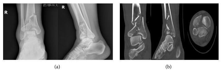
A multi-panel radiological image displaying preoperative diagnostic imaging of a Pilon fracture. Section (a) contains anteroposterior (AP) and lateral X-ray views of the right ankle, showing a complex, displaced distal tibial fracture extending into the articular surface (tibial plafond). Disruption of the distal fibula is also suggested. Section (b) provides high-resolution CT scans in coronal, sagittal, and axial planes. These CT images reveal the extensive comminution and fragmentation of the distal tibia, clearly demonstrating the involvement of the articular surface and the degree of displacement among bone fragments. The images illustrate a high-energy traumatic injury, with the CT providing critical detail for preoperative planning by characterizing the specific fracture patterns and intra-articular extension that are less distinct on the initial radiographs. This visual serves as an educational example of advanced orthopedic trauma imaging for medical students and surgical residents specializing in traumatology.
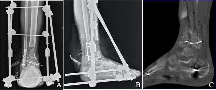
This composite medical image displays postoperative diagnostic studies of a complex distal tibia fracture, specifically a type C3.3 pilon fracture with significant bone loss. Panels A and B provide anteroposterior (AP) and lateral X-ray views showing a large, 6 cm segmental defect in the distal tibial metaphysis following debridement and bone resection. The limb is stabilized by a spanning triangular external fixator involving the tibia and foot. Internal fixation is visible at the distal articular surface, consisting of multiple Kirschner wires (K-wires) and a lateral fibular plate with screws to maintain the distal syndesmosis and fibular length. Panel C presents a sagittal computed tomography (CT) reconstruction of the ankle joint, demonstrating successful articular reduction. The CT confirms joint surface congruence with minimal displacement, specifically showing a step-off or gap of less than 2 mm. These images illustrate the first stage of a multi-stage surgical protocol for treating high-energy open fractures, focusing on debridement, joint reconstruction, and temporary stabilization before secondary bone transport procedures.
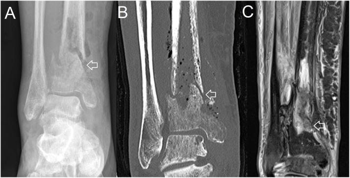
This composite clinical image displays a pathological fracture of the distal tibia through three imaging modalities: (A) plain X-ray, (B) Computed Tomography (CT), and (C) Magnetic Resonance Imaging (MRI). The plain X-ray shows a radiolucent fracture line and periosteal reaction in the distal tibial shaft above the ankle joint. The coronal CT scan provides superior bony detail, revealing cortical disruption, sequestration, and small gas bubbles within the medullary cavity and surrounding soft tissue, highly suggestive of chronic osteomyelitis with secondary fracture. The MRI (STIR or T2-weighted sequence) highlights the extent of the inflammatory process, showing hyperintense signals indicative of significant marrow edema and soft tissue phlegmon or abscess formation. This comparison demonstrates the multi-modal diagnostic approach to bone and joint infections (BJI), illustrating how CT excels at identifying structural bone destruction while MRI is most sensitive for detecting early inflammatory changes and soft tissue involvement. The location and appearance are characteristic of a complication resulting from severe osteomyelitis.
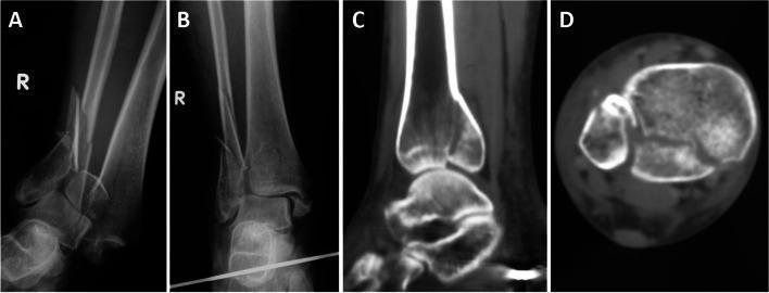
This composite diagnostic image illustrates an 'Open Logsplitter' injury of the right ankle, characterized by a severe fracture-dislocation with proximal talar displacement into the distal tibiofibular syndesmosis. Panel A (anteroposterior X-ray) shows the initial injury with significant talar displacement and a comminuted fibular fracture above the syndesmotic level. Panel B shows the ankle after closed reduction, with the talus repositioned within the mortise, although fracture lines in the fibula and distal tibia remain evident. Panels C and D provide computed tomography (CT) detail. The coronal CT reconstruction (C) demonstrates the longitudinal split and articular surface disruption of the distal tibia. The axial CT scan (D) confirms a posterior pilon fracture, showing a posterior compression fragment and cortical irregularity of the distal tibia along with widening of the syndesmotic space. The case highlights a complex high-energy trauma involving syndesmotic disruption and articular impaction, requiring careful radiological assessment for surgical planning.
"pilon fracture" AND treatment
Pilon Fracture (Tibial Plafond Fracture)
Definition and Terminology
- Rockwood and Green's Fractures in Adults, 10th Ed 2025
Anatomy
- Medial malleolus - with anterior and posterior colluli separated by the intercollicular groove
- Anterior tibial process - forms the tubercle of Chaput
- Posterior malleolus - extends more distally
- Fibular groove - bordered by the anterior and posterior tibial tubercles, forming the syndesmotic recess
- The fibula is fractured in 85% of pilon fractures (Campbell's Operative Orthopaedics 2026)
- Strongest cancellous bone is at the subchondral plate, extending ~3 cm from the joint
Mechanism of Injury
| Type | Mechanism | Characteristics |
|---|---|---|
| Low-energy | Rotational forces (e.g., skiing, twisting) | Minimal comminution, limited soft-tissue injury, favorable prognosis |
| High-energy | Axial compression (MVA, falls from height) | Articular/metaphyseal comminution, severe soft-tissue injury, poor prognosis |
Classification
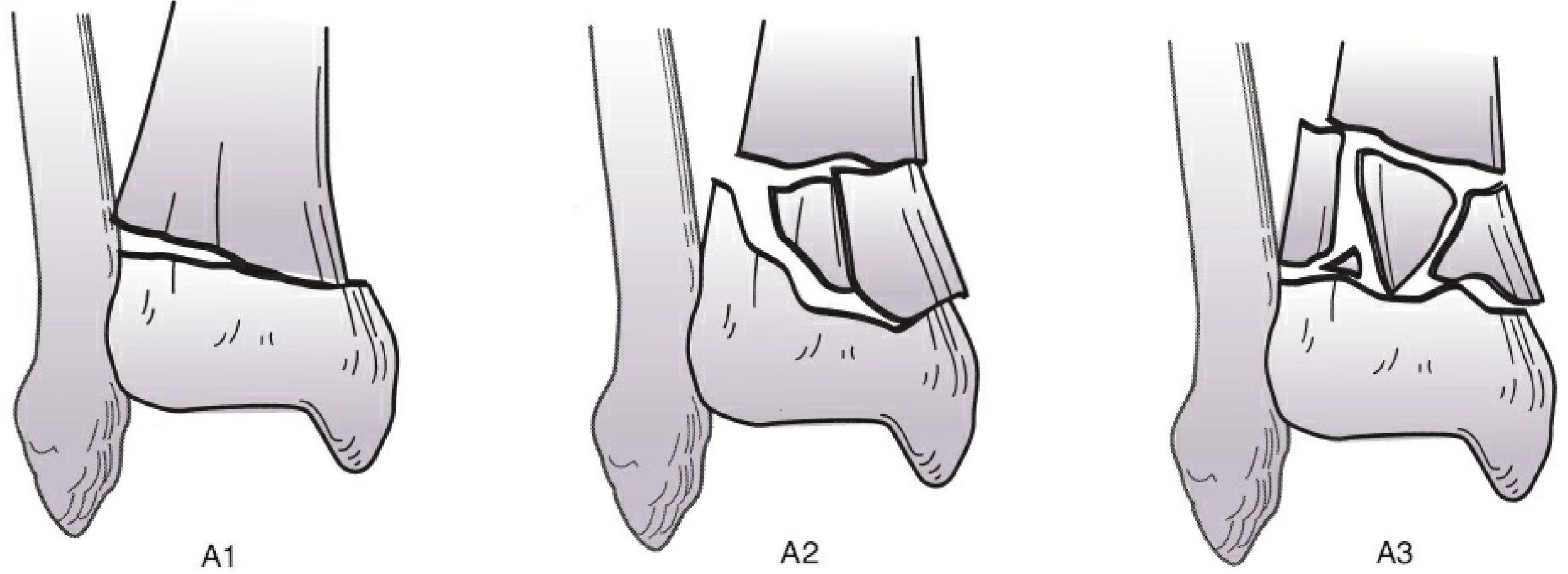
AO/OTA Classification (AO 43)
| Type | Description |
|---|---|
| 43A | Extra-articular - metaphyseal fracture only |
| 43B | Partial articular - part of the articular surface retains continuity with the shaft |
| 43C | Complete articular - entire articular surface is dissociated from the shaft |
Rüedi-Allgöwer Classification (older, still referenced)
- Type I: Non-displaced, cleavage fracture of the plafond
- Type II: Displaced with minimal comminution
- Type III: Displaced with significant comminution and impaction
Imaging
Plain Radiographs
- AP, lateral, and mortise views of the ankle
- Full-length tibia films to identify diaphyseal extension
CT Scan (Mandatory)
- Characterizes articular comminution, fragment displacement, impaction
- Identifies the key fragments: posterior (P), medial die-punch (M), anterior (A), and lateral (L) - seen in the 3D reconstruction below
Soft Tissue Assessment
- Open injuries: classify with Gustilo system
- Closed injuries: use Tscherne classification
- Blood-filled fracture blisters indicate more extensive cutaneous damage than clear-fluid blisters
- Check for: vascular injury, compartment syndrome, swelling, closed degloving
- Patient factors that worsen outcome: smoking, alcoholism, peripheral vascular disease, diabetes mellitus
Management: The Staged Approach
Historical context
- 1980s: Early ORIF became standard; however, a 40% complication rate was reported (McFerran 1992), including deep sepsis and osteomyelitis
- Early 1990s: Shift to external fixation, which later proved to cause malunion, nonunion, pin-tract infections, and slower return to function
- 1999: Sirkin et al. described the staged approach - 3% major complication rate in closed fractures, 11% in open injuries - which became the standard of care
Current Standard: "Span, Scan, and Plan"
- Spanning tibiotalar external fixator (proximal tibial Schanz pin + transcalcaneal pin)
- Fibular ORIF if anatomic reduction can be achieved through limited dissection (preferred: intramedullary fibular nail for length-stable injuries)
- Wounds managed (debridement if open)
- Limb elevation, monitoring for compartment syndrome
- Conversion to definitive ORIF after resolution of swelling, fracture blisters, and soft tissue inflammation
- Articular reconstruction first, then metaphyseal stabilization
- Multiple limited approaches preferred over large extensile exposures
- Low-profile, anatomically contoured periarticular plates
- Bone grafting for metaphyseal defects if needed
- Goal: articular step-off < 2 mm
Surgical Approaches for Definitive ORIF
- Anterolateral - for anterior/lateral fragments (Chaput tubercle)
- Anteromedial - for medial die-punch fragments
- Posterolateral - for posterior malleolar fragments (most common posterior approach)
- Posteromedial - for posteromedial fragments
Complications
| Complication | Notes |
|---|---|
| Wound dehiscence/infection | Most common after premature ORIF; superficial vs. deep |
| Deep wound infection / osteomyelitis | Requires debridement ± hardware removal |
| Nonunion | More common with >50% comminution, high-energy injury |
| Malunion | Risk with external fixation alone |
| Posttraumatic arthritis | Near-universal with 43C injuries; may require ankle fusion or arthroplasty |
| Compartment syndrome | Must be monitored acutely |
Prognosis
- High-energy fractures heal in an average of 6 months vs. ~4 months for low-energy
- Fragment contact after reduction of 50-90% is associated with faster healing
- Displacement > 50% of tibial width = significant risk of nonunion
- Long-term: posttraumatic ankle arthritis is the most impactful sequela, particularly in 43C injuries
- Patient-reported outcomes are consistently lower than the general population, even with anatomic reduction