
Who classification and advances in epithelial ovarian tumor and small cell carcinoma of ovary, complete classification of ovarian tumors Pathogenesis morphology md pathology 10 marks
ovarian serous cystadenocarcinoma gross microscopy histology
ovarian tumor gross morphology serous mucinous cystic
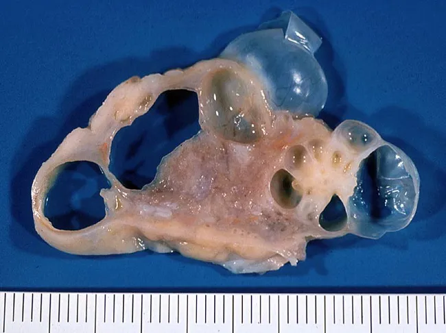
This is a gross pathology photograph of an ovarian tumor. The specimen represents a unilateral ovarian mass with multilocular cystic architecture. The internal content consists of clear, colorless serous fluid within multiple thin-walled cysts. The cyst walls are smooth and translucent, without obvious mural nodules or solid components on gross examination. The external surface is pale, smooth, and without frank hemorrhage or necrosis. The overall appearance is consistent with a benign serous cystadenoma of ovarian origin. The lesion is located in the pelvis, arising from the ovarian surface or cortical epithelium, and demonstrates a predominately cystic, noninfiltrative growth pattern. The patient is a 38-year-old female; serous cystadenomas are common epithelial ovarian neoplasms and typically present as enlarging adnexal masses or incidental findings. Diagnostic significance: gross features favor a benign process, but histopathologic confirmation is essential to exclude serous cystadenocarcinoma or mucinous variants. Differential considerations include mucinous cystadenoma/cystadenocarcinoma, serous borderline tumors, and paraovarian cysts. Clinical correlation: complete surgical excision with intact capsule provides definitive treatment and favorable prognosis. Potential clinical use cases: educational reference for ovarian tumor morphology, gross-pathology catalogs, radiologic-pathologic correlation studies, and machine learning datasets for ovarian neoplasms. This image supports differential diagnosis and educational training in gynecologic pathology.
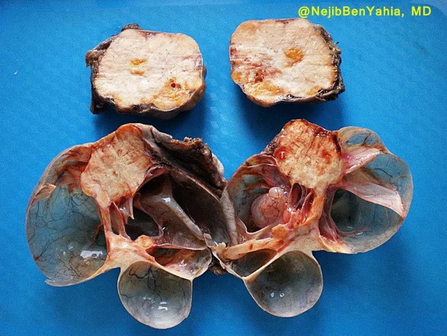
Gross pathology image of bilateral ovarian neoplasms from a surgical resection specimen. The top ovary displays a Brenner tumor with a yellow-white solid cut surface, a feature consistent with fibrous stroma and epithelial nests seen on histology but evident here in gross form. The bottom ovary shows a mucinous cystadenoma manifesting as a multilocular cystic mass with a gleaming, mucoid inner surface. A conspicuous solid yellowish nodule is noted within the mucinous component, adding a focal solid element to the otherwise cystic lesion. The external surfaces show typical ovarian morphology; cross-sectioning reveals distinct tumor components in a single image field, illustrating a concurrent benign Brenner tumor and benign mucinous cystadenoma in two ovaries. This composite ovarian tumor presentation is recognized in gynecologic pathology as an ovarian collision or coexistence of histologically distinct neoplasms, with Brenner tumor arising from transitional-type epithelium in fibrous stroma and mucinous cystadenoma arising from mucinous epithelium. Clinically, these lesions are usually benign; definitive classification requires histopathology, but gross appearance supports benign behavior. Potential differential diagnoses include mucinous cystadenocarcinoma, serous cystadenoma/carcinoma, and urothelial-like (transitional) lesions. The image is intended for educational purposes and correlates with gross anatomy, tumor morphology, adnexal pathology, and surgical pathology workflows, enhancing recognition of mixed ovarian neoplasm patterns in teaching datasets and libraries.
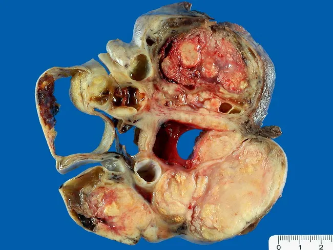
This gross pathology image depicts a unilateral ovarian tumor measuring 12 cm, displaying both solid and cystic components. Cysts are filled with serous fluid and are partially lined by papillary proliferations; scattered necrotic foci are evident within the parenchyma. The specimen demonstrates classic features of a malignant epithelial ovarian tumor of serous type, specifically serous papillary cystadenocarcinoma, as described in the accompanying diagnosis. The outer surface is irregular with friable, yellow-tan tissue and focal hemorrhagic areas; cross-sectional areas reveal multilocular cystic spaces interspersed with solid nodules. In a 69-year-old female, these macroscopic findings correlate with advanced-age onset ovarian carcinoma and may reflect high-grade disease. This image is suitable for educational illustrating the gross morphology of serous ovarian carcinoma, including papillary architecture, cystic dilation, necrosis, and a mix of solid and cystic tumor burden. The specimen highlight underscores the importance of comprehensive macroscopic examination as part of surgical pathology workflows, aiding staging and guiding histologic sampling. Pathoanatomic correlation with microscopic serous papillary carcinoma features—papillary fronds, atypia, mitotic activity—requires histology. Used clinically, such gross images support tumor identification, differential diagnosis from serous cystadenoma or mucinous tumors, and are valuable for medical education and tumor registries. Image credit: pathorama.ch.
Ovarian Tumors: WHO Classification, Epithelial Tumors, and Small Cell Carcinoma
(MD Pathology - 10 Marks)
I. WHO CLASSIFICATION OF OVARIAN NEOPLASMS (Table 22.3 - Robbins, Cotran & Kumar)
- Surface/fallopian tube epithelium and endometriosis
- Germ cells (from yolk sac, pluripotent)
- Stromal cells (sex cord cells, endocrine precursors)
A. SURFACE EPITHELIAL-STROMAL TUMORS (~65% of all ovarian tumors)
| Tumor Type | Benign | Borderline | Malignant |
|---|---|---|---|
| Serous tumors | Cystadenoma, cystadenofibroma | Serous borderline tumor | Low-grade & high-grade serous adenocarcinoma |
| Mucinous tumors (endocervical-like & intestinal type) | Cystadenoma, cystadenofibroma | Mucinous borderline tumor | Mucinous adenocarcinoma |
| Endometrioid tumors | Cystadenoma, cystadenofibroma | Endometrioid borderline tumor | Endometrioid adenocarcinoma |
| Clear cell tumors | Benign | Borderline | Clear cell adenocarcinoma |
| Brenner tumors | Benign Brenner tumor | Borderline Brenner tumor | Malignant Brenner tumor |
| Epithelial-stromal | - | - | Adenosarcoma, Carcinosarcoma |
B. SEX CORD-STROMAL TUMORS (~8% of ovarian tumors)
- Granulosa cell tumors
- Fibromas / Fibrothecomas / Thecomas
- Sertoli-Leydig cell tumors
- Steroid (lipid) cell tumors
C. GERM CELL TUMORS (~15-20% of ovarian tumors)
- Teratoma: Immature | Mature (solid / cystic - dermoid cyst) | Monodermal (struma ovarii, carcinoid)
- Dysgerminoma
- Yolk sac tumor
- Mixed germ cell tumors
D. METASTATIC TUMORS TO OVARY (~5%)
- Colonic, appendiceal
- Gastric (Krukenberg tumor - signet ring cell type)
- Breast, endometrial, cervical
II. FREQUENCY OF MAJOR OVARIAN TUMORS (Table 22.4 - Robbins)
| Type | % of Malignant Ovarian Tumors | % Bilateral |
|---|---|---|
| Serous | 47% | Benign 25%, Borderline 30%, Malignant 65% |
| Mucinous | 3% | Benign 5%, Borderline 10%, Malignant <5% |
| Endometrioid carcinoma | 20% | 40% |
| Undifferentiated carcinoma | 10% | - |
| Clear cell carcinoma | 6% | 40% |
| Granulosa cell tumor | 5% | 5% |
| Teratoma | 1% | Rare (benign) |
| Metastatic | 5% | >50% |
III. EPITHELIAL OVARIAN TUMORS - DETAILED CLASSIFICATION & PATHOGENESIS
General Features
- Differentiation of neoplastic epithelium (serous, mucinous, endometrioid)
- Extent of epithelial proliferation (benign, borderline, malignant)
Pathogenesis - Molecular Advances (Type I vs Type II Paradigm)
| Feature | Type I (Low-grade) | Type II (High-grade) |
|---|---|---|
| Precursor | Borderline tumor | STIC (serous tubal intraepithelial carcinoma) in fallopian tube |
| Behavior | Slow-growing, indolent | Aggressive, rapid |
| Examples | Low-grade serous, mucinous, clear cell, endometrioid, Brenner | High-grade serous carcinoma (HGSC), carcinosarcoma |
| Key mutations | KRAS, BRAF, ERBB2, PIK3CA, PTEN, ARID1A | TP53 (>96%), BRCA1/BRCA2 |
| Chromosomal instability | Low | High |
- High-grade serous carcinoma (HGSC): TP53 mutations in virtually all cases; BRCA1/BRCA2 germline or somatic mutations in 15-25%; most arise from the fallopian tube fimbria (STIC), not the ovarian surface
- Low-grade serous: KRAS/BRAF mutations; arise from serous borderline tumors
- Endometrioid and clear cell: ARID1A, PIK3CA mutations; arise in a background of endometriosis
- Mucinous: KRAS mutations; often arise from Brenner tumors or teratomas
A. SEROUS TUMORS (Most Common Epithelial Type - 47%)
Pathogenesis
- Benign/borderline: arise from ovarian surface epithelium invaginations (inclusion cysts) or directly from Müllerian-type epithelium; modern evidence points to fallopian tube origin for many
- Malignant (HGSC): now considered to originate primarily from the distal fallopian tube fimbria as STIC lesions, secondarily involving the ovary
Morphology
- Most common ovarian neoplasm
- Grossly: thin-walled, unilocular or multilocular cyst filled with clear serous fluid; smooth inner lining
- Microscopically: single layer of ciliated tubal-type epithelium lining the cyst; minimal atypia
- 25% are bilateral
- Grossly: papillary projections on internal surface; partially solid
- Microscopically: epithelial stratification, nuclear atypia, mitoses, but NO stromal invasion - the key diagnostic criterion
- 30% bilateral
- Favorable prognosis; rare progression to carcinoma
- Most common cause of ovarian cancer death
- Grossly: partly solid, partly cystic with extensive papillary excrescences; friable, yellow-tan with necrosis and hemorrhage; frequently bilateral (65%)
- Microscopically: papillary, glandular, or solid architecture; highly pleomorphic cells; numerous mitoses; psammoma bodies (concentric calcifications - characteristic); TP53 mutation present in >96%
- Spreads transcoelomically to peritoneum, omentum (omental cake), bowel serosa
B. MUCINOUS TUMORS (3% of malignant ovarian tumors)
Morphology
- Grossly: large, multilocular cysts filled with thick, viscid mucinous material; smooth capsule; can reach enormous size (up to 50 kg reported)
- Microscopically: lined by tall columnar mucin-secreting cells resembling endocervical (Müllerian type) or intestinal epithelium (goblet cells)
- 80% benign; 5% bilateral
- Microscopically: epithelial stratification, tufting, nuclear atypia; NO invasion
- Grossly: partly solid areas within multiloculated cysts
- Microscopically: glandular structures with obvious stromal invasion; malignant mucinous cells
- Must be distinguished from metastatic colonic adenocarcinoma (Krukenberg) - CK7+/CK20- favors primary ovarian
C. ENDOMETRIOID TUMORS (20% of malignant ovarian tumors)
- Pathogenesis: strongly associated with endometriosis; ARID1A mutations, PIK3CA mutations
- 15-20% occur simultaneously with endometrial carcinoma (synchronous primaries)
- Grossly: partly solid and cystic; brown "chocolate" cyst contents if arising from endometrioma
- Microscopically: glands resembling proliferative endometrium; tubular glands lined by stratified non-mucinous cells; often squamous differentiation (squamous metaplasia)
- 40% bilateral
D. CLEAR CELL CARCINOMA (6% of malignant tumors)
- Pathogenesis: arises from endometriosis; ARID1A mutations
- Grossly: partly solid, partly cystic
- Microscopically: cells with abundant clear (glycogen-rich) cytoplasm arranged in solid sheets, tubular or papillary patterns; "hobnail" cells projecting into cystic spaces
- Relatively resistant to platinum chemotherapy; worse prognosis than endometrioid
E. BRENNER TUMOR (Transitional Cell Tumor)
- Mostly benign; rare borderline and malignant forms
- Grossly: solid, fibrous, whitish-grey appearance; often small, incidental
- Microscopically: nests of transitional (urothelial-like) epithelium embedded in dense fibrous stroma; cells resemble Walthard cell rests; characteristic "coffee bean" nuclei (nuclear grooves)
- Often coincides with mucinous cystadenoma in the same or contralateral ovary
IV. ADVANCES IN EPITHELIAL OVARIAN TUMORS
-
Fallopian tube origin of HGSC: The current consensus is that most HGSC arise from serous tubal intraepithelial carcinoma (STIC) in the fimbria, not the ovarian surface epithelium. This has changed surgical prophylaxis strategy - bilateral salpingectomy is now considered for risk reduction even in average-risk women.
-
Dualistic carcinogenesis model (Type I vs Type II - see above): completely revised understanding of pathogenesis; Type I low-grade tumors have a stepwise progression through borderline stages with specific driver mutations; Type II HGSC has massive chromosomal instability from the outset.
-
BRCA1/BRCA2 testing: Germline or somatic BRCA1/BRCA2 mutations are found in 15-25% of HGSC. This predicts sensitivity to PARP inhibitors (olaparib, niraparib, rucaparib) - now used as maintenance therapy after platinum-based chemotherapy.
-
Homologous recombination deficiency (HRD) is a broader biomarker; tumors with HRD respond to platinum and PARP inhibitors even without BRCA mutations.
-
Bevacizumab (anti-VEGF): approved for frontline and recurrent ovarian cancer, exploiting the angiogenic dependency of HGSC.
-
Molecular subtypes of HGSC: The TCGA (The Cancer Genome Atlas) identified four molecular subtypes of HGSC - immunoreactive, differentiated, proliferative, and mesenchymal - with prognostic implications.
V. SMALL CELL CARCINOMA OF THE OVARY (SCCO)
A. Hypercalcemic Type (SCCOHT) - More Common
- Average age at diagnosis: 24 years (range 2-46 years); young women and adolescents
- Typically unilateral
- ~2/3 have paraendocrine hypercalcemia (accounts for 50% of all hypercalcemia associated with ovarian tumors)
- ~50% have spread beyond the ovary at diagnosis
- Characterized by germline or somatic mutations in SMARCA4 (SWI/SNF chromatin remodeling gene)
- Similar SMARCA4 mutations found in rhabdoid tumors at other sites
- It is now proposed that SCCOHT be reclassified as ovarian rhabdoid tumors and treated as such
- Grossly: large solid unilateral mass, often with areas of hemorrhage and necrosis
- Microscopically: small cells with scanty cytoplasm and hyperchromatic nuclei in diffuse sheets; variable large cell component with prominent nucleoli in ~50%; IHC shows loss of SMARCA4 (BRG1) expression - a diagnostic marker
- IHC distinguishes from lymphoma, leukemia, sarcoma
- Surgery + platinum-based chemotherapy ± radiation
- Aggressive combination chemotherapy regimens (as used for rhabdoid tumors) may be beneficial
- Hypercalcemia managed with aggressive hydration, loop diuretics, bisphosphonates, or calcitonin
B. Pulmonary Type SCCO
- Resembles small cell carcinoma of the lung (neuroendocrine)
- Occurs in postmenopausal women
- Associated with surface epithelial carcinoma (often endometrioid)
- IHC: positive for neuroendocrine markers (synaptophysin, chromogranin, NSE)
- Not associated with hypercalcemia
- SMARCA4 mutations NOT present
Summary Table: Key Differentiating Features
| Feature | SCCOHT | Pulmonary Type |
|---|---|---|
| Age | Young (~24 yr) | Postmenopausal |
| Hypercalcemia | ~66% | No |
| SMARCA4 mutation | Yes (IHC: BRG1 loss) | No |
| Neuroendocrine markers | Negative | Positive |
| Associated tumor | None | Endometrioid carcinoma |
| Proposed reclassification | Ovarian rhabdoid tumor | Neuroendocrine carcinoma |
- Robbins, Cotran & Kumar - Pathologic Basis of Disease (Robbins 10th Ed.), Chapter 22 - Ovarian Tumors, pp. 940-950
- Berek & Novak's Gynecology, Chapter on Small Cell Carcinoma, p. 2406
Please include latest WHO classification
WHO 2020 5th edition classification ovarian tumors female genital tract complete list
WHO classification ovarian tumors 2020 female genital tract
https://www.mdpi.com/2075-4418/11/4/697
PMID: 34996131
https://schaberg.faculty.ucdavis.edu/wp-content/uploads/site…
WHO 2020 ovarian tumors classification changes seromucinous removed mesonephric-like added SCCOHT SMARCA4 complete table
WHO 2020 Classification of Ovarian Tumors + Advances in Epithelial Ovarian Tumors & SCCO
MD Pathology - 10 Marks
I. WHO 2020 (5th EDITION) COMPLETE CLASSIFICATION OF OVARIAN TUMORS
A. SURFACE EPITHELIAL TUMORS
1. Serous Tumors
| Grade | Entities |
|---|---|
| Benign | Serous cystadenoma, serous adenofibroma, serous cystadenofibroma |
| Borderline | Serous borderline tumor (atypical proliferative serous tumor) - typical and micropapillary/cribriform variants |
| Malignant | Low-grade serous carcinoma (LGSC) - separate entity from 4th ed. |
| Malignant | High-grade serous carcinoma (HGSC) |
Key 2020 change: LGSC and HGSC are now firmly recognized as two completely separate diseases with different origins, mutations, and treatment responses.
2. Mucinous Tumors
- Benign: Mucinous cystadenoma, mucinous adenofibroma
- Borderline: Mucinous borderline tumor (intestinal type; endocervical-type seromucinous borderline tumor moved separately)
- Malignant: Mucinous carcinoma
3. Endometrioid Tumors
- Benign: Endometrioid cystadenoma, endometrioid adenofibroma
- Borderline: Endometrioid borderline tumor
- Malignant: Endometrioid carcinoma (now includes former "seromucinous carcinoma" as a subtype)
4. Clear Cell Tumors
- Benign: Clear cell cystadenoma, clear cell adenofibroma
- Borderline: Clear cell borderline tumor
- Malignant: Clear cell carcinoma
5. Brenner Tumors (Transitional Cell)
- Benign Brenner tumor
- Borderline Brenner tumor
- Malignant Brenner tumor
6. Seromucinous Tumors (Retained for benign/borderline; carcinoma removed)
- Benign: Seromucinous cystadenoma, seromucinous adenofibroma
- Borderline: Seromucinous borderline tumor
- Seromucinous carcinoma REMOVED from 2020 edition - reclassified as endometrioid carcinoma
7. NEW in 2020: Mesonephric-like Adenocarcinoma
- New entity added; associated with endometriosis; KRAS mutations; IHC: TTF1+, GATA3+, ER-/PR-/WT1-
8. Undifferentiated and Dedifferentiated Carcinoma
- Undifferentiated: no specific line of differentiation
- Dedifferentiated: undifferentiated component with abrupt transition to differentiated (usually endometrioid) component
9. Mixed Carcinoma (Reintroduced in 2020)
- Composed of two or more distinct histologic types, each comprising ≥10% of the tumor
10. Carcinosarcoma (Malignant Mixed Mesodermal Tumor / MMMT)
- Reclassified as a dedifferentiated carcinoma (not a mixed tumor); arises via epithelial-mesenchymal transition (EMT)
B. SEX CORD-STROMAL TUMORS
- Fibroma (NOS; cellular fibroma)
- Thecoma (NOS; luteinized thecoma)
- Fibrothecoma
- Sclerosing stromal tumor
- Microcystic stromal tumor (new recognition)
- Leydig cell tumor
- Steroid cell tumor NOS
- Fibrosarcoma (rare, malignant)
- Adult granulosa cell tumor (AGCT) - FOXL2 mutation (C134W) pathognomonic
- Juvenile granulosa cell tumor (JGCT)
- Sertoli cell tumor
- Sertoli-Leydig cell tumors (SLCT) - DICER1 mutations in ~60%
- Gynandroblastoma
- Sex cord tumor with annular tubules (SCTAT) - associated with Peutz-Jeghers syndrome
C. GERM CELL TUMORS
| Tumor | Key Feature |
|---|---|
| Dysgerminoma | Ovarian counterpart of seminoma; OCT3/4+, NANOG+, KIT mutations; isochromosome 12p |
| Yolk sac tumor (endodermal sinus tumor) | AFP+, SALL4+; Schiller-Duval bodies |
| Embryonal carcinoma | CD30+, OCT4+; rare in pure form |
| Non-gestational choriocarcinoma | β-hCG+; very rare pure ovarian |
| Immature teratoma | Graded 1-3 by quantity of immature neuroepithelium |
| Mature teratoma (dermoid cyst) | Most common ovarian tumor overall |
| Monodermal teratoma | Struma ovarii (thyroid tissue); carcinoid |
| Mixed germ cell tumor | Two or more germ cell components |
D. MISCELLANEOUS TUMORS (Category reorganized in 2020)
- Small cell carcinoma of the ovary, hypercalcemic type (SCCOHT) - SMARCA4 mutations; now under miscellaneous, not epithelial
- Small cell carcinoma of the ovary, pulmonary type - neuroendocrine
- Wilms tumor (rare)
- Paraganglioma
- Solid pseudopapillary neoplasm
- Hepatoid carcinoma
- Primary ovarian melanoma
Key 2020 change: SCCOHT is now placed under Miscellaneous tumors, not epithelial tumors, reflecting its rhabdoid/SMARCA4-mutant identity. [McCluggage et al., Histopathology 2022, PMID 34996131]
E. MESOTHELIAL TUMORS
- Adenomatoid tumor
- Mesothelioma (rare; peritoneal origin)
F. SOFT TISSUE TUMORS
- Myxoma
- Leiomyoma (rare)
G. LYMPHOID AND MYELOID TUMORS
- Lymphomas (diffuse large B-cell most common)
- Myeloid sarcoma
H. METASTATIC TUMORS TO THE OVARY (~5-6% of all ovarian tumors)
| Primary Site | Features |
|---|---|
| Stomach (Krukenberg tumor) | Bilateral; signet ring cells; mucin-secreting cells in cellular stroma |
| Colorectal / Appendiceal | Bilateral; mucinous; CK7-/CK20+ |
| Breast | Bilateral; lobular > ductal; ER/PR+ |
| Endometrial / Tubal | Direct spread; endometrioid type |
| Cervix | Rare (<1%) |
| Appendix | Pseudomyxoma peritonei |
II. KEY CHANGES: WHO 2020 vs WHO 2014 (4th Edition)
| Feature | WHO 2014 (4th Ed) | WHO 2020 (5th Ed) |
|---|---|---|
| Serous carcinoma split | Recognized but less defined | LGSC and HGSC firmly = two separate diseases |
| Seromucinous carcinoma | Listed as distinct entity | Removed - subsumed into endometrioid carcinoma |
| Mesonephric-like adenocarcinoma | Not recognized | Newly added |
| Mixed carcinoma | Not separately listed | Reintroduced (≥2 components, each ≥10%) |
| SCCOHT | Listed under epithelial tumors/miscellaneous | Firmly placed in Miscellaneous, linked to SMARCA4/rhabdoid |
| Molecular data | Limited | Integrated into diagnostic criteria |
| Site of origin for HGSC | Ovary | Fallopian tube is primary site for most HGSCs (via STIC) |
| Carcinosarcoma | Mixed tumor | Reclassified as dedifferentiated carcinoma (EMT-derived) |
| IHC algorithms | Limited | Detailed IHC panels for each histotype |
III. FIVE MAIN TYPES OF OVARIAN CARCINOMA (WHO 2020 Emphasis)
| Type | Frequency | Origin | Key Mutation | IHC Profile |
|---|---|---|---|---|
| HGSC | 70% | Fallopian tube fimbria (STIC) | TP53 (>96%), BRCA1/2 (15-25%) | PAX8+, WT1+, p53 mutant, ER+/- |
| Endometrioid | 10% | Endometriosis | ARID1A, PIK3CA, CTNNB1 | PAX8+, WT1-, p53 wt, PR+ |
| Clear cell | 6-10% | Endometriosis | ARID1A, PIK3CA | PAX8+, NapsinA+, WT1-, HNF1β+ |
| LGSC | 5% | Serous borderline tumor | KRAS, BRAF, NRAS | PAX8+, WT1+, p53 wt, ER+ |
| Mucinous | 3-4% | Brenner tumor/teratoma/de novo | KRAS | CK20+, CDX2+, WT1-, ER- |
IV. ADVANCES IN EPITHELIAL OVARIAN TUMORS (2020 onwards)
-
Fallopian tube origin confirmed for HGSC: WHO 2020 introduced specific criteria for site assignment (tubal, ovarian, tubo-ovarian, peritoneal). By these criteria, 80% of HGSC are of tubal origin. Peritoneal primary requires absence of STIC and no ovarian involvement.
-
Molecular-morphologic integration: Each histotype now has a defined IHC panel and molecular profile (see table above) as part of routine diagnosis - not just research.
-
PARP inhibitors: Olaparib, niraparib, rucaparib approved as maintenance in BRCA-mutated/HRD-positive HGSC after platinum response. Bevacizumab (anti-VEGF) for frontline and recurrent disease.
-
TCGA molecular subtypes of HGSC - Immunoreactive, Differentiated, Proliferative, Mesenchymal - with prognostic/predictive implications.
-
Seromucinous carcinoma reclassification: Abolished as a distinct entity; now endometrioid carcinoma subtype.
-
Mesonephric-like carcinoma: Newly recognized; arises from Müllerian epithelium with mesonephric differentiation; associated with endometriosis; IHC: TTF1+, GATA3+, CD10 (luminal), PAX8+; ER/PR/WT1 negative.
V. SMALL CELL CARCINOMA OF THE OVARY (SCCO) - WHO 2020 Update
A. Hypercalcemic Type (SCCOHT) - Under Miscellaneous in 2020 WHO
| Feature | Detail |
|---|---|
| WHO 2020 placement | Miscellaneous tumors (not epithelial) |
| Age | Mean 24 years (range 2-46 years) |
| Laterality | Usually unilateral |
| Hypercalcemia | ~66% (paraendocrine) |
| Molecular hallmark | Germline or somatic SMARCA4 mutations (SWI/SNF chromatin remodeling) |
| IHC | Loss of BRG1 (SMARCA4) expression - diagnostic; INI1 retained |
| Relationship | Proposed reclassification as ovarian rhabdoid tumor |
| Prognosis | Poor; 5-year OS ~16%; most die within 2 years |
| Treatment | Surgery + platinum ± radiation; rhabdoid tumor regimens under investigation |
B. Pulmonary Type SCCO
| Feature | Detail |
|---|---|
| Age | Postmenopausal |
| Association | Often with surface epithelial carcinoma (endometrioid) |
| IHC | Synaptophysin+, Chromogranin+, NSE+ |
| SMARCA4 | Normal (retained BRG1) |
| Hypercalcemia | Absent |
VI. QUICK SUMMARY TABLE: COMPLETE OVARIAN TUMOR CLASSIFICATION (WHO 2020)
OVARIAN TUMORS (WHO 2020, 5th Edition)
│
├── A. SURFACE EPITHELIAL TUMORS
│   ├── Serous (benign / borderline / LGSC / HGSC)
│   ├── Mucinous (benign / borderline / carcinoma)
│   ├── Endometrioid (benign / borderline / carcinoma) [includes former seromucinous Ca]
│   ├── Clear cell (benign / borderline / carcinoma)
│   ├── Brenner/Transitional (benign / borderline / malignant)
│   ├── Seromucinous (benign / borderline only — no carcinoma)
│   ├── Mesonephric-like adenocarcinoma [NEW 2020]
│   ├── Undifferentiated / Dedifferentiated carcinoma
│   ├── Mixed carcinoma [NEW 2020]
│   └── Carcinosarcoma (reclassified as dedifferentiated Ca)
│
├── B. SEX CORD-STROMAL TUMORS
│   ├── Pure stromal: Fibroma, Thecoma, Sclerosing stromal tumor, Leydig cell, Steroid cell
│   ├── Pure sex cord: AGCT, JGCT, Sertoli cell tumor
│   └── Mixed: SLCT, Gynandroblastoma, SCTAT
│
├── C. GERM CELL TUMORS
│   ├── Dysgerminoma
│   ├── Yolk sac tumor
│   ├── Embryonal carcinoma
│   ├── Non-gestational choriocarcinoma
│   ├── Teratoma (Immature / Mature / Monodermal)
│   └── Mixed germ cell tumor
│
├── D. MISCELLANEOUS TUMORS
│   ├── SCCOHT (SMARCA4-mutant) [moved from epithelial]
│   ├── SCCO, pulmonary type
│   ├── Wilms tumor, paraganglioma, hepatoid Ca, etc.
│
├── E. MESOTHELIAL TUMORS
├── F. SOFT TISSUE TUMORS
├── G. LYMPHOID AND MYELOID TUMORS
└── H. METASTATIC TUMORS (Krukenberg, colorectal, breast, etc.)
- WHO Classification of Tumours of the Female Genital Tract, 5th ed. (IARC, Lyon, 2020)
- McCluggage WG et al. - Key changes in WHO 5th edition, Histopathology 2022 (PMID 34996131)
- De Leo A et al. - New WHO 2020 ovarian carcinoma classification, Diagnostics 2021 (PMID 33919741)
- Robbins, Cotran & Kumar - Pathologic Basis of Disease (10th Ed.), Chapter 22
Who latest classification and advances in epithelial ovarian tumor and small cell carcinoma of ovary, complete classification of ovarian tumors Pathogenesis morphology md pathology 10 marks
Ovarian Tumors: WHO 2020 Classification, Epithelial Tumors & Small Cell Carcinoma
MD Pathology — 10 Marks
I. WHO 2020 (5th EDITION) — COMPLETE CLASSIFICATION OF OVARIAN TUMORS
- Surface/Müllerian epithelium (and endometriosis)
- Germ cells (yolk sac-derived, pluripotent)
- Sex cord-stromal cells (endocrine precursors)
A. SURFACE EPITHELIAL TUMORS (~65%)
1. Serous Tumors
- Benign: Serous cystadenoma, serous adenofibroma, serous cystadenofibroma
- Borderline: Serous borderline tumor (typical and micropapillary/cribriform variants)
- Malignant:
- Low-grade serous carcinoma (LGSC)
- High-grade serous carcinoma (HGSC)
LGSC and HGSC are now firmly recognized as two entirely separate diseases in WHO 2020.
2. Mucinous Tumors
- Benign: Mucinous cystadenoma, mucinous adenofibroma
- Borderline: Mucinous borderline tumor
- Malignant: Mucinous carcinoma
3. Endometrioid Tumors
- Benign: Endometrioid cystadenoma, endometrioid adenofibroma
- Borderline: Endometrioid borderline tumor
- Malignant: Endometrioid carcinoma (now includes former "seromucinous carcinoma")
4. Clear Cell Tumors
- Benign: Clear cell cystadenoma, clear cell adenofibroma
- Borderline: Clear cell borderline tumor
- Malignant: Clear cell carcinoma
5. Brenner (Transitional Cell) Tumors
- Benign Brenner tumor
- Borderline Brenner tumor
- Malignant Brenner tumor
6. Seromucinous Tumors (benign/borderline retained; carcinoma REMOVED)
- Benign: Seromucinous cystadenoma, seromucinous adenofibroma
- Borderline: Seromucinous borderline tumor
Seromucinous carcinoma— Abolished in 2020; reclassified as endometrioid carcinoma
7. Mesonephric-like Adenocarcinoma (NEW in 2020)
- New entity; tubular/glandular/slit-like architecture; associated with endometriosis
- IHC: TTF1+, GATA3+, CD10 (luminal)+; ER/PR/WT1 negative; KRAS mutations
8. Undifferentiated and Dedifferentiated Carcinoma
- Undifferentiated: no specific line of differentiation
- Dedifferentiated: undifferentiated component with abrupt transition to differentiated (endometrioid) component
9. Mixed Carcinoma (Reintroduced 2020)
- Two or more distinct histologic types, each comprising ≥10% of the tumor
10. Carcinosarcoma (MMMT)
- Reclassified as dedifferentiated/metaplastic carcinoma derived via epithelial-mesenchymal transition (EMT); not a true mixed tumor
B. SEX CORD-STROMAL TUMORS (~8%)
- Fibroma (NOS; cellular fibroma), Thecoma (NOS; luteinized), Fibrothecoma
- Sclerosing stromal tumor, Microcystic stromal tumor
- Leydig cell tumor, Steroid (lipid) cell tumor NOS, Fibrosarcoma
- Adult granulosa cell tumor (AGCT) — FOXL2 C134W mutation pathognomonic
- Juvenile granulosa cell tumor (JGCT)
- Sertoli cell tumor
- Sertoli-Leydig cell tumor (SLCT) — DICER1 mutations ~60%
- Gynandroblastoma
- Sex cord tumor with annular tubules (SCTAT) — associated with Peutz-Jeghers syndrome
C. GERM CELL TUMORS (~15-20%)
| Tumor | Key Feature |
|---|---|
| Dysgerminoma | OCT3/4+, NANOG+, KIT mutations; i(12p); ovarian counterpart of seminoma |
| Yolk sac tumor | AFP+, SALL4+; Schiller-Duval bodies |
| Embryonal carcinoma | CD30+, OCT4+; rare in pure form |
| Non-gestational choriocarcinoma | β-hCG+; very rare |
| Immature teratoma | Graded 1-3 by immature neuroepithelium |
| Mature teratoma (dermoid cyst) | Most common ovarian tumor overall |
| Monodermal teratoma | Struma ovarii (thyroid); primary ovarian carcinoid |
| Mixed germ cell tumor | Two or more components |
D. MISCELLANEOUS TUMORS (Reorganized in 2020)
- Small cell carcinoma of the ovary, hypercalcemic type (SCCOHT) — SMARCA4-mutant; moved here from epithelial category
- Small cell carcinoma of the ovary, pulmonary type
- Wilms tumor (rare)
- Paraganglioma, hepatoid carcinoma, solid pseudopapillary neoplasm
E. MESOTHELIAL TUMORS
- Adenomatoid tumor
- Mesothelioma (peritoneal)
F. SOFT TISSUE TUMORS
- Myxoma, Leiomyoma (rare)
G. LYMPHOID AND MYELOID TUMORS
- Lymphomas (DLBCL most common), Myeloid sarcoma
H. METASTATIC TUMORS TO THE OVARY (~5-6%)
| Primary Site | Distinctive Feature |
|---|---|
| Stomach (Krukenberg tumor) | Bilateral; signet ring cells in cellular stroma |
| Colorectal / Appendiceal | Bilateral; CK7-/CK20+; pseudomyxoma peritonei (appendix) |
| Breast | Lobular > ductal; bilateral; ER/PR+ |
| Endometrium / Fallopian tube | Direct spread; endometrioid type |
| Cervix | Rare (<1%) |
II. KEY CHANGES: WHO 2020 vs 2014 (4th Edition)
| Feature | WHO 2014 | WHO 2020 |
|---|---|---|
| Seromucinous carcinoma | Distinct entity | Removed → subsumed into endometrioid Ca |
| Mesonephric-like adenocarcinoma | Not recognized | Newly added |
| Mixed carcinoma | Not separately listed | Reintroduced (each component ≥10%) |
| LGSC vs HGSC | Recognized but less emphasized | Two completely separate diseases |
| SCCOHT | Listed under various categories | Firmly in Miscellaneous; SMARCA4 identity |
| Carcinosarcoma | Mixed tumor | Dedifferentiated carcinoma (EMT-derived) |
| Site of origin for HGSC | Ovary | Fallopian tube fimbria (STIC) — 80% of cases |
| Molecular data | Research-level | Integrated into routine diagnostic criteria |
III. EPITHELIAL OVARIAN TUMORS — PATHOGENESIS & MORPHOLOGY
General Features
- ~80% of ovarian tumors are benign; malignant tumors peak at 45-65 years
- Ovarian cancer = 3% of all female cancers; 5th commonest cancer death in women
- Most arise from Müllerian-type epithelium (surface epithelium and endometriosis)
DUALISTIC (TYPE I / TYPE II) CARCINOGENESIS MODEL
| Feature | TYPE I (Low-grade) | TYPE II (High-grade) |
|---|---|---|
| Growth | Slow, indolent | Rapid, aggressive |
| Precursor | Borderline tumor / endometriosis | STIC (serous tubal intraepithelial carcinoma) |
| Chromosomal instability | Low (microsatellite instability) | High (CIN) |
| Key mutations | KRAS, BRAF, ERBB2, PIK3CA, PTEN, ARID1A, CTNNB1 | TP53 (>96%), BRCA1/BRCA2 |
| Tumors included | LGSC, mucinous Ca, endometrioid Ca, clear cell Ca, Brenner | HGSC (70%), carcinosarcoma |
| Response to platinum | Moderate | Good initially; then resistance |
A. SEROUS TUMORS — MORPHOLOGY
Benign Serous Cystadenoma
- Gross: Thin-walled, unilocular or multilocular; smooth inner lining; clear serous fluid; unilateral (25% bilateral)
- Micro: Single layer of ciliated columnar tubal-type (fallopian tube-like) epithelium; minimal atypia
Serous Borderline Tumor
- Gross: Papillary projections on inner cyst surface
- Micro: Epithelial stratification (2-3 layers), nuclear atypia, mitoses, papillary tufting — NO stromal invasion (key criterion)
- Micropapillary/cribriform variant has slightly higher risk of peritoneal implants
Low-Grade Serous Carcinoma (LGSC)
- Pathogenesis: Stepwise progression from cystadenoma → borderline tumor → LGSC; KRAS/BRAF/NRAS mutations; TP53 wild-type
- Micro: Micropapillary architecture; small uniform cells with mild atypia; rare mitoses; psammoma bodies common; TP53 normal
- Relatively resistant to platinum chemotherapy; MEK inhibitors (trametinib) show activity
High-Grade Serous Carcinoma (HGSC)
- Pathogenesis: Arises from STIC in the fallopian tube fimbria (not the ovarian surface); TP53 mutation earliest event; BRCA1/BRCA2 in 15-25%; massive chromosomal instability
- Gross: Partly solid, partly cystic; papillary excrescences; friable, tan-yellow with hemorrhage and necrosis; 65% bilateral
- Micro: Complex papillary, glandular, or solid architecture; severely pleomorphic cells; numerous atypical mitoses; psammoma bodies (concentric calcifications, characteristic); TP53 mutation in >96% (IHC: diffuse strong positive or complete null)
- IHC: PAX8+, WT1+, p53 mutant pattern, ER+/- (40%)
- Spreads transcoelomically → peritoneum, omentum ("omental cake"), bowel serosa
B. MUCINOUS TUMORS — MORPHOLOGY
- Gross: Large (often enormous — largest benign ovarian tumors), multilocular; thick viscid mucin; smooth inner lining; 80% benign; 5% bilateral
- Micro: Tall columnar mucin-secreting cells resembling endocervical (Müllerian) or intestinal epithelium with goblet cells
- Micro: Glands with obvious stromal invasion; malignant columnar cells; must be distinguished from metastatic colonic adenocarcinoma (IHC: CK7+/CK20- favors primary ovarian; CK7-/CK20+ favors metastatic colorectal)
- Pseudomyxoma peritonei: Massive mucinous ascites — almost always arises from appendiceal mucinous neoplasm, not primary ovarian
C. ENDOMETRIOID CARCINOMA — MORPHOLOGY & PATHOGENESIS
- Pathogenesis: Arises from endometriosis (endometrioma → atypical endometriosis → carcinoma); ARID1A mutations (loss of BAF250a), PIK3CA, CTNNB1 (β-catenin), PTEN; microsatellite instability in Lynch syndrome
- 15-20% associated with synchronous endometrial carcinoma (most are independent primaries)
- Gross: Partly cystic, partly solid; chocolate-colored contents if arising from endometrioma; 40% bilateral
- Micro: Tubular/glandular structures resembling proliferative endometrium; stratified non-mucinous columnar cells; squamous metaplasia/morules (common); WT1 negative (unlike HGSC), PR+ (80%)
D. CLEAR CELL CARCINOMA — MORPHOLOGY
- Pathogenesis: Endometriosis → atypical endometriosis → clear cell carcinoma; ARID1A, PIK3CA mutations; relative resistance to platinum
- Gross: Partly solid, partly cystic
- Micro: Large cells with abundant clear cytoplasm (glycogen); "hobnail cells" projecting into cystic spaces; tubular, papillary, or solid architecture
- IHC: NapsinA+, HNF1β+, PAX8+; WT1- and ER- (distinguishes from HGSC)
E. BRENNER TUMOR — MORPHOLOGY
- Gross: Solid, firm, whitish-grey, usually small and incidental; commonly associated with ipsilateral mucinous cystadenoma
- Micro: Nests of transitional (urothelial-like) epithelium embedded in dense fibrous stroma; characteristic longitudinal nuclear grooves ("coffee-bean" nuclei); cells resemble Walthard cell rests; cystic spaces filled with mucinous or eosinophilic secretion
IV. ADVANCES IN EPITHELIAL OVARIAN TUMORS (2020 Onwards)
-
Fallopian tube as primary site of HGSC: WHO 2020 formalized criteria for site assignment. ~80% of HGSC now classified as tubal in origin via STIC. Risk-reducing bilateral salpingectomy (even in average-risk women) is now a clinical strategy.
-
Molecular diagnostic integration: Each histotype has a validated IHC panel + molecular signature used in routine diagnosis:
| Histotype | PAX8 | WT1 | p53 | NapsinA | PR |
|---|---|---|---|---|---|
| HGSC | 95% | 95% | Mutant (>95%) | 1% | 40% |
| LGSC | 95% | 99% | Wild-type | 0% | 60% |
| Endometrioid | 80% | 10% | Wild-type (15% mutant) | 5% | 80% |
| Clear cell | 95% | 1% | Wild-type | 95% | 5% |
| Mucinous | 40% | 0% | 60% mutant | 0% | 0% |
-
PARP inhibitors: Olaparib, niraparib, rucaparib approved as maintenance therapy in BRCA-mutated/HRD-positive HGSC after platinum response — targeted therapy based on WHO 2020 molecular classification.
-
HRD (Homologous Recombination Deficiency): Broader biomarker beyond BRCA mutations; HRD-positive tumors (even BRCA wild-type) respond to platinum + PARP inhibitors.
-
TCGA molecular subtypes of HGSC: Immunoreactive, Differentiated, Proliferative, and Mesenchymal — prognostic and predictive significance.
-
Bevacizumab (anti-VEGF): Approved frontline + recurrent HGSC maintenance.
-
Seromucinous carcinoma abolished: Now classified as endometrioid carcinoma based on IHC (ER+, PR+, PAX8+, WT1-) and molecular similarity (ARID1A, PIK3CA).
-
DICER1 syndrome: Germline DICER1 mutations predispose to SLCT, cystic nephroma, pleuropulmonary blastoma — included in WHO 2020.
V. SMALL CELL CARCINOMA OF THE OVARY (WHO 2020)
A. Hypercalcemic Type (SCCOHT) — Now under Miscellaneous
| Feature | Detail |
|---|---|
| WHO 2020 category | Miscellaneous tumors (NOT epithelial) |
| Age | Mean 24 years (range 2-46 yrs) |
| Laterality | Unilateral |
| Hypercalcemia | ~66% (paraendocrine — PTHrP-like mechanism) |
| Spread at diagnosis | ~50% beyond ovary |
| Molecular hallmark | Germline or somatic SMARCA4 mutation (SWI/SNF chromatin remodeling complex) |
| IHC | Loss of BRG1 (SMARCA4) expression; INI1 (SMARCB1) retained |
| Proposed reclassification | Ovarian rhabdoid tumor (similar SMARCA4 mutations to rhabdoid tumors at other sites) |
| Prognosis | Poor: 5-year OS ~16%; most die within 2 years |
- Surgery (aggressive staging)
- Platinum-based chemotherapy ± radiation
- Rhabdoid tumor-based aggressive regimens under investigation (given SMARCA4 identity)
- Hypercalcemia control: hydration, loop diuretics, bisphosphonates, calcitonin
B. Pulmonary Type SCCO
| Feature | Hypercalcemic Type | Pulmonary Type |
|---|---|---|
| Age | Young (~24 yr) | Postmenopausal |
| Hypercalcemia | ~66% | Absent |
| SMARCA4/BRG1 | Mutated/lost | Normal |
| Neuroendocrine markers | Negative | Synaptophysin+, Chromogranin+, NSE+ |
| Associated tumor | None | Endometrioid carcinoma |
| WHO 2020 category | Miscellaneous | Miscellaneous |
| Proposed identity | Ovarian rhabdoid tumor | Neuroendocrine carcinoma |
VI. FREQUENCY TABLE (Robbins/Cotran 10th Ed.)
| Type | % of Malignant Tumors | % Bilateral |
|---|---|---|
| Serous (HGSC + LGSC) | 47% | Malignant 65%, Borderline 30%, Benign 25% |
| Endometrioid | 20% | 40% |
| Undifferentiated | 10% | - |
| Clear cell | 6-10% | 40% |
| Mucinous | 3% | <5% |
| Granulosa cell | 5% | 5% |
| Teratoma | 1% | Rare |
| Metastatic | 5% | >50% |
QUICK MEMORY AID — WHO 2020 NEW ADDITIONS/CHANGES
"SMART MD"
- Seromucinous carcinoma — Removed
- Mesonephric-like adenocarcinoma — Added
- All serous carcinomas — LGSC and HGSC as two separate diseases
- Reclassified Carcinosarcoma — dedifferentiated carcinoma (EMT)
- Tubal origin of HGSC — STIC confirmed as precursor
- Mixed carcinoma — Reintroduced
- Dedifferentiated carcinoma — separate entity
- WHO Classification of Tumours of the Female Genital Tract, 5th Ed. (IARC, Lyon, 2020)
- McCluggage WG, Singh N, Gilks CB - Key changes in WHO 5th edition, Histopathology 2022 (PMID 34996131)
- De Leo A et al. - Integrated morphologic & molecular analysis per WHO 2020, Diagnostics 2021 (PMID 33919741)
- Robbins, Cotran & Kumar — Pathologic Basis of Disease, 10th Ed., Chapter 22