
Midline neck swellings
midline neck swelling thyroglossal cyst dermoid

A clinical photograph of an adult male depicting a prominent midline neck mass, consistent with a thyroglossal duct cyst. The patient's neck is slightly extended, revealing a well-circumscribed, ovoid, and globular swelling located in the anterior midline of the neck, specifically at the level of the hyoid bone and inferior to the submental region. The overlying skin is intact and appears unremarkable, without signs of erythema, inflammation, ulceration, or fistulous tracts. This visual presentation is characteristic of a congenital thyroglossal duct anomaly, which typically manifests as a painless, mobile midline mass that often moves superiorly with tongue protrusion or swallowing. The image serves as a clinical example for Otolaryngology and General Surgery education, emphasizing the anatomical localization and physical characteristics necessary for the differential diagnosis of midline neck swellings, such as dermoid cysts or ectopic thyroid tissue.
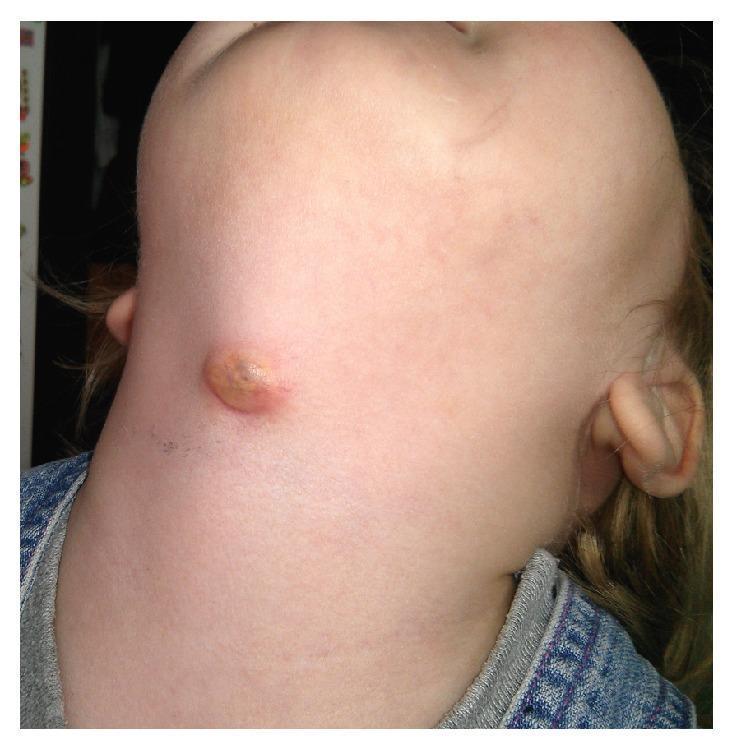
This clinical photograph displays a 2-year-old female patient with a prominent anterior neck swelling, characteristic of an infected thyroglossal duct cyst (TGDC). The lesion is located near the midline in the thyrohyoid region, slightly below the mandibular level. The swelling is approximately 1-2 cm in diameter, presenting as a raised, nodular mass with a circular to oval morphology. The surface exhibits a yellowish-brown crusting and erythema of the surrounding skin, suggesting an inflammatory or infectious process, such as an abscess or a draining fistula. This image serves as a clinical example of a common congenital midline neck mass in pediatric otolaryngology, illustrating the typical presentation of a thyroglossal duct cyst when secondary infection occurs. It is an essential visual reference for differential diagnosis of pediatric neck masses, distinguishing TGDC from other entities like dermoid cysts or lymphadenopathy based on its characteristic anatomical position.
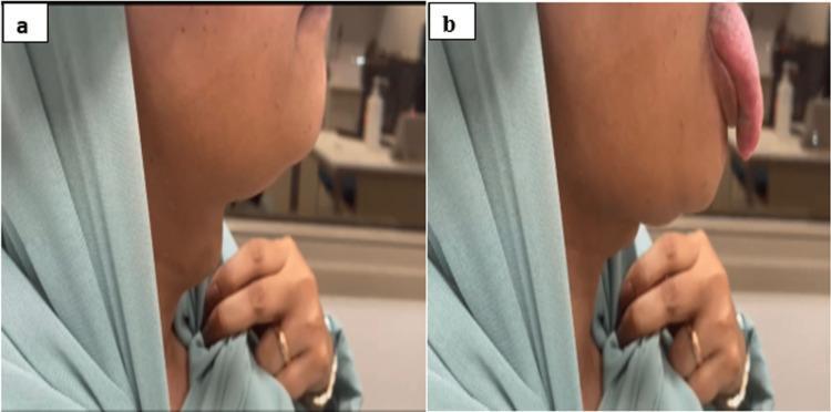
A two-panel clinical photograph illustrating a physical examination finding of a midline neck mass. Image (a) shows the lateral profile of a patient's neck at rest, revealing a smooth, rounded, subcutaneous swelling located in the midline near the hyoid region. Image (b) shows the same patient during tongue protrusion; the midline mass moves superiorly in coordination with the tongue movement. This specific physical sign (movement with tongue protrusion) is a classic clinical indicator of a thyroglossal duct cyst, reflecting its embryological attachment to the base of the tongue via the foramen cecum. The images are relevant for medical education in head and neck examination, surgical pathology, and endocrinology, specifically for differentiating midline neck masses such as dermoid cysts, ectopic thyroid tissue, and thyroglossal duct cysts.
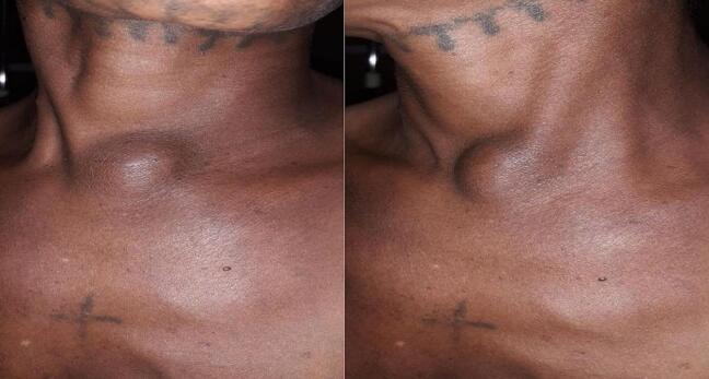
Clinical photograph (preoperative) showing the anterior neck and upper chest of a patient. The central focus is a prominent, well-defined, round cystic swelling located in the midline at the suprasternal notch. The mass is approximately 5-6 cm in diameter and protrudes significantly from the surrounding skin surface. The overlying skin appears intact without erythema or ulceration. This presentation is characteristic of a thyroglossal duct cyst or other congenital midline neck masses like dermoid cysts. Anatomical landmarks include the sternocleidomastoid muscles and the clavicular heads. Incidental findings include decorative tattoos on the submental region and the upper chest, along with a small melanocytic nevus on the left upper chest. The image demonstrates the typical clinical manifestation of a low-lying midline neck cyst, which is an important diagnostic consideration in pediatric and adult surgical pathology.
thyroglossal duct cyst ultrasound imaging
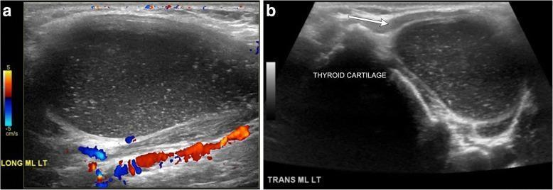
This dual-panel ultrasound image displays a thyroglossal duct cyst in the anterior neck. Panel (a) is a long-axis grayscale ultrasound with color Doppler showing a well-circumscribed, hypoechoic, oval structure. The interior contains numerous tiny, hyperechoic foci, consistent with proteinaceous or inflammatory debris. The color Doppler overlay confirms the absence of internal vascularity, while posterior acoustic enhancement (through transmission) is visible, confirming the cystic nature of the mass. Panel (b) shows a transverse grayscale ultrasound at the midline (ML LT), identifying the lesion's paramidline position. The cyst is located adjacent to the hyperechoic thyroid cartilage and is positioned deep to the strap muscles (indicated by the white arrow). This appearance is characteristic of an infrahyoid thyroglossal duct cyst, with internal complexity suggesting a history of prior infection or high protein content. The imaging is diagnostic for identifying midline congenital neck masses and differentiating them from solid lesions.
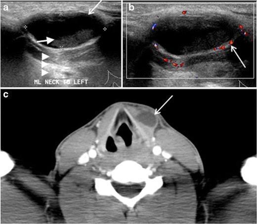
This composite figure displays diagnostic imaging of a long-standing infected thyroglossal duct cyst in a 29-year-old male. (a) Transverse greyscale ultrasound shows an oval, well-defined cystic lesion at the midline neck. It features low-level internal echoes (white arrows) and posterior acoustic enhancement (arrowheads), consistent with complex fluid. (b) Transverse color Doppler ultrasound demonstrates increased peripheral vascularity around the cyst wall (white arrow), indicating an inflammatory process. (c) Axial contrast-enhanced CT scan at the level of the thyroid cartilage reveals a well-circumscribed, homogeneous, hypoattenuating cystic lesion. The lesion is located slightly off-midline, embedded within the left strap muscle. It exhibits a prominent peripherally enhancing rim (white arrow) without internal calcifications or solid enhancing components. These findings are characteristic of an infected thyroglossal duct cyst, a common midline developmental remnant that may deviate from the midline when located caudally near the thyroid gland.
Midline Neck Swellings
Classification: Midline Swellings from Above Downwards
| Region | Swelling |
|---|---|
| Submental (above hyoid) | Ludwig's angina, submental lymph nodes, sublingual dermoid cyst, lipoma |
| Hyoid level | Thyroglossal cyst, subhyoid bursitis |
| Infrahyoid / thyroid | Goitre (thyroid isthmus, pyramidal lobe), pretracheal lymph nodes |
| Suprasternal | Enlarged nodes and lipoma in the space of Burns, retrosternal goitre, thymic swelling |
| Anywhere in midline | Dermoid cyst |
Key Causes in Detail
1. Thyroglossal Duct Cyst (TDC)
- 65% below the hyoid bone
- 15% at the level of the hyoid bone
- 75% in the midline; the rest up to 2 cm off midline
- Painless, smooth, cystic midline mass - seen in children and young adults
- Moves upward on swallowing AND on tongue protrusion - the classic sign, due to attachment to the hyoid and hence the tongue base
- Can become infected - causing pain, redness, sinus/fistula formation
- Ultrasound: anechoic cystic mass with posterior wall enhancement; can appear pseudosolid if proteinaceous/infected
- CT/MRI: high T2 signal on MRI; important to confirm normal thyroid in its proper location (ectopic thyroid can masquerade as TDC)
- Always check for a 1% incidence of papillary thyroid carcinoma within TDC
2. Dermoid Cyst
- Can occur anywhere in the midline neck
- Usually asymptomatic, may become infected
- Does NOT move with tongue protrusion - key differentiator from thyroglossal cyst
- Contents: keratin-filled (vs. mucus in thyroglossal)
- Treatment: simple cystectomy (no need to remove hyoid bone)
3. Subhyoid Bursitis
- Rare swelling at the level of the hyoid bone
- May mimic thyroglossal cyst
- Arises from the bursa beneath the hyoid bone
4. Goitre (Thyroid Swellings)
- Enlargement of the thyroid isthmus or pyramidal lobe presents in the midline
- Moves upward on swallowing (tethered to larynx/trachea)
- Pyramidal lobe goitre is a common midline lump just above the isthmus
- Refer to thyroid section for full classification (diffuse, nodular, toxic, malignant)
5. Pretracheal Lymph Nodes
- Lie anterior to the trachea in the midline
- May move with swallowing if fixed to the larynx/trachea
- Look for a primary source (thyroid carcinoma, laryngeal malignancy)
6. Ludwig's Angina
- Cellulitis of the floor of the mouth spreading to the submental region
- An acute, painful, indurated swelling in the submental midline
- Bilateral submandibular and submental space involvement
- Medical emergency due to risk of airway obstruction
7. Submental Lymph Nodes
- Drain the tip of tongue, lower lip, floor of mouth, and the lower central incisor region
- Enlargement may be reactive, tuberculous, or metastatic
8. Retrosternal Goitre / Thymic Swelling
- Low midline swellings at or below the suprasternal notch
- Retrosternal goitre: lower pole of thyroid descends into chest, visible/palpable at sternal notch, moves with swallowing
- Thymic cyst/mass: rare, in the superior mediastinum extending to the neck
9. Laryngocele
- Air-containing diverticulum from herniation of mucous membrane through the thyrohyoid membrane
- Resonant on percussion; becomes prominent when the patient blows their nose or strains
- Associated with trumpet players, glass blowers, and chronic cough
Useful Clinical Distinctions
| Feature | Thyroglossal Cyst | Dermoid Cyst |
|---|---|---|
| Position | Midline, near hyoid | Midline, any level |
| Moves with swallowing | Yes | Variable |
| Moves with tongue protrusion | Yes | No |
| Contents | Mucus | Keratin |
| Treatment | Sistrunk's op | Simple cystectomy |
Imaging Summary
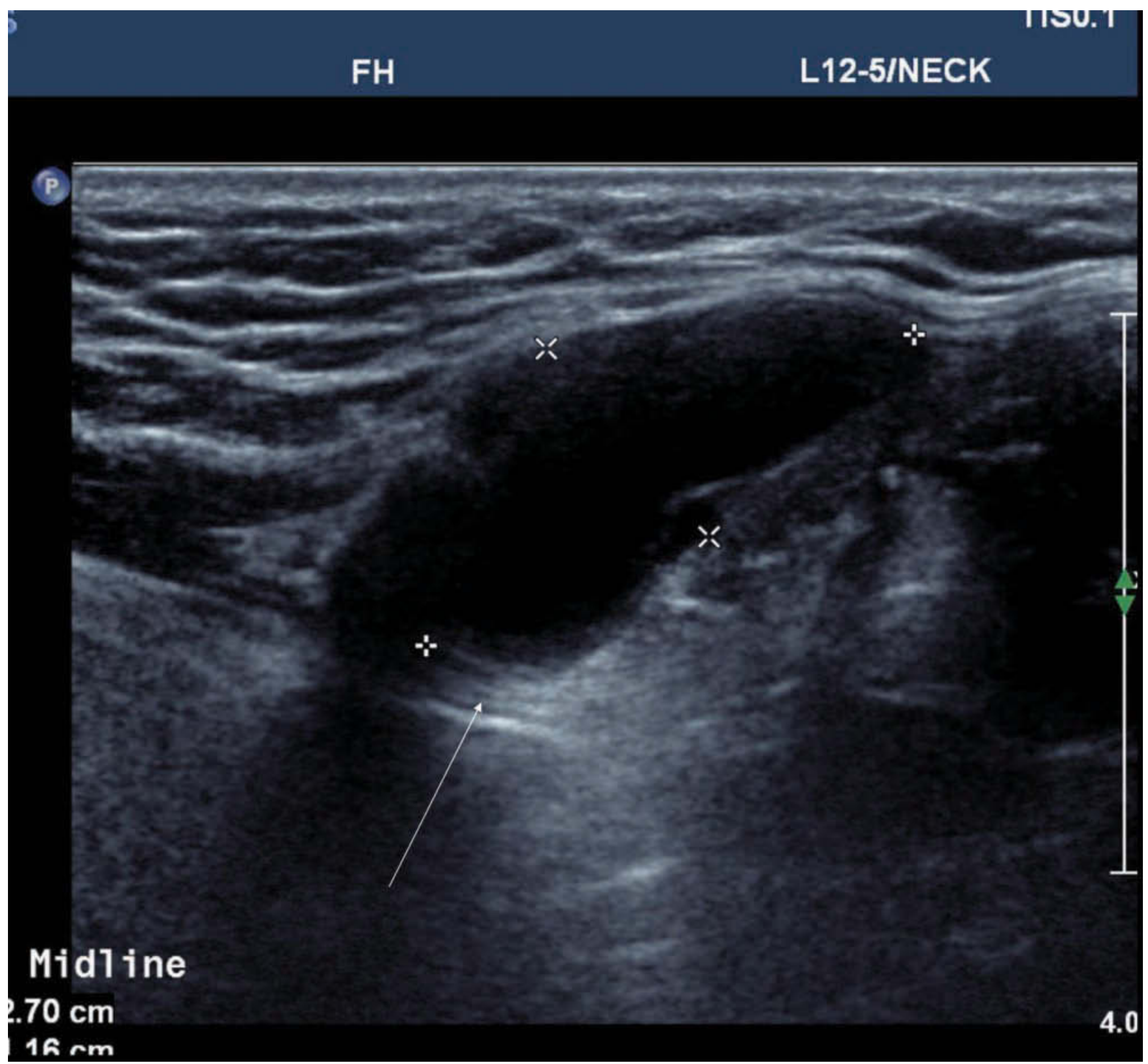
Key Rule to Remember
An upper midline cystic neck lesion in a toddler is a thyroglossal duct cyst until proven otherwise. - Sabiston Textbook of Surgery