
Gustafson changes in teeth
Reading File
Gustafson's method uses six age-related dental changes to estimate age at death in forensic odontology. The six parameters are: attrition, secondary dentine formation, periodontosis, cementum apposition, root resorption, and root transparency.
Searching sources...
Finding Sources
Finding Sources
Good - I have three relevant textbooks. Let me read the detailed content from each.
Reading File
Reading File
Reading File
I have rich content. Let me also read a bit more from Essentials for the scoring formula and fetch the diagram image.
Reading File
Reading File
Loading Image
I now have comprehensive information from three forensic medicine textbooks plus the classic Gustafson diagram. Here is the full answer:
Gustafson's Method of Age Estimation from Teeth
Gustafson (1950) described a method of estimating age in adults by microscopically examining a longitudinal ground section through the central part of a tooth, assessing six progressive age-related changes. The mnemonic APSRTC (or variations thereof) helps recall them.
The method is applicable from 25 to 60 years of age and is used in forensic odontology, primarily on dead bodies.
The Six Gustafson Changes
Each change is graded 0 to 3 (0 = absent; 1 = beginning; 2 = obvious; 3 = maximum/severe).
| # | Parameter | Abbreviation | Description |
|---|---|---|---|
| 1 | Attrition | A | Wear of the occlusal (biting) surface due to mastication friction. Proceeds from enamel → dentine → pulp exposure. |
| 2 | Periodontosis | P | Regression and recession of gums and periodontal tissue, gradually exposing the root. Poor oral hygiene accelerates it. |
| 3 | Secondary dentine | S | Deposition of secondary dentine within the pulp cavity, reducing its size. In mandibular teeth it starts from above; in maxillary teeth from below. |
| 4 | Root resorption | R | Decaying change affecting cementum and dentine, showing sharp grooves. Begins at the apex and extends upwards. |
| 5 | Transparency of root | T | Dentinal tubules become progressively filled with mineral, making dentine translucent. Occurs from apex upwards (lower jaw) and from above downwards (upper jaw). Not seen before ~30 years. |
| 6 | Cementum apposition | C | Progressive thickening of cementum, especially near the root apex, forming incremental lines. |
Grading Details
Attrition (A)
- A0 - No attrition
- A1 - Attrition within enamel only
- A2 - Attrition within dentine
- A3 - Attrition exposing soft pulp
Periodontosis (P)
- P0 - No periodontosis / no root exposure
- P1 - Exposure of less than 1/3 of root length
- P2 - Exposure of 1/3 to 2/3 of root length
- P3 - Periodontosis beyond 2/3 of root length
Secondary Dentine (S)
- S0 - No deposition
- S1 - Deposition in the upper part of pulp cavity
- S2 - Deposition in half the pulp cavity from above
- S3 - Almost the whole pulp cavity involved
Root Resorption (R)
- R0 - No root resorption
- R1 - Seen only in some parts
- R2 - Seen over a large area
- R3 - Involves both cementum and dentine
Cementum Apposition (C)
- C0 - Only normal layer present
- C1 - Slightly greater than normal
- C2 - Thick layer has formed
- C3 - Heavy layer present
Transparency (T)
- T0 - No transparency
- T1 - Transparency mainly over apical region
- T2 - Transparency up to 1/3 from apex
- T3 - Transparency up to 2/3 of root length from apex
Diagram of Gustafson's Stages
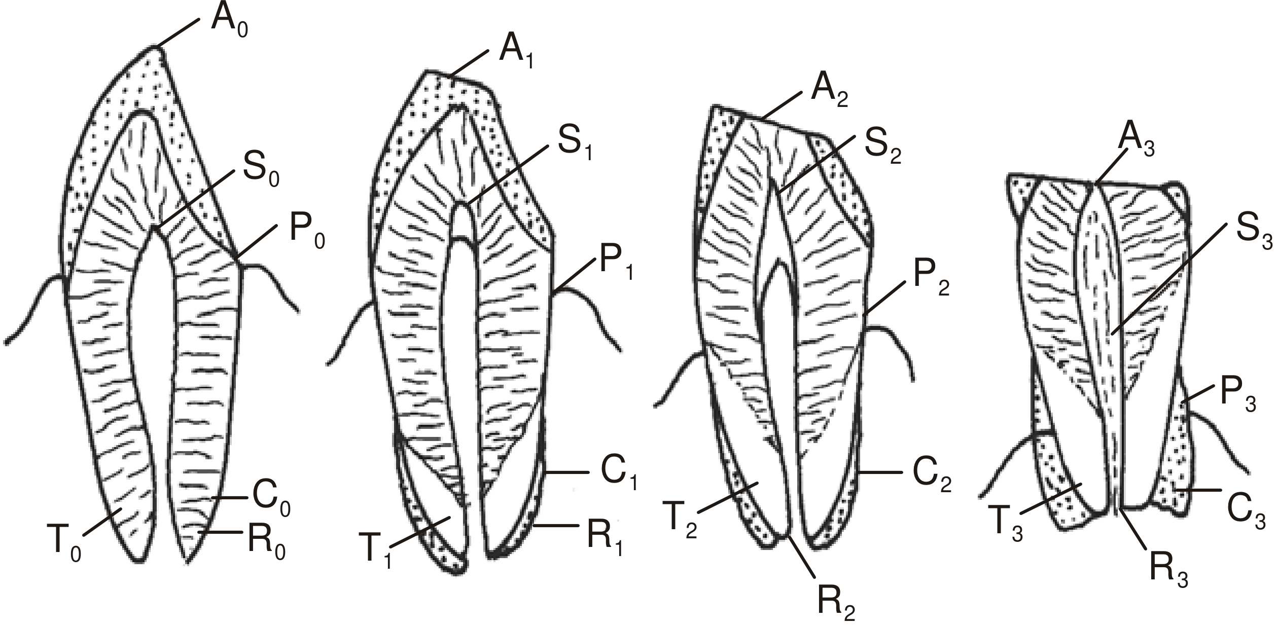
(The four sections show increasing age changes from left to right, labeled A = attrition, S = secondary dentine, P = periodontosis, C = cementum, T = transparency, R = root resorption)
Procedure
- Before extraction - estimate the degree of periodontosis by direct inspection.
- Extract the tooth.
- Ground section - grind the tooth on a glass slab to ~1 mm thickness to assess transparency.
- Further grinding to ~0.25 mm for microscopic examination of all remaining parameters.
- Score each of the six criteria (0-3 points each); maximum total = 18 points.
- Plot total points on a regression line graph (total points vs. known-age reference teeth).
Key Points
- Most reliable criterion: Transparency of the root - it is considered the most consistent and accurate of all six changes.
- Most suitable teeth: Anterior teeth (incisors > canines > premolars). Third molars are quite unsuitable.
- Changes are absent at 15 years of age.
- Error margin: ±3.5 to ±7 years (error increases above 50 years of age).
- Johanson's modification of Gustafson's method is considered more accurate.
- Pathological conditions (caries, periodontitis) can independently alter individual criteria and introduce error.
Sources: P.C. Dikshit Textbook of Forensic Medicine and Toxicology, p. 68-69; The Essentials of Forensic Medicine and Toxicology 36th ed., p. 83; Parikh's Textbook of Medical Jurisprudence, p. 88
This is a shared conversation. Sign in to Orris to start your own chat.