
Acute and chronic bronchitis
Reading File
Now I'll search the medical library database in parallel for both acute and chronic bronchitis.
Finding Sources
Searching PubMed
acute bronchitis chronic bronchitis management
Good. Now I'll read the most relevant sections in parallel - Fishman's (best pulmonary text), Family Medicine (acute), and Robbins (pathology).
Reading File
Reading File
Reading File
Reading File
Now let me get the Robbins full pathology on chronic bronchitis and the Tintinalli section on acute bronchitis, plus fetch the histology image.
Reading File
Reading File
Loading Image
Now let me get the chronic bronchitis morphology image from Robbins and read the clinical features section from Tintinalli, plus the Reid index image.
Reading File
Reading File
Now let me get the Reid index / morphology image from Robbins, and check if there are chronic bronchitis images.
Finding Sources
I now have rich content from multiple authoritative textbooks. Let me compile the comprehensive response.
Acute and Chronic Bronchitis
ACUTE BRONCHITIS
Definition and Epidemiology
Acute bronchitis is inflammation of the large airways without evidence of pneumonia. About 5% of adults develop it in a given year, generating ~100 million ambulatory care visits in the US annually. It is the ninth most common ED diagnosis in the US and one of the ten most common outpatient diagnoses worldwide. - Fishman's Pulmonary Diseases and Disorders
Etiology / Causative Agents
Viral (up to 90% of cases):
- Rhinovirus, coronavirus, parainfluenza virus, RSV, human metapneumovirus, influenza A & B, adenovirus
Bacterial (6-15.5% of cases):
- Bordetella pertussis, Mycoplasma pneumoniae, Chlamydia pneumoniae - most common in immunocompetent adults
- Moraxella catarrhalis, H. influenzae, S. pneumoniae - more common in COPD patients and smokers
- Atypical organisms (Mycoplasma, Chlamydia, Bordetella) are always a consideration
Incidence is higher in fall and winter, reflecting the seasonal pattern of respiratory viruses. - Fishman's Pulmonary Diseases and Disorders
Pathophysiology
Epithelial infection of the bronchi causes inflammation and thickening of the bronchial and tracheal mucosa, leading to:
- Airflow obstruction
- Bronchial hyperresponsiveness
- Reversible decrease in FEV1
- Cough, wheezing, and dyspnea
Sputum discoloration (yellow/green) results from cellular debris and microorganisms - not a reliable indicator of bacterial infection. - Tintinalli's Emergency Medicine
Clinical Features
| Feature | Details |
|---|---|
| Hallmark | Cough (with or without sputum production) |
| Duration | Typically 10-20 days; can exceed 1 month |
| Associated symptoms | Headache, rhinorrhea, mild constitutional symptoms, fever, mild dyspnea |
| Sputum color | Poor predictor of bacterial cause |
| PFTs | Reversible FEV1 reduction (bronchial hyperresponsiveness) |
Symptoms of acute upper respiratory infection and acute bronchitis are indistinguishable during the first few days. Cough persisting beyond 5 days and up to 3-4 weeks is the distinguishing marker. - Tintinalli's Emergency Medicine
Diagnosis
- Clinical diagnosis - no routine sputum cultures needed
- Diagnosed when acute cough (dry or productive) persists >5 days AND pneumonia, acute asthma, and COPD exacerbation are excluded
- If pneumonia is suspected: chest radiograph - pneumonia is unlikely if there is no fever, tachycardia, tachypnea, hypoxia, or abnormal auscultatory findings
- Cough persisting >3 weeks should prompt consideration of alternative diagnoses
- Procalcitonin and rapid point-of-care molecular viral testing have not been shown in large RCTs to reduce unnecessary antibiotic prescribing - Tintinalli's Emergency Medicine
Treatment
Key principle: supportive management; antibiotics are NOT indicated for uncomplicated acute bronchitis.
| Intervention | Evidence / Recommendation |
|---|---|
| Antibiotics | Not recommended (CDC, IDSA, Choosing Wisely). Minimal benefit (~0.5 day shorter cough), significant adverse effects |
| Beta-2 agonists | Avoid routinely; use only if measurable airflow obstruction/wheezing is present |
| Oral corticosteroids | Not recommended (no evidence in those without COPD/asthma) |
| Antitussives | Limited evidence; benzonatate and guaifenesin may give modest relief |
| Humidification, antipyretics | Supportive care |
"Red flag" symptoms requiring urgent re-evaluation: hemoptysis, worsening dyspnea, weight loss, dysphagia, persistent fever. - Fishman's Pulmonary Diseases and Disorders
Despite guidelines, antibiotic prescription rates for acute bronchitis in US outpatient settings remain around 71% - a persistent challenge highlighted by the IDSA's Choosing Wisely campaign. - Fishman's Pulmonary Diseases and Disorders
CHRONIC BRONCHITIS
Definition
Chronic bronchitis is defined clinically as a persistent productive cough for at least 3 consecutive months in at least 2 consecutive years, in the absence of any other identifiable cause. (Compare: emphysema is defined anatomically.) It is part of the COPD spectrum. - Robbins & Cotran Pathologic Basis of Disease
Epidemiology and Risk Factors
- 90% of those affected are smokers (40 pack-years or more is a common threshold)
- Other air pollutants: sulfur dioxide, nitrogen dioxide, grain dust, cotton dust, silica
- Common in urban dwellers in smog-ridden cities
Pathogenesis
Four main mechanisms drive chronic bronchitis: - Robbins & Cotran Pathologic Basis of Disease
-
Mucus hypersecretion - the earliest feature. Enlargement of submucosal glands in the trachea and bronchi, increased goblet cells in small airways. Driven by inflammatory mediators (histamine, IL-13, neutrophil elastase). Both changes are thought to be protective reactions to irritants that become pathological over time.
-
Acquired CFTR dysfunction - smoking leads to acquired CFTR dysfunction, producing abnormal dehydrated mucus that worsens disease severity.
-
Inflammation - inhalants cause cellular damage, eliciting acute and chronic inflammatory responses involving neutrophils, lymphocytes, and macrophages (notably, eosinophils are NOT seen - differentiating it from asthma). Persistent inflammation and fibrosis of small airways (bronchi and bronchioles <2-3 mm) drives airway obstruction.
-
Infection - does not initiate chronic bronchitis but maintains it and triggers acute exacerbations. Cigarette smoke impairs ciliary action, preventing mucus clearance and increasing infection susceptibility.
The airflow obstruction in chronic bronchitis results primarily from small airway disease (chronic bronchiolitis) - not from the large airway mucus hypersecretion. - Robbins & Kumar Basic Pathology
Morphology (Pathological Changes)
Gross:
- Hyperemia, swelling, and edema of mucous membranes
- Excessive mucinous or mucopurulent secretions
- Heavy casts of secretions and pus in bronchi/bronchioles
Microscopic:
- Chronic inflammation (lymphocytes and macrophages predominantly)
- Bronchiolar wall thickening: smooth muscle hypertrophy, extracellular matrix deposition, peribronchial fibrosis
- Goblet cell hyperplasia in small airways
- Enlargement of mucus-secreting glands (most striking change)
Reid Index:
- The Reid index = ratio of mucous gland layer thickness to total bronchial wall thickness (epithelium to cartilage)
- Normal: ≤ 0.4
- Elevated in chronic bronchitis, increasing with severity and duration of disease
- Severe cases: mucus plugging + inflammation + fibrosis = bronchiolar lumen obliteration (bronchiolitis obliterans)
Emphysematous changes frequently coexist. - Robbins & Cotran Pathologic Basis of Disease
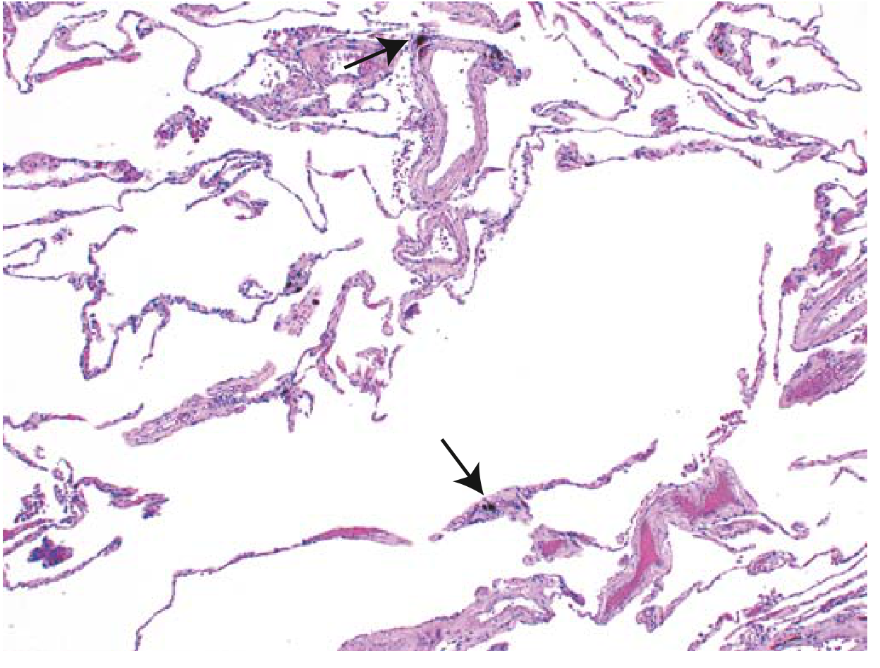
Below is a histological image of pulmonary emphysema showing enlarged air spaces with destroyed alveolar septa, which often coexists with chronic bronchitis (arrows indicate anthracotic pigment):
Clinical Features: "Blue Bloater" vs "Pink Puffer"
The COPD spectrum includes two classic phenotypes: - Robbins & Cotran Pathologic Basis of Disease
| Feature | Chronic Bronchitis ("Blue Bloater") | Emphysema ("Pink Puffer") |
|---|---|---|
| Age | 40-45 years | 50-75 years |
| Dyspnea | Mild; late onset | Severe; early onset |
| Cough | Early; copious sputum | Late; scanty sputum |
| Infections | Common | Occasional |
| Cor pulmonale | Common | Uncommon (end-stage) |
| Airway resistance | Increased | Normal or slightly increased |
| Elastic recoil | Normal | Low |
| CXR | Prominent vessels; large heart | Hyperinflation; normal heart size |
| Appearance | Cyanotic | Well oxygenated |
| Blood gases | Hypoxemia + hypercapnia | Relatively normal at rest |
Symptoms are typically worse in the morning. Most real patients have mixed features.
Spirometry: FEV1/FVC ratio < 0.7 confirms airflow obstruction.
Complications
Long-standing chronic bronchitis can lead to:
- Pulmonary hypertension
- Cor pulmonale (right heart failure from pulmonary hypertension)
- Acute respiratory failure from superimposed infection
- Increased all-cause mortality from mucus plugs
Treatment
| Approach | Details |
|---|---|
| Smoking cessation | Most important intervention |
| Long-acting bronchodilators (LABA/LAMA) | Cornerstone of maintenance therapy |
| Inhaled corticosteroids (ICS) | Combined with bronchodilators in moderate-severe disease |
| Antibiotics | For acute exacerbations with bacterial triggers |
| Oxygen therapy | In hypoxemic patients |
| Pulmonary rehabilitation | Physical therapy and exercise |
| Surgical | Bullectomy, lung volume reduction surgery, or lung transplantation in selected patients |
Quick Comparison: Acute vs. Chronic Bronchitis
| Acute Bronchitis | Chronic Bronchitis | |
|---|---|---|
| Definition | Airway inflammation without pneumonia | Productive cough ≥3 months/year for ≥2 years |
| Main cause | Viral (90%) | Cigarette smoking (90%) |
| Pathology | Mucosal inflammation, transient | Mucous gland hypertrophy, goblet cell metaplasia, fibrosis |
| Duration | Self-limited (10-20 days) | Chronic, progressive |
| Key investigation | Clinical diagnosis; CXR to rule out pneumonia | Spirometry (FEV1/FVC <0.7), Reid index >0.4 |
| Antibiotics | NOT indicated routinely | For exacerbations only |
| Management | Supportive care | Smoking cessation, bronchodilators, ICS |
Sources: Fishman's Pulmonary Diseases and Disorders, 5e; Robbins, Cotran & Kumar Pathologic Basis of Disease, 10e; Robbins & Kumar Basic Pathology, 11e; Tintinalli's Emergency Medicine, 9e; Textbook of Family Medicine, 9e
This is a shared conversation. Sign in to Orris to start your own chat.