
Development of ear and congenital anomalies of external ear in detail for 20 marker long answer for unive ms ent pg exam in simplified manner
Reading File
Searching sources...
Finding Sources
Finding Sources
Searching Images
development of ear embryology branchial arch
Finding Sources
Finding Sources
Finding Sources
Reading File
Reading File
Reading File
Finding Sources
Finding Sources
Reading File
Reading File
Reading File
Loading Image
Finding Sources
Reading File
Reading File
Loading Image
Reading File
I now have comprehensive material from three authoritative sources. Let me compile the complete answer.
Development of Ear and Congenital Anomalies of the External Ear
(University MS ENT PG Long Answer - 20 Marks)
INTRODUCTION
The ear is divided into three anatomical compartments: external ear, middle ear, and internal ear. Each develops from different embryological structures - a critical fact because it explains why anomalies of one part do not always accompany anomalies of another.
PART I: EMBRYOLOGICAL DEVELOPMENT OF THE EAR
A. Periods of Development (Scott-Brown's)
Development spans three stages:
- Pre-embryonic: Days 1-21
- Embryonic: Days 22-56 (most organ formation occurs here)
- Foetal: Day 56 to birth (growth, shape refinement)
The key structures of the ear are formed during the embryonic period, so teratogens acting during this window cause the most severe anomalies.
B. Branchial (Pharyngeal) Arches - The Foundation
The mesenchyme around the primitive pharynx differentiates into 5-6 branchial arches (numbered 1 to 5, head to tail). Between each arch on the outside is a branchial groove (ectodermal); on the inside is a pharyngeal pouch (endodermal).
Each arch contains:
- A bar of cartilage
- A group of muscles
- An artery
- A cranial nerve (post-trematic nerve)
| Arch | Nerve | Relevance to Ear |
|---|---|---|
| 1st (Meckel's) | CN V (Trigeminal) | Malleus, incus, tragus, EAM, tympanic membrane |
| 2nd (Reichert's) | CN VII (Facial) | Stapes superstructure, bulk of pinna |
| 3rd | CN IX | Stapes footplate (some) |
C. Development of the External Ear
The external ear has three components: the auricle (pinna), the external auditory meatus (EAM), and the tympanic membrane.
1. Development of the Auricle (Pinna)
The auricle begins developing during week 5 of gestation (some sources say week 6). It develops from six mesenchymal swellings called the Hillocks of His (auricular hillocks), which arise around the margins of the first pharyngeal groove (the groove between arches 1 and 2):
- Hillocks 1, 2, 3 - from the 1st pharyngeal arch (mandibular arch)
- Hillocks 4, 5, 6 - from the 2nd pharyngeal arch (hyoid arch)
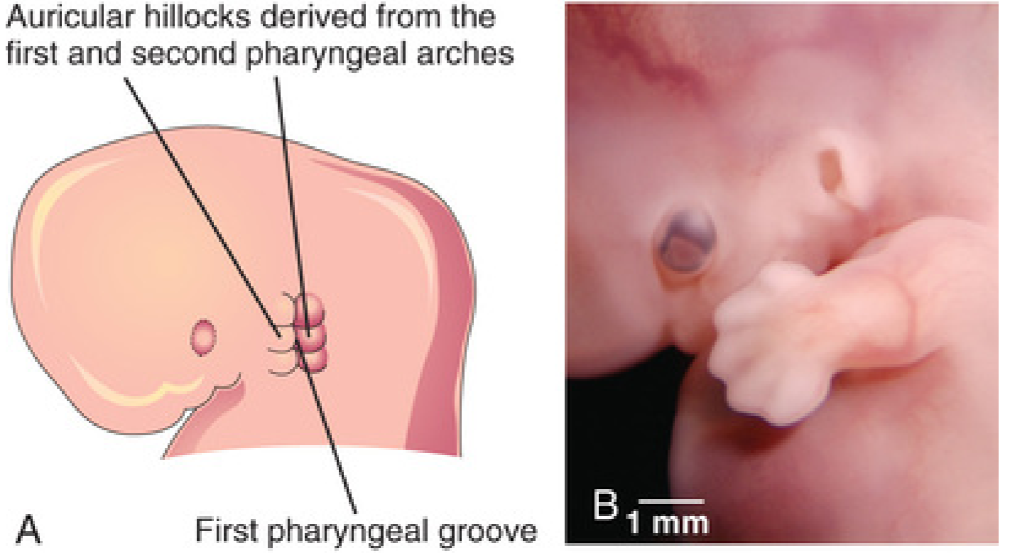
Contributions of each hillock/arch:
- The 2nd arch hillocks form the bulk of the pinna (helix, antihelix, antitragus, lobule)
- The 1st arch contribution progressively reduces; it forms the tragus and part of the helical root
- Recent research (Cummings) indicates tissue caudal to the 2nd arch hillocks gives rise to the free ear fold (helix + scapha)
- The lobule is the last part of the auricle to develop and is the least vulnerable to teratogens
Migration: The auricular complex initially forms in the anterior neck region (base of neck) at weeks 5-6, then migrates dorsally and cephalad as the mandible develops during weeks 8-12. This explains why in mandibular hypoplasia (e.g., Treacher Collins syndrome), the ears appear low-set.
Key gene: HoxA2 is critical to auricle development.
The rudimentary pinna is formed by 60 days and reaches its adult structure by 22 weeks. The auricle continues to grow throughout life.
Innervation of the auricle (follows arch derivation):
- Parts from 1st arch: mandibular branch of CN V
- Parts from 2nd arch: Greater auricular and lesser occipital nerves (cervical plexus)
- The facial nerve (nerve of 2nd arch) does not supply the skin of the 2nd arch derivatives - an exception explained by migration of cervical neural crest cells
2. Development of the External Auditory Meatus (EAM)
- Develops from the dorsal part of the first pharyngeal groove (ectoderm)
- Ectodermal cells at the base proliferate to form a solid epithelial plate: the meatal plug
- Late in foetal life (around week 28), the central cells of the meatal plug degenerate and the cavity formed becomes the internal (deep) part of the EAM
- The meatus is relatively short at birth and attains adult length by approximately 9 years of age
- Failure of meatal plug canalization causes congenital EAM atresia (meatal atresia)
3. Development of the Tympanic Membrane
The tympanic membrane is derived from three sources (a classic exam point):
| Layer | Embryological Source |
|---|---|
| Outer (lateral) layer | Ectoderm of the 1st pharyngeal groove |
| Middle (fibrous) layer | Mesenchyme of 1st and 2nd arches (differentiates into collagenous fibres) |
| Inner (medial) layer | Endoderm of the tubotympanic recess (1st pharyngeal pouch) |
D. Development of the Middle Ear
- The tubotympanic recess develops from the first pharyngeal pouch (endoderm)
- This recess expands to form:
- Eustachian tube (medial part)
- Tympanic cavity (lateral expanded part)
- Mastoid antrum (further expansion)
- The Eustachian tube lumen and middle ear spaces are formed by 8 months' gestation
- The epitympanum and mastoid antrum develop by birth
- Mastoid air cells are mostly absent at birth (filled with amniotic fluid); 90% of air cell development is complete by age 6, with remainder by age 18
Ossicles:
- Develop from the dorsal ends of arch cartilages, beginning at 4 weeks
- Malleus head + incus body: 1st arch (Meckel's cartilage)
- Stapes superstructure: 2nd arch (Reichert's cartilage)
- Stapes footplate: contributes from the otic capsule (lateral wall of the labyrinth)
- Adult shape, size, and ossification is present by 25 weeks' gestation
- Tensor tympani = 1st arch (CN V); Stapedius = 2nd arch (CN VII)
E. Development of the Internal Ear
- At day 22-23, ectodermal thickening forms on the sides of the head: the otic placode
- The placode deepens to form the otic pit, which closes off to become the otocyst (otic vesicle) by week 4
- The otocyst divides into:
- Dorsal utricular part → utricle, semicircular ducts, endolymphatic duct
- Ventral saccular part → saccule and cochlear duct; cochlear duct gives rise to the organ of Corti
- The bony labyrinth develops from the mesenchyme adjacent to the membranous labyrinth (otic capsule)
- Neural crest cells cluster near the otocyst to become the spiral (auditory), vestibular (Scarpa's), and geniculate (facial) ganglia
- Because the labyrinth is phylogenetically ancient, labyrinthine anomalies are rare
PART II: CONGENITAL ANOMALIES OF THE EXTERNAL EAR
Key Clinical Principle
"The auricle is formed early. Therefore, malformation of the auricle implies malformation of the middle ear, mastoid, and VII nerve. On the other hand, a normal auricle with canal atresia indicates abnormal development during the 28th week, by which time the ossicles and middle ear are already formed." - KJ Lee's Essential Otolaryngology
1. Microtia (Underdevelopment of the Auricle)
Definition: A spectrum of auricular underdevelopment ranging from minor shape abnormalities to complete absence.
Incidence: Approximately 1 in 20,000 births (some sources quote 1:10,000-1:20,000). Unilateral:bilateral ratio = 3-4:1; right side more commonly affected.
Etiology (multifactorial):
- Genetic factors: HOXA2, other genes
- Neural crest cell disturbance
- Vascular disruption (stapedial artery)
- Teratogens: retinoic acid (days 20-22), rubella, alcohol, thalidomide
- Living at altitude
- Associations: Hemifacial microsomia (Goldenhar/oculo-auricular-vertebral spectrum), Treacher Collins syndrome, Nager syndrome
Classification of Microtia (Marx/Modified - 4 grades, Cummings)
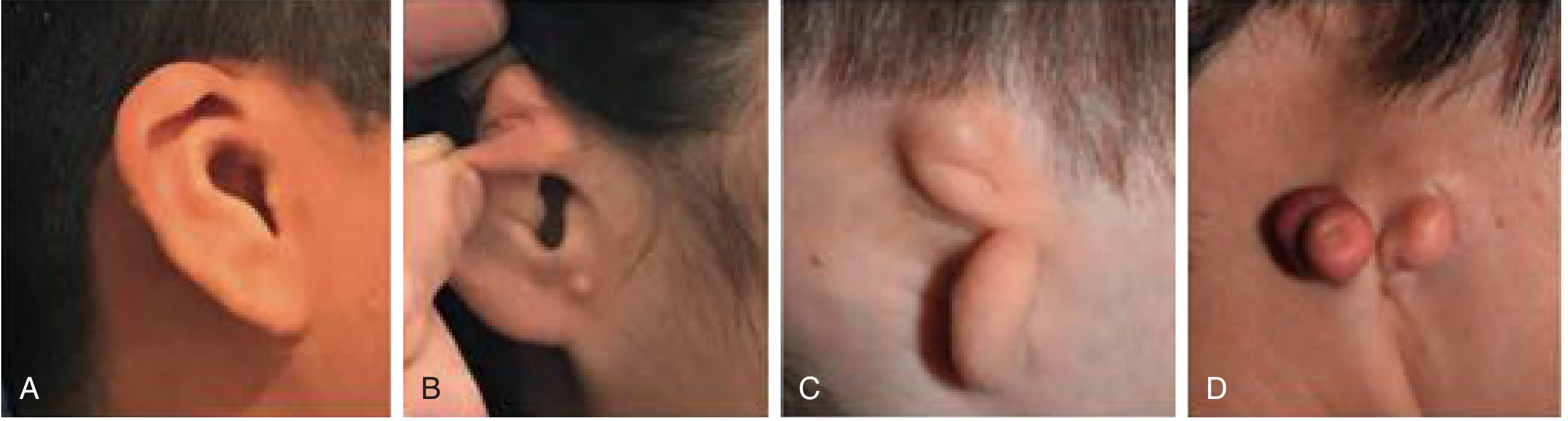
| Grade | Description |
|---|---|
| Grade I | All structures identifiable; ear smaller than normal (constricted ear) |
| Grade II (Conchal type) | Some superior structures absent; concha still identifiable |
| Grade III (Lobular type) | Only the lobule is present; no recognizable structures |
| Grade IV | Anotia - complete absence of all auricular structures |
The lobule is almost always present even in severe forms (least vulnerable tissue).
Management of Microtia:
- Observation
- Prosthesis (adhesive or implant-retained)
- Single-stage reconstruction with alloplastic framework (MedPor/SuPor with temporoparietal fascia flap)
- Staged autologous costochondral reconstruction (gold standard) - Brent technique (3-4 stages) or Nagata technique (2 stages)
- Future: Tissue engineering + 3D printing
Important points:
- Age-appropriate hearing assessment is mandatory in all microtia patients
- EAC atresia repair is performed after auricular reconstruction
- Rib cartilage harvest is possible once ear reaches ~85% of adult size (around age 6-8)
2. Anotia
Complete absence of the auricle. Grade IV of the microtia spectrum. Most severe anomaly.
3. Congenital EAM Atresia (Meatal Atresia)
Pathology: Failure of canalization of the meatal plug (which normally degenerates around week 28). May be:
- Bony atresia (most common): solid bone occludes the canal
- Membranous/soft tissue atresia: soft tissue band occludes canal
Clinical significance:
- A normal auricle with canal atresia suggests the insult occurred around 28th week - ossicles and middle ear are likely normal
- Audiological evaluation and CT temporal bone (high-resolution) are essential
- Grading by Jahrsdoerfer score helps assess candidacy for surgical repair (canaloplasty)
- Usually managed by canaloplasty + meatoplasty or with bone-anchored hearing aids (BAHA)
4. Preauricular Sinus / Cyst / Fistula
Pathology: Improper fusion/incomplete coalescence of the auricular hillocks during development results in epithelium-lined tracts/sinuses.
- Preauricular sinus: blind-ended tract (most common), opens in front of the tragus
- Preauricular cyst: enclosed remnant, no external opening
- First branchial arch fistula (Collaural fistula): tract between the EAC and the skin of the cheek, runs through the parotid gland, often between branches of the facial nerve
Clinical features:
- Small pit anterior to the tragus
- Intermittent discharge, recurrent infections
- 20% are bilateral
- May be asymptomatic for years
Management:
- Observe if asymptomatic
- Surgical excision (complete tract removal) - preferably when infection-free
- Collaural fistula: requires careful dissection around facial nerve
5. Accessory Auricle (Polyotia/Ear Tags)
- Small soft-tissue or cartilaginous tags anterior to the tragus or along the line from tragus to angle of mouth
- Represent supernumerary auricular hillocks or failure of regression of arch tissue
- Associated with other first and second arch anomalies
- Treatment: surgical excision
6. Prominent/Bat Ear (Prominent Auricle)
- Most common congenital auricle deformity
- Due to failure of normal folding of the antihelical fold
- The conchoscaphal angle is >90° (normal: 90°)
- Concha may be enlarged (conchal hypertrophy)
- Treatment: Pinnaplasty/Otoplasty - ideally before school age (5-6 years)
- Mustardé suture technique (antihelical folding)
- Converse technique
- Furnas suture (conchomastoid fixation)
7. Darwin's Tubercle
- A small cartilaginous nodule on the helix
- Represents a vestigial remnant (phylogenetically, the tip of the pointed animal ear)
- Common, no clinical significance
8. Low-Set Ears
- Normal position: upper pole of pinna at the level of the lateral canthus of the eye
- Low-set ears: pinna positioned below this line
- Associated with chromosomal abnormalities, especially trisomy 13 (Patau) and trisomy 18 (Edwards)
- Also seen in Turner syndrome (45, XO)
9. Treacher Collins Syndrome (Mandibulofacial Dysostosis)
Autosomal dominant (TCOF1 gene - treacle protein). First arch syndrome.
Features relevant to the ear:
- Bony meatal atresia
- Malformed incus and malleus
- Short mandible
- Antimongoloid slant of eyes (outward-downward)
- Notched lower eyelid
- Fishmouth deformity
- Absent malar bones
- When maxilla is also malformed alongside the first arch features = Treacher Collins syndrome
10. Goldenhar Syndrome (Hemifacial Microsomia / OAV Spectrum)
- Unilateral facial underdevelopment
- Microtia + EAC atresia
- Epibulbar dermoids
- Vertebral anomalies
- Mandibular asymmetry
- Renal/cardiac anomalies
Summary Table: Embryological Origins and Associated Anomalies
| Structure | Embryological Origin | Anomaly if Disrupted |
|---|---|---|
| Auricle | Hillocks of His (arches 1+2) | Microtia, anotia, accessory auricles, bat ear |
| EAM | 1st pharyngeal groove ectoderm | Meatal atresia/stenosis |
| Tympanic membrane | Groove + pouch + mesenchyme | Absent/perforated TM |
| Tympanic cavity | 1st pharyngeal pouch (endoderm) | Middle ear anomalies |
| Eustachian tube | 1st pharyngeal pouch | ET dysfunction |
| Malleus head, incus | Meckel's cartilage (arch 1) | Ossicular abnormalities |
| Stapes superstructure | Reichert's cartilage (arch 2) | Stapes fixation/absence |
| Membranous labyrinth | Otic placode → otocyst | Sensorineural hearing loss |
| Bony labyrinth | Otic capsule mesenchyme | Labyrinthine dysplasia (rare) |
Key Exam Points to Remember
- Six hillocks of His → from 1st (hillocks 1-3) and 2nd (hillocks 4-6) arches
- Tragus = only 1st arch contribution to adult pinna
- Tympanic membrane = 3 layers from 3 sources (groove, pouch, mesenchyme)
- Otic placode → otic pit → otocyst at day 22-23
- Microtia + middle ear anomaly = insult before week 5-6 (early)
- Normal auricle + canal atresia = insult around week 28 (late)
- Preauricular sinus = improper fusion of hillocks; 20% bilateral
- Collaural fistula = tract between EAC and cheek skin, traverses parotid, near facial nerve
- Low-set ears = associated with trisomy 13 and trisomy 18
- Labyrinthine anomalies are rare because the labyrinth is phylogenetically ancient
- Mastoid process not formed until end of 2nd year; mastoid air cells mostly complete by age 6
- Ossicles reach adult shape and ossification by 25 weeks' gestation
Sources: The Developing Human - Moore & Persaud (9th ed.); Scott-Brown's Otorhinolaryngology Head & Neck Surgery Vol. 2; Cummings Otolaryngology Head & Neck Surgery; KJ Lee's Essential Otolaryngology
This is a shared conversation. Sign in to Orris to start your own chat.