
Nephritic vs nephrotic syndrome
Reading File
Now let me search the library in parallel for both syndromes.
Finding Sources
Finding Sources
Reading File
Reading File
Finding Sources
Finding Sources
Reading File
Finding Sources
Finding Sources
Reading File
Reading File
Loading Image
I now have comprehensive data from multiple authoritative textbooks. Here is the full comparison:
Nephritic vs Nephrotic Syndrome
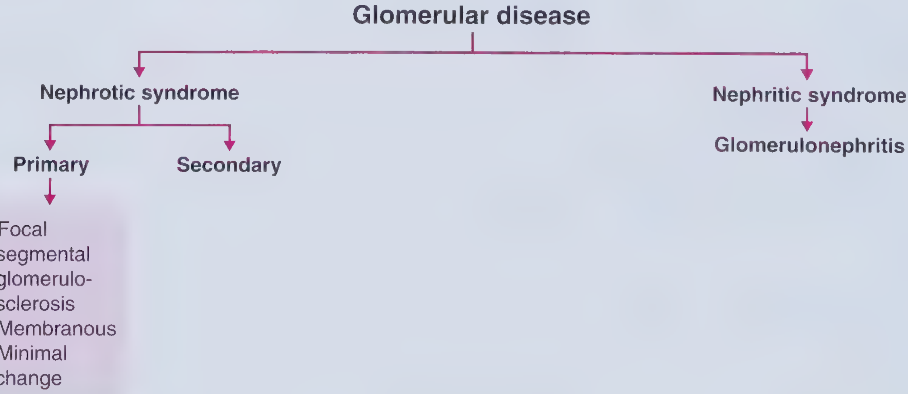
Core Pathophysiology
| Nephritic | Nephrotic | |
|---|---|---|
| Mechanism | Glomerular inflammation - proliferation of glomerular cells + leukocyte infiltration, injuring capillary walls | Increased permeability of glomerular capillary wall to proteins (structural/physicochemical defect in GBM/podocytes) |
| Primary defect | Capillary wall disruption - blood leaks into urine | Protein leaks across the filtration barrier |
Clinical Features at a Glance
| Feature | Nephrotic | Nephritic |
|---|---|---|
| Onset | Insidious | Abrupt |
| Proteinuria | Massive (>3.5 g/day in adults) ++++ | Sub-nephrotic range ++ |
| Hematuria | May or may not occur | +++ (hallmark) |
| RBC casts | Absent | Present (hallmark) |
| Serum albumin | Low (<3 g/dL) | Normal or slightly reduced |
| Edema | Massive, pitting, periorbital/dependent (++++) | Mild-moderate (++) |
| Blood pressure | Normal | Raised |
| JVP | Normal/low | Raised |
| Azotemia/oliguria | Absent or mild | Common (reduced GFR) |
| Lipids | Hyperlipidemia + lipiduria | Normal |
(Table adapted from Comprehensive Clinical Nephrology, 7e, Table 16.4)
Nephrotic Syndrome - Key Details
Diagnostic criteria: Proteinuria >3.5 g/day + hypoalbuminemia + generalized edema + hyperlipidemia/lipiduria
Pathophysiology cascade:
- Glomerular barrier damage → protein leaks (massive proteinuria)
- Protein loss > liver synthesis → hypoalbuminemia
- Low oncotic pressure → generalized edema (periorbital, ascites, pleural effusions)
- Sodium/water retention (aldosterone, sympathetic activation, reduced ANP) worsens edema
- Liver compensatory lipoprotein synthesis + reduced catabolism → hyperlipidemia
- Lipoproteins leak → lipiduria (oval fat bodies, free fat in urine)
Complications unique to nephrotic:
- Thrombosis/thromboembolism - loss of antithrombin III and other anticoagulants in urine; renal vein thrombosis is especially associated with membranous nephropathy
- Infections - loss of immunoglobulins (esp. staphylococcal and pneumococcal)
Causes by prevalence:
| Cause | Children | Adults |
|---|---|---|
| Minimal change disease | 75% | 8% |
| Focal segmental glomerulosclerosis (FSGS) | 10% | 35% |
| Membranous nephropathy | 3% | 30% |
| Membranoproliferative GN | 10% | 10% |
| Secondary (DM, amyloid, SLE, drugs) | rare | common |
(Robbins & Cotran, Table 20.7)
Nephritic Syndrome - Key Details
Core features: Hematuria (RBCs + RBC casts) + subnephrotic proteinuria + azotemia + oliguria + hypertension
Pathophysiology: Inflammatory proliferation damages capillary walls → blood enters urine + reduced GFR → fluid retention → hypertension (also from renin release from ischemic kidneys)
Causes:
| Disease | Key feature |
|---|---|
| Post-streptococcal GN | Subepithelial immune deposits; types 1, 4, 12 group A strep; latent period 1-4 weeks |
| IgA nephropathy (Berger's) | Mesangial IgA deposits; most common GN worldwide |
| RPGN/Crescentic GN | Rapid kidney function loss over days-weeks; crescents in Bowman's space |
| Anti-GBM (Goodpasture's) | Type I RPGN; linear IgG deposits on GBM |
| ANCA-associated vasculitis | GPA, MPA, EGPA; pauci-immune |
| Lupus nephritis | Immune complex-mediated; can also cause nephrotic |
| Cryoglobulinemia | Often hepatitis C-associated |
| Infective endocarditis | Immune complex deposition |
Mixed Presentations
Some diseases sit in both categories - notably membranoproliferative GN (MPGN) and lupus nephritis, which can present with nephrotic syndrome, nephritic syndrome, or a combination of both.
Urine Sediment: The Key Differentiator
- Nephritic: "Active sediment" - dysmorphic RBCs, RBC casts, granular casts. These are virtually pathognomonic of glomerular inflammation.
- Nephrotic: "Bland sediment" - fatty casts, oval fat bodies, waxy casts. Little to no hematuria.
Memory Hook
Nephr-OTic = Oedema + Three point five grams protein. Leaky barrier, not inflamed.
Nephr-ITic = IT (inflammation) = hematuria + hypertension. Inflamed, broken capillaries.
Sources: Robbins & Cotran Pathologic Basis of Disease; Comprehensive Clinical Nephrology, 7e; Goldman-Cecil Medicine; Frameworks for Internal Medicine
This is a shared conversation. Sign in to Orris to start your own chat.