
For how many days we give amphotericin b in cerebral mucormycosis
mucormycosis amphotericin B treatment duration
cerebral mucormycosis amphotericin B duration days weeks IDSA guidelines
Amphotericin B Duration in Cerebral Mucormycosis
The Key Point: No Fixed Number of Days
What the Guidelines Say
- For CNS involvement specifically: Liposomal amphotericin B at 10 mg/kg/day for the initial 28 days (Grade A, Level III evidence)
- After that initial 28-day induction phase, therapy continues until:
- Clinical improvement
- Radiographic resolution (weekly imaging used for response assessment)
- Correction of underlying immunologic defects
- Step-down to an oral azole (posaconazole or isavuconazole) is used once the patient is stable
- Doses of 10 mg/kg/day are specifically suggested for CNS infections
- Duration is determined on an individual basis, but therapy "usually continues for at least 6-8 weeks"
- Step-down to posaconazole or isavuconazole is considered after "2 to 3 weeks or more of initial induction therapy" once source control and clinical improvement are achieved
- "Therapy should continue until..." clinical and radiographic resolution
Practical Summary
| Parameter | Recommendation |
|---|---|
| Preferred drug | Liposomal amphotericin B |
| Dose for CNS | 10 mg/kg/day (higher than the 5 mg/kg/day for non-CNS) |
| Initial induction phase | 28 days (per ECMM for CNS involvement) |
| Total minimum duration | 6-8 weeks (per ECIL-3) |
| Overall duration | Until clinical improvement + radiographic resolution + immune recovery |
| Step-down therapy | Posaconazole or isavuconazole once patient is stable |
Important Notes
- Full dose from Day 1 - dose escalation is not recommended; give the full daily dose from the start
- Surgery is co-required - antifungal therapy alone is rarely sufficient; surgical debridement is a cornerstone of management
- Treat the underlying condition - reverse immunosuppression, control diabetes, stop deferoxamine
- The "28 days" figure specifically applies to CNS mucormycosis induction per ECMM guidelines; total treatment extends far beyond this
- The optimal duration remains undefined in the literature; treatment can extend to months in severe or refractory cases
Management of post lscs pneumonia
Management of Post-LSCS Pneumonia
1. Immediate Assessment and Diagnosis
- Fever >38°C, productive cough, tachypnea, reduced SpO2
- Pleuritic chest pain, dullness to percussion, bronchial breathing
- Onset usually within 48-72 hrs post-op (aspiration) or 3-7 days (HAP)
- Chest X-ray (PA view)
- CBC, CRP, procalcitonin
- Blood cultures x 2 sets (before antibiotics)
- Sputum Gram stain and culture
- ABG if SpO2 <94% or respiratory distress
- Blood urea, creatinine (guides antibiotic dosing)
- HRCT chest if CXR inconclusive
2. Risk Stratification: Is this Low-Risk or High-Risk HAP?
| Risk Category | Criteria |
|---|---|
| Low risk (no MDR concern) | No prior IV antibiotics in 90 days; short hospital stay; not in septic shock |
| MRSA risk | Prior IV antibiotics + local MRSA prevalence >20% in ICU |
| MDR risk (VAP) | Prior IV antibiotics + septic shock + ARDS + ≥5 days in hospital + RRT |
| High mortality risk | Septic shock, need for mechanical ventilation |
3. Empiric Antibiotic Therapy
A. Low-risk HAP (no MRSA risk, no MDR risk) - MONOTHERAPY:
| Drug | Dose |
|---|---|
| Piperacillin-tazobactam | 4.5 g IV q6h (extended infusion preferred) |
| OR Cefepime | 2 g IV q8h |
| OR Levofloxacin | 750 mg IV/PO once daily |
| OR Imipenem / Meropenem | Reserved for higher-risk |
B. MRSA Risk - ADD:
- Vancomycin 15-20 mg/kg IV q8-12h (target trough 15-20 mg/L) OR
- Linezolid 600 mg IV/PO q12h (preferred if renal impairment)
C. High Mortality Risk / MDR Concern - DUAL gram-negative coverage + MRSA cover:
| Component | Options |
|---|---|
| Anti-pseudomonal β-lactam | Pip-tazo OR Cefepime OR Meropenem |
| PLUS second gram-negative | Aminoglycoside (amikacin/tobramycin) OR ciprofloxacin/levofloxacin |
| PLUS MRSA cover | Vancomycin OR Linezolid |
4. De-escalation (Critical Step)
- Narrow the spectrum to organism-directed therapy
- If MSSA confirmed: switch to oxacillin, nafcillin, or cefazolin
- If MRSA: continue vancomycin or linezolid
- If Pseudomonas: monotherapy based on susceptibility (once septic shock resolves)
- Total duration: 7 days (evidence shows 7 days = 14 days for HAP/VAP; longer courses increase resistance) - Harrison's 22nd ed.
5. Respiratory Support
| Severity | Support |
|---|---|
| SpO2 94-98%, mild distress | Supplemental O2 via face mask (FiO2 titrated to SpO2 >94%) |
| Moderate hypoxemia (SpO2 <94%) | High-flow nasal cannula (HFNC) - preferred in post-op patients |
| Failure of HFNC | Non-invasive ventilation (NIV/BiPAP) - use cautiously post-laparotomy |
| SpO2 <88% on high FiO2, ARDS criteria | Intubation + invasive mechanical ventilation |
6. Supportive Management
- Analgesia: Adequate pain control is essential - pain from the LSCS wound inhibits deep breathing and coughing. Use paracetamol + NSAID (ketorolac) +/- opioids carefully
- Physiotherapy: Chest physiotherapy, incentive spirometry, early ambulation - core to recovery
- Positioning: Head-of-bed elevation 30-45° (reduces aspiration risk and shown to reduce VAP by ~70%)
- DVT prophylaxis: Post-LSCS patients are already at high VTE risk; pneumonia further immobilizes them - continue LMWH
- Fluid management: Avoid overhydration (worsens pulmonary edema); target euvolemia
- Nutritional support: Early enteral feeding if able to swallow safely
- Bronchodilators: Nebulized salbutamol if significant bronchospasm
7. Consider Specific Scenarios
| Scenario | Action |
|---|---|
| Aspiration pneumonia (within 48h, witnessed aspiration) | Add metronidazole for anaerobic cover; consider amoxicillin-clavulanate |
| Aspiration pneumonitis | Supportive only initially; antibiotics only if fever persists >48h |
| Community-acquired pneumonia in post-LSCS (e.g., COVID-19, influenza, atypical) | Treat as CAP: amoxicillin-clavulanate + azithromycin; add oseltamivir if influenza suspected |
| Worsening despite 72h of antibiotics | Re-culture, HRCT chest, consider MDR organisms, fungal infection, or non-infectious cause (PE, ARDS, pulmonary edema) |
8. Special Note on Antibiotics and Breastfeeding (Post-LSCS)
- Cephalosporins, penicillins: Safe
- Levofloxacin: generally avoided (theoretical cartilage risk in infant), short courses acceptable
- Vancomycin: minimal oral absorption in infant, generally safe
- Linezolid: use with caution; monitor infant
- Aminoglycosides: poor oral absorption - generally acceptable for short courses
Key References
- Current Surgical Therapy, 14th ed. - HAP/VAP management table
- Harrison's Principles of Internal Medicine, 22nd ed. (2025) - VAP management, 7-day duration evidence
- Fishman's Pulmonary Diseases (ATS/IDSA 2016 guideline principles)
- Miller's Anesthesia, 10th ed. - Postoperative pulmonary complications
Syp Alex in pregnancy
Syrup Alex composition ingredients bromhexine guaifenesin pregnancy safety
Syp Alex in bread feeding mother
Syrup safe in breastfeeding
cough syrup safe breastfeeding mother dextromethorphan guaifenesin WHO LactMed
chlorpheniramine phenylephrine breastfeeding LactMed safety milk supply inhibit
Syrup Alex in Breastfeeding - and Safe Alternatives
First: Syrup Alex Composition (Glenmark)
Component-by-Component Analysis (LactMed / Evidence-Based)
1. Chlorpheniramine (Antihistamine) - ⚠️ USE WITH CAUTION
- Small occasional doses (2-4 mg) are acceptable during breastfeeding
- Larger doses or prolonged use may:
- Decrease milk supply (especially when combined with a sympathomimetic like phenylephrine)
- Cause infant sedation, irritability, colicky symptoms
- Particularly risky before lactation is well established (first few weeks postpartum)
- Better alternatives: Cetirizine, loratadine, fexofenadine (non-sedating, preferred)
- InfantRisk Center rating: L3 - "heavily sedating, not recommended"
2. Phenylephrine (Decongestant) - ⚠️ AVOID if possible
- Oral bioavailability is negligible, so direct drug transfer to infant is unlikely
- However, oral/IV phenylephrine may decrease milk production (animal data; comparable to pseudoephedrine which is well-documented to reduce milk supply)
- Nasal spray/eye drops are safer alternatives if decongestion is needed
- Alternate drug recommended: Oxymetazoline nasal spray (topical, minimal systemic absorption)
3. Dextromethorphan (Antitussive) - ✅ RELATIVELY SAFE
- Amounts in breast milk are very low - not expected to affect the nursing infant
- Watch for occasional infant drowsiness or poor feeding
- InfantRisk rating: L3 - "safest of the antitussives, unlikely to transfer into milk"
- Avoid formulations with high alcohol content
Overall Verdict for Syrup Alex in Breastfeeding
| Component | Breastfeeding Safety | Key Concern |
|---|---|---|
| Chlorpheniramine | ⚠️ Cautious use | Sedation in infant, may reduce milk supply |
| Phenylephrine | ⚠️ Avoid if possible | May reduce milk production |
| Dextromethorphan | ✅ Relatively safe | Very low milk transfer |
| Alex as a combination | ❌ Not recommended | Chlorpheniramine + phenylephrine together worsen milk supply risk |
Safe Cough Syrups for Breastfeeding Mothers
For Dry Cough (Antitussive):
- Dextromethorphan alone (e.g., Delsym, Benylin DM) - L3, safest antitussive
- Avoid codeine and other opioid antitussives (can cause neonatal sedation/apnea)
For Productive Cough / Congestion (Expectorant):
- Guaifenesin alone (e.g., Mucinex, Robitussin plain) - L2, no reported adverse effects in infants; encourage adequate hydration alongside
For Allergic Symptoms / Runny Nose:
- Loratadine (e.g., Clarityn) - L1, antihistamine of choice while breastfeeding; does not inhibit lactation
- Cetirizine (e.g., Zyrtec) - L2, preferred second choice
- Avoid first-generation antihistamines (chlorpheniramine, diphenhydramine) unless no alternative - they inhibit lactation and sedate infant
For Nasal Congestion:
- Oxymetazoline nasal spray (e.g., Otrivin, Nasivion) - topical, negligible systemic absorption, safe for short-term use (max 3-5 days)
- Saline nasal spray - completely safe, first choice
Safe Combinations in Breastfeeding:
| Product type | Safe combination |
|---|---|
| Dry cough + congestion | Dextromethorphan + Guaifenesin (e.g., Mucinex DM) |
| Runny nose + cough | Loratadine + Dextromethorphan |
| Nasal block only | Oxymetazoline nasal spray |
Practical Tips
- Time feeds - take medications immediately after breastfeeding to minimize infant exposure before next feed
- Use lowest effective dose for shortest duration
- Single-ingredient preparations are always safer than combinations
- Avoid all decongestants (oral phenylephrine, pseudoephedrine) if lactation is not yet well established or milk supply is borderline
- Honey + warm water - evidence shows honey is effective for cough symptom relief (as effective as some OTC drugs); safe for the mother, never give directly to the infant
Sources: LactMed - Chlorpheniramine, LactMed - Phenylephrine, LactMed - Dextromethorphan, InfantRisk Center, Rosen's Emergency Medicine 10th ed.
Tab Montair lc in breastfeeding
montelukast levocetirizine breastfeeding LactMed safety infant milk
Tab Montair LC in Breastfeeding
Composition of Montair LC (Sun Pharma)
Component-by-Component Analysis
1. Montelukast (Leukotriene Receptor Antagonist) - ✅ SAFE
| Parameter | Data |
|---|---|
| Milk levels | Very low - average peak milk concentration 9.74 mcg/L at 4 hrs after dose |
| Relative Infant Dose (RID) | 0.68% of weight-adjusted maternal dose (very safe - anything <10% is acceptable) |
| Infant exposure | Daily infant dose ~0.79 mcg/kg |
| Expected adverse effects | None expected |
2. Levocetirizine (Second-Generation Antihistamine) - ✅ ACCEPTABLE
| Parameter | Data |
|---|---|
| Milk levels | Peak milk concentration 19.3 mcg/L at ~2 hrs; average 7.2 mcg/L |
| Relative Infant Dose (RID) | 1.9% (worst-case 3.4%) - well within safe threshold |
| Infant half-life in milk | 6.2 hours |
| Reported adverse effects | No adverse effects reported in one nursing infant on levocetirizine 5 mg daily |
Overall Verdict: Tab Montair LC in Breastfeeding
| Drug | LactMed Category | Breastfeeding Safety |
|---|---|---|
| Montelukast | ✅ Safe | Very low RID (0.68%), approved in infants directly |
| Levocetirizine | ✅ Acceptable | Low RID (1.9%), preferred over 1st-gen antihistamines |
| Montair LC (combination) | ✅ Can be used | Both components have favorable profiles |
Tab Montair LC can be used in breastfeeding mothers when clinically indicated (allergic rhinitis, asthma, urticaria).
Practical Advice
- Timing of dose: Take the tablet immediately after a feed - this maximizes the time interval before the next feed, allowing drug levels in milk to fall
- Monitor the infant for excessive drowsiness, poor feeding, or irritability (mainly due to levocetirizine component)
- If lactation is not yet established (first 2-3 weeks postpartum) - use with extra caution; prefer loratadine if antihistamine alone is sufficient
- If only antihistamine is needed and leukotriene antagonist is not required, loratadine alone is the preferred antihistamine in breastfeeding (L1 rating, highest safety)
- No need to pump and discard milk after a dose
Sources: LactMed - Montelukast, LactMed - Levocetirizine, ERS/TSANZ Task Force 2020, Fishman's Pulmonary Diseases
Jaundice with transaminitis of 1500 in young girl differential
acute liver injury causes differential diagnosis hepatitis algorithm
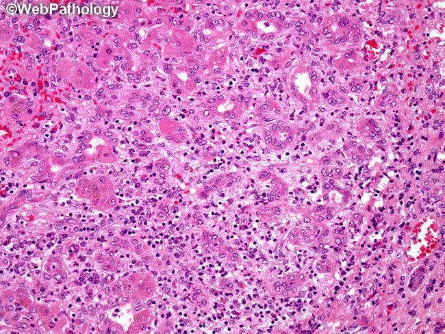
Light microscopic liver histology from a biopsy in acute hepatitis B shows portal areas (portal triads) with dense mononuclear inflammatory infiltrates, predominantly T lymphocytes, often with plasma cells. Inflammation extends from the portal tract into adjacent parenchyma, producing interface activity and mild lobular disarray. Hepatocytes exhibit cytoplasmic ballooning degeneration and scattered apoptotic bodies (Councilman bodies) in degenerating hepatocytes. Focal hepatocellular necrosis may be present, with preserved overall lobular architecture in early disease. Bile ducts within portal tracts are typically preserved; there is no marked cholestasis or fibrotic scarring at this stage. The pattern is compatible with acute viral hepatitis, most commonly hepatitis B, though similar features can appear in other viral or drug-induced liver injuries. Correlation with serology (HBsAg positivity, anti-HBc IgM, HBV DNA) supports the diagnosis. Clinically, the histology explains transaminase elevation and jaundice associated with acute hepatic injury. Diagnostic significance lies in recognizing portal-centric inflammation, interface hepatitis, and hepatocellular injury as a viral hepatitis pattern; differential diagnoses include acute hepatitis A and C, HDV co-infection, drug-induced liver injury, or autoimmune hepatitis. This image is valuable for medical education, pathology teaching, and radiology–pathology correlation discussions. These features emphasize portal inflammation and hepatocyte injury as diagnostic hallmarks for education purposes.
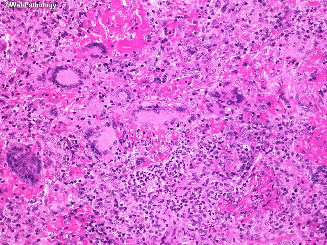
This is a hematoxylin and eosin stained liver biopsy prepared for bright-field light microscopy. Observed tissue shows hepatic parenchyma with preserved lobular architecture disrupted by a diffuse to focal inflammatory infiltrate, predominately mononuclear (lymphocytes and plasma cells) with scattered neutrophils. Inflammatory cells concentrate around portal tracts and within the hepatic lobules, consistent with portal and lobular hepatitis. The hepatocytes display variable cytoplasmic eosinophilia and occasional vacuolization; subtle hepatocellular injury is suggested but conspicuous massive necrosis is not evident at this magnification. Extracellular pink matrix outlines sinusoids and occasional endothelial cell changes; no clear well-formed granulomas are readily apparent, though small aggregates cannot be excluded. The overall pattern raises a differential diagnosis including viral hepatitis (A, B, C, E), autoimmune hepatitis, drug-induced liver injury, alcoholic hepatitis, and less likely granulomatous hepatitis or cholestatic injury. Clinically, such histology can accompany acute or chronic liver inflammation and guides further serology, imaging, and management. The findings emphasize the need for correlation with liver function tests, serology, viral panels, autoantibodies, drug history, and clinical presentation to refine diagnosis and treatment strategy. This image is valuable for education on inflammatory liver disease and differential diagnosis. It remains compatible with multiple pathologies and contributes to teaching differential histology in practice.
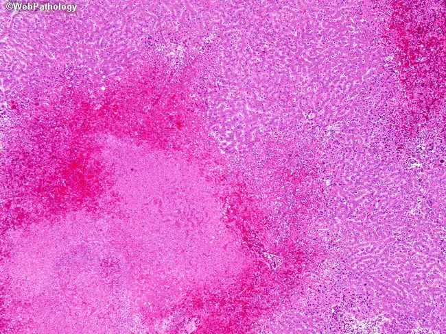
This image depicts a liver biopsy stained with Hematoxylin and Eosin, examined under light microscopy. The hepatic parenchyma exhibits irregular, patchy coagulative necrosis with adjacent hemorrhage and a surprisingly sparse inflammatory response, a histologic hallmark of herpes simplex virus (HSV) hepatitis. The necrotic zones disrupt normal lobular architecture and are conspicuously hemorrhagic, creating mottled pale-to-dark pink areas on the slide. Within viable portal tracts and residual cords, hepatocytes show minimal inflammatory cells, reinforcing the non-destructive inflammatory pattern typical of HSV-related liver injury. Infected hepatocytes may display nuclear viral inclusions (Cowdry type A) and occasional multinucleation, reflecting HSV cytopathic effect. The gross correlation often shows mottling and hemorrhagic liver surface. HSV hepatitis is an uncommon but severe complication of adult HSV-1 or HSV-2 infection and can occur in immunocompetent hosts. Clinically, presentation is nonspecific until acute liver failure ensues; thus histology plays a critical diagnostic role, prompting confirmatory HSV PCR or immunohistochemistry. Early antiviral therapy with acyclovir significantly improves prognosis. This image is educational for pathology education, differential diagnosis in acute liver injury, and recognition of viral cytopathic effects in hepatic tissue. Useful for medical students, residents, and researchers studying viral hepatitis, transplant pathology, and fulminant hepatic failure. Educational utility emphasized.
Jaundice + Transaminitis ~1500 U/L in a Young Girl - Differential Diagnosis
Priority Differentials (Most Important First)
1. Wilson's Disease ⭐ MUST NOT MISS in Young Patients
- Presents in the 2nd decade of life (occasionally up to 40s)
- Can present acutely with jaundice + hemolytic anemia + rapidly rising bilirubin
- Diagnostic clues:
- Low alkaline phosphatase (ALP:Bilirubin ratio <4) - hallmark - ALP is paradoxically LOW despite severe liver injury
- AST:ALT ratio >2.2 - distinguishes Wilson from other ALF causes
- Coombs-negative hemolytic anemia (copper release causes RBC destruction)
- Kayser-Fleischer rings on slit-lamp (usually present in acute Wilson's)
- Low serum ceruloplasmin, high 24-hr urine copper
- Accounts for up to 25% of ALF cases in the young - Sleisenger & Fordtran
- Can deteriorate rapidly to fulminant hepatic failure
2. Acute Viral Hepatitis
| Virus | Key Features |
|---|---|
| Hepatitis A (HAV) | Commonest acute viral hepatitis in India; feco-oral; prodrome of nausea/vomiting/fever; self-limiting; IgM anti-HAV |
| Hepatitis E (HEV) | Common in endemic regions; worse in pregnancy (30% mortality); IgM anti-HEV |
| Hepatitis B (HBV) | Blood/sexual route; HBsAg + IgM anti-HBc; can rarely cause ALF |
| Hepatitis C (HCV) | Rarely causes acute icteric illness; anti-HCV + HCV RNA |
| EBV (Infectious Mononucleosis) | Young patients; pharyngitis + lymphadenopathy + splenomegaly; heterophile antibodies; transaminases usually <500 but can be higher |
| CMV | Immunocompromised or primary infection; CMV IgM/PCR |
| HSV Hepatitis | Rare but severe; fever + skin vesicles absent in 50%; ALF pattern; acyclovir responsive; HSV PCR |
| Dengue | Thrombocytopenia + myalgia; relevant in tropical regions |
3. Autoimmune Hepatitis (AIH) ⭐ CRITICAL in Young Women
- Classic demographic: young to middle-aged women
- Can present as acute fulminant hepatitis mimicking viral hepatitis
- Features: arthralgia, other autoimmune conditions, menstrual irregularity
- ANA, anti-smooth muscle antibody (ASMA), anti-LKM1 positive
- Raised serum IgG
- Biopsy: interface hepatitis, rosetting, plasma cell infiltrate
- Treatable - responds dramatically to corticosteroids
- Important: low-titer autoantibodies can appear non-specifically in any ALF - don't over-diagnose without full criteria
4. Drug/Toxin-Induced Liver Injury (DILI)
| Category | Examples |
|---|---|
| Prescription drugs | Isoniazid, rifampicin (TB treatment), methotrexate, statins, valproate, nitrofurantoin |
| Herbal/Ayurvedic | Very common in India; Kalonji, Brahmi, various churnas |
| Over-the-counter | Paracetamol (even therapeutic doses in malnourished patients), NSAIDs |
| Recreational | Alcohol (acute alcoholic hepatitis), MDMA/ecstasy |
| Supplements | Weight-loss products, gym supplements |
| Mushroom poisoning | Amanita phalloides - severe diarrhea → liver failure 4-5 days later |
5. Ischemic Hepatitis ("Shock Liver")
- Transaminases can reach 1000-5000+ U/L - often the highest values seen
- Due to acute hypoperfusion: cardiac failure, hypotension, PE, sepsis
- Rises sharply within 1-2 days of ischemic event, falls rapidly with recovery
- LDH disproportionately elevated (LDH > AST suggests ischemic etiology)
- Look for: low BP episode, tachycardia, signs of cardiac failure
6. Pregnancy-Related (if applicable)
- Acute Fatty Liver of Pregnancy (AFLP): 3rd trimester; microvesicular steatosis; hypoglycemia, coagulopathy
- HELLP Syndrome: Hemolysis + Elevated Liver enzymes + Low Platelets; hypertension
- Severe pre-eclampsia/eclampsia
7. Budd-Chiari Syndrome
- Hepatic vein thrombosis - young women on OCP especially at risk
- Hepatomegaly (liver enlargement in ALF = unusual → think Budd-Chiari)
- Ascites + RUQ pain + hepatomegaly triad
- Doppler ultrasound is diagnostic
8. Less Common But Important
- Leptospirosis (Weil's disease): fever + myalgia + jaundice + renal failure; raised CK
- Malaria (especially P. falciparum): high fever, thrombocytopenia, smear/RDT
- Lymphoma/malignant infiltration: hepatomegaly; consider if no other cause
- Celiac disease: rare association with transaminitis
- Thyrotoxicosis: rarely causes transaminitis
Diagnostic Approach - Investigations to Order
Tier 1 - Screen in ALL:
| Test | Targets |
|---|---|
| LFT full (AST, ALT, ALP, GGT, bilirubin, albumin) | Pattern of injury |
| PT/INR + aPTT | Severity (coagulopathy = ALF) |
| CBC + peripheral smear | Hemolysis (Wilson's), thrombocytopenia (dengue/HELLP) |
| Blood glucose | Hypoglycemia in ALF |
| Serum creatinine, urea, electrolytes | Hepatorenal involvement |
| IgM anti-HAV, IgM anti-HEV | Commonest viral causes in India |
| HBsAg + IgM anti-HBc + HBV DNA | Acute HBV |
| ANA, ASMA, anti-LKM1 + serum IgG | Autoimmune hepatitis |
| Serum ceruloplasmin + 24-hr urine copper | Wilson's disease |
| Urine/blood for paracetamol levels | Drug toxicity |
| USG abdomen + Doppler | Liver size, Budd-Chiari, biliary pathology |
Tier 2 - If suspected:
| Test | Targets |
|---|---|
| Slit-lamp examination | Kayser-Fleischer rings (Wilson's) |
| Monospot/EBV IgM, CMV IgM | Viral (EBV/CMV) |
| HCV RNA (not antibody - too early) | Acute HCV |
| HSV PCR | Herpes hepatitis |
| NS1 antigen, dengue IgM | Dengue |
| Urine pregnancy test + uric acid | AFLP if pregnant |
| LDH | Disproportionate elevation → ischemic hepatitis |
| Echocardiogram | Cardiac cause of ischemia |
Key Differentiating Clues
| Finding | Points Toward |
|---|---|
| Low ALP + hemolytic anemia + young age | Wilson's disease |
| Female + high IgG + ANA positive | Autoimmune hepatitis |
| Fever + feco-oral exposure + jaundice | Hepatitis A/E |
| Drug history (TB drugs, herbal) | DILI |
| OCP use + hepatomegaly + ascites | Budd-Chiari |
| Very high LDH + hypotension episode | Ischemic hepatitis |
| Pregnant + 3rd trimester + hypoglycemia | AFLP |
| Fever + thrombocytopenia + myalgia | Dengue / Leptospirosis |
Sources: Sleisenger & Fordtran's GI and Liver Disease; Goldman-Cecil Medicine 22nd ed.; Rosen's Emergency Medicine