
analyse_media
The media analysis returned encoded data rather than the text. I can read the handwritten notes directly from the image. Let me now consult the medical library for this case.
Quick summary from the image:
This is a 15-year-old male from Kalyan with a 2-month history of bilateral lower limb weakness, insidious onset, gradually progressive, with later difficulty maintaining balance. He visited a nearby hospital (treatment noted as DNA - did not attend/no response). He then went to another hospital where investigations on 3/7/26 included a Nerve Conduction Study (NCS) showing sensorimotor demyelinating with secondary axonal polyneuropathy affecting bilateral upper and lower limbs. MRI Dorso-lumbar spine showed subtle T2 hyperintense signal intensity in the posterior aspect of the thoracic cord T5-T11 without contrast enhancement or cord expansion - suggestive of myelopathy.
Searching sources...
Reading File
Finding Sources
Finding Sources
Searching PubMed
demyelinating polyneuropathy myelopathy adolescent
Reading File
Reading File
Finding Sources
Reading File
Finding Sources
Finding Sources
Reading File
Finding Sources
Loading Image
Finding Sources
I now have sufficient information to provide a thorough clinical analysis of this case. Let me compile the response.
Clinical Case Analysis
Transcribed Clinical Notes
Present Medical History:
- Patient: 15-year-old male, resident of Kalyan
- Presenting complaint: 2 months history of weakness in bilateral lower limbs
- Onset: Insidious, gradually progressive
- After a few days (due to this): difficulty maintaining balance while walking
- Went to a nearby hospital; doctor gave medicine (DNA - did not attend/no response/DNE noted), no improvement
- Went to another hospital for investigation on 3/7/26
Investigations:
-
Nerve Conduction Study (NCS): Evidence of sensorimotor demyelinating with secondary axonal polyneuropathy affecting bilateral upper and lower limbs
-
MRI Dorso-lumbar Spine: Subtle T2 hyperintense signal intensity in the posterior aspect of thoracic cord T5 to T11, without contrast enhancement or cord expansion - findings suggest myelopathy
Clinical Analysis
Dual Pathology: Combined Myelopathy + Polyneuropathy
This 15-year-old presents with a striking combination of:
- Peripheral: Sensorimotor demyelinating polyneuropathy with secondary axonal involvement (NCS proven, bilateral upper and lower limbs)
- Central: T2 hyperintensity in posterior thoracic cord T5-T11 without enhancement or cord expansion (MRI-proven myelopathy)
This combination - simultaneous central myelopathy + peripheral demyelinating neuropathy - in a teenager is a classic diagnostic setup. The key differential diagnoses are:
Priority Differential Diagnoses
1. Subacute Combined Degeneration (SCD) - Vitamin B12 Deficiency (TOP PRIORITY)
This is the most likely unifying diagnosis.
Why it fits:
- Posterior column T2 hyperintensity on MRI (T5-T11, no enhancement, no expansion) - exactly matching the "inverted V" or posterior column signal described in SCD
- Combined myelopathy + peripheral demyelinating polyneuropathy - SCD causes both simultaneously
- Gradually progressive course over 2 months
- Adolescent with possible nutritional deficiency (vegetarian diet common in this region)
- NCS showing demyelinating polyneuropathy is consistent - B12 deficiency demyelinates both central and peripheral myelin
As noted in Bradley and Daroff's Neurology: "Subacute combined degeneration represents the most common form of metabolic myelopathy. Vitamin B12 deficiency results in demyelination and eventually degeneration of the lateral and dorsal columns of the spinal cord... T2-weighted images show hyperintense signal change, typically involving the dorsal columns... there is no enhancement after gadolinium."
The MRI image below (from a confirmed B12-deficiency SCD case) shows the same posterior column T2 signal - nearly identical to what this patient's MRI demonstrates:
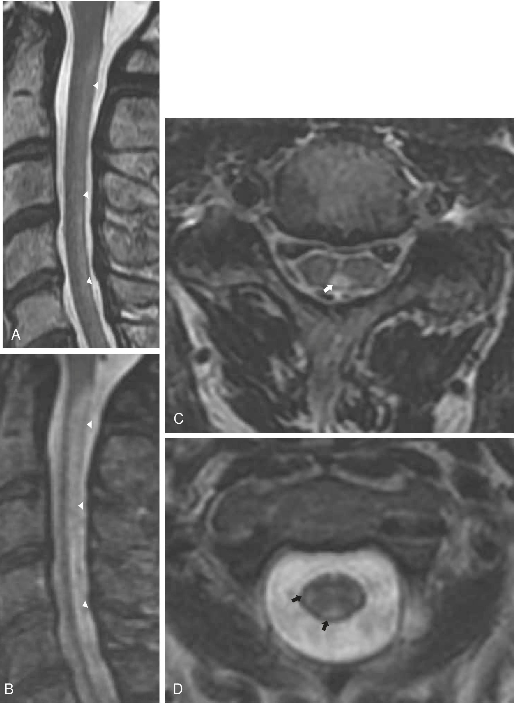
A/B: Sagittal T2 - longitudinal hyperintense signal in posterior columns. C/D: Axial T2 - posterior and lateral column hyperintensity (arrows).
2. CIDP (Chronic Inflammatory Demyelinating Polyneuropathy) + Concurrent CNS Demyelination
- CIDP explains the NCS findings perfectly (sensorimotor demyelinating, bilateral, with secondary axonal change)
- However, CIDP typically does NOT involve the spinal cord on MRI; CNS involvement would suggest an overlap syndrome (e.g., CIDP + MS or NMO)
- Less likely in a 15-year-old as a primary explanation for both findings
3. Neuromyelitis Optica Spectrum Disorder (NMOSD) + Peripheral Neuropathy
- NMOSD can cause longitudinally extensive transverse myelitis, but typically with cord expansion and enhancement
- T5-T11 is a long segment - NMO-IgG (anti-AQP4 antibody) should be tested
- Peripheral demyelinating NCS findings are unusual for NMOSD
4. Copper Deficiency Myelopathy
- Produces identical MRI picture to SCD (posterior column T2 signal)
- Can also cause peripheral neuropathy
- Less common but must be considered, especially if B12 is normal
5. Infectious / Parainfectious (HTLV-1, HIV, Lyme)
- HIV-associated myelopathy + neuropathy is a classic combination
- HTLV-1 myelopathy (tropical spastic paraparesis) - endemic in some parts of India
- Check serology
6. Hereditary/Genetic (Adrenomyeloneuropathy, Friedreich Ataxia)
- Both can cause myelopathy + neuropathy in young patients
- Usually more slowly progressive; genetic testing warranted if metabolic causes excluded
Recommended Next Workup
| Investigation | Rationale |
|---|---|
| Serum Vitamin B12 | Rule out/confirm SCD - first priority |
| Serum Homocysteine + Methylmalonic acid | Functional B12 deficiency markers (more sensitive) |
| Serum Copper + Ceruloplasmin | Copper deficiency myelopathy |
| CBC + Peripheral smear | Macrocytic anaemia with B12 deficiency |
| Anti-AQP4 antibody (NMO-IgG) | NMOSD |
| Anti-MOG antibody | MOG-antibody disease |
| HIV, HTLV-1 serology | Infectious myelopathy+neuropathy |
| CSF analysis | Elevated protein in CIDP (>100 mg/dL); oligoclonal bands for MS/demyelination |
| Nerve biopsy | If NCS pattern unclear or CIDP diagnosis uncertain |
| VLCFA (Very Long Chain Fatty Acids) | Adrenomyeloneuropathy in young males |
| MRI Brain | Look for supratentorial/juxtacortical lesions (MS/NMO) |
| MRI entire spine with contrast | Map full extent of myelopathy |
Key Diagnostic Clues from MRI Description
The MRI finding - posterior aspect T2 hyperintensity T5-T11, no enhancement, no expansion - is the classic signature of:
- Subacute combined degeneration (B12 / copper deficiency) - posterior > lateral columns, long segment, no enhancement
- Distinguishes from inflammatory myelitis (which typically shows enhancement in acute phase and cord swelling)
- Distinguishes from MS plaques (which are typically smaller, patchy, asymmetric)
Most Likely Working Diagnosis
Subacute combined degeneration of the spinal cord (Vitamin B12 deficiency) with peripheral demyelinating polyneuropathy - pending serum B12, homocysteine, and MMA levels.
If B12 and copper are normal, CIDP with concurrent CNS demyelination (MS/NMO overlap) moves to the top. Anti-AQP4 and CSF studies are then key.
Urgency: Check B12 immediately - treatment with intramuscular B12 (hydroxocobalamin 1 mg IM daily for 1 week, then weekly for 4 weeks, then monthly for life) can halt and partially reverse deficiency-related myelopathy if started early.
Sources: Bradley and Daroff's Neurology in Clinical Practice; Adams and Victor's Principles of Neurology, 12th Ed; Harrison's Principles of Internal Medicine 22E (2025, McGraw Hill)
This is a shared conversation. Sign in to Orris to start your own chat.