
why women affected by thyroid disease more rhan man always
thyroid autoimmunity sex differences women estrogen
why are women more susceptible to thyroid disease than men biological reasons
thyroid autoimmune disease Hashimoto Graves female sex difference diagram
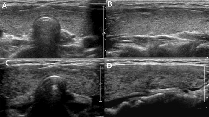
Diagnostic ultrasound comparison of the thyroid gland in a 16-year-old female patient, demonstrating the progression of autoimmune thyroid disease over two years. The image consists of four panels: transverse (A) and longitudinal (B) views from the initial scan, and transverse (C) and longitudinal (D) views from the two-year follow-up. Initial images (A, B) show a diffusely enlarged thyroid gland with mild heterogeneity involving less than one-third of the parenchyma (Grade 2), maintaining relatively normal echogenicity. The follow-up images (C, D) demonstrate marked progression (Grade 4), characterized by severe diffuse heterogeneity and pronounced hypoechogenicity, where the thyroid tissue appears darker than the adjacent strap muscles. These visual changes correlate with the clinical transition from a euthyroid state to overt hyperthyroidism. The series serves as an educational example of how ultrasonographic features like echogenicity and parenchymal texture can be used to grade the severity of thyroid dysfunction in conditions such as Graves' disease or Hashimoto's thyroiditis.
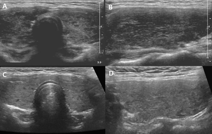
Diagnostic ultrasound images of the thyroid gland in a 14-year-old female patient, demonstrating longitudinal and transverse planes across two time points. Panel A (transverse) and B (longitudinal) represent the initial assessment, showing an enlarged thyroid with Grade 4 (G4) changes characterized by marked hypoechogenicity and diffuse heterogeneity compared to the anterior strap muscles. The parenchyma appears coarse with irregular, low-echo areas. Panels C (transverse) and D (longitudinal) show a follow-up ultrasound one year later. These images demonstrate improvement to Grade 3 (G3), where the thyroid exhibits reduced heterogeneity and improved echogenicity (becoming more isoechoic to the strap muscles) with a smoother, more uniform parenchymal texture. The study illustrates the radiological progression and correlation between ultrasound grading of autoimmune thyroid disease (such as Hashimoto's thyroiditis or Graves' disease) and clinical thyroid function improvement from overt hypothyroidism to euthyroidism.
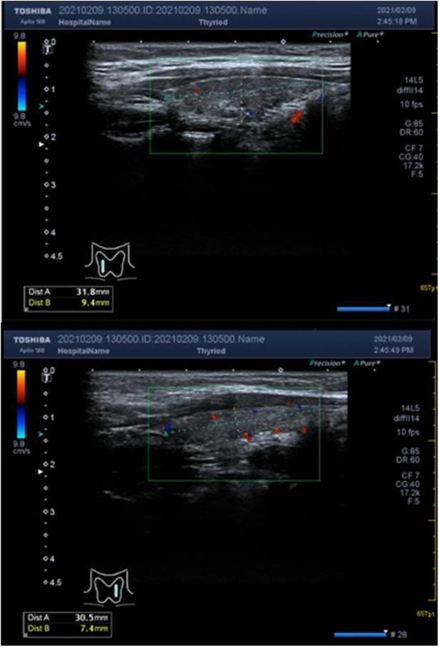
This diagnostic image consists of two sagittal-view ultrasound frames of the thyroid gland utilizing color Doppler. The imaging depicts the right and left lobes of the thyroid, showing a characteristic hypoechoic and heterogeneous parenchymal echogenicity. The texture of the gland appears non-uniform and darker relative to adjacent musculature, which is a common sonographic finding in autoimmune thyroiditis (Hashimoto's thyroiditis). Annotations and dashed measurement lines indicate the dimensions of the lobes, with the superior-inferior diameters measuring approximately 31.8 mm and 30.5 mm respectively. The color Doppler overlays (red and blue signals within the green sampling box) demonstrate normal vascularity without evidence of pathological hypervascularity (the 'thyroid inferno' pattern). There are no discrete solid or cystic nodules, masses, or occupancy areas visible in either lobe. This visual presentation is educationally significant for identifying parenchymal changes in chronic thyroid inflammation and differentiating normal blood flow from the hypervascularity seen in Graves' disease.
Why Women Get Thyroid Disease Far More Than Men
1. Women Have Stronger Immune Systems (Double-Edged Sword)
- Higher antibody production
- More robust T-helper cell (Th1/Th2) activation
- Stronger innate immune signaling
2. Estrogen Actively Amplifies Autoimmunity
- Activates B cells and T cells, promoting antibody production
- Stimulates neutrophil immunometabolism
- Increases thyroid-binding globulin (TBG) levels in the blood, altering thyroid hormone transport and metabolism
3. The X Chromosome - Double Dose of Immune Genes
- Women have two X chromosomes (XX); men have one (XY)
- Many critical immune-regulation genes sit on the X chromosome, including:
- TLR-7 (Toll-like receptor 7) - recognizes self-RNA and triggers type I interferons
- TLR-8
- IRAK-1
- In normal cells, one X chromosome is inactivated (a process called X-inactivation or lyonization). But this inactivation is incomplete and sometimes skewed - some immune cells end up expressing both copies of TLR-7
- Double TLR-7 expression means the immune system is more easily triggered by self-antigens, increasing autoimmune risk
4. Pregnancy and Postpartum Effects
- The immune system must tolerate a genetically foreign fetus
- Fetal cells can cross into the mother's circulation and persist for years - a phenomenon called fetal microchimerism
- These foreign cells may trigger autoimmune responses after delivery
- Postpartum thyroiditis (thyroid inflammation after childbirth) affects up to 5-10% of women and is essentially an immune rebound after the immune suppression of pregnancy
5. Specific Thyroid Diseases and Their Female Predominance
| Disease | Female:Male Ratio |
|---|---|
| Hashimoto's thyroiditis | 10:1 to 20:1 (Robbins Pathology) |
| Graves' disease | 8:1 (Sabiston Textbook of Surgery) |
| Toxic multinodular goiter | 5:1 |
| Autoimmune thyroid disease overall | 5-10:1 |
Thyroid Ultrasound - What the Damage Looks Like
Summary Table
| Mechanism | How It Raises Risk in Women |
|---|---|
| Stronger immune response | More antibody production, more T-cell activation |
| Estrogen effect | Activates B/T cells, suppresses tolerance |
| Low androgens | No natural immune suppression |
| Double X chromosome (XX) | Extra TLR-7/TLR-8 gene copies, incomplete X-inactivation |
| Pregnancy/postpartum | Fetal microchimerism, postpartum immune rebound |
| Hormonal transitions | Puberty, menopause as trigger points |